Cephalometrics
Dr.Omar .S.M.J.AliIntroduction
Origin: ‘Cephalo’ means head and ‘Metric’ is measurementDiscovery of X-rays measurement of the head from shadows of bony and soft tissue landmarks on the image
Definitions
“The scientific measurement of the bones of the cranium and face, utilizing a fixed, reproducible position for lateral radiographic exposure of skull and facial bones” -- Moyers“ A scientific study of the measurements of the head with relation to specific reference points; used for evaluation of facial growth and development, including soft tissue profile” -- Grabers
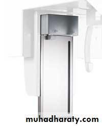
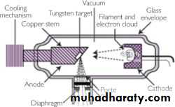
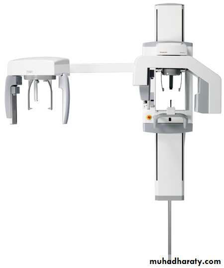
Cephalometric Imaging System
X- ray apparatusAn image receptor
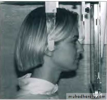
Cephalostat
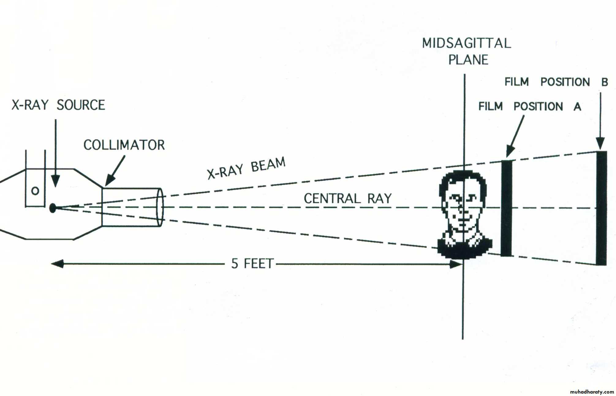

15 cm
Uses of cephalometricIn orthodontic diagnosis & treatment planning
In classification of skeletal & dental abnormalities
In establishing facial types
In evaluation of treatment results
In predicting growth related changes & changes associated with surgical treatment
Valuable aid in research work involving the cranio-dentofacial region
-- Moyers
Principle of Cephalometric analysis
To compare the patient with a normal reference group, so that differences between the patient’s actual dentofacial relationships and those expected for his/her racial or ethnic groups are revealed-- Jacobson
Goals of CephalometricsTo evaluate the relationships, both horizontally and vertically, of the five major functional components of the face:
The cranium and the cranial base
The skeletal maxilla
The skeletal mandible
The maxillary dentition and the alveolar process
The mandibular dentition and the alveolar process
-- Jacobson
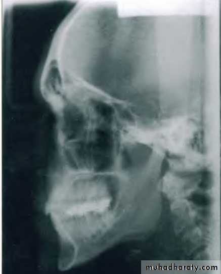
Types of cephalogramsLateral cephalometric
Taken with head in a standardized reproducible position at a specific distance from X-ray source
Uses :
Important in orthodontic growth analysisDiagnosis & Treatment planning
Monitoring of therapy
Evaluation of final treatment outcome
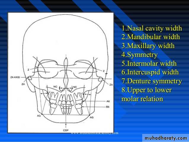
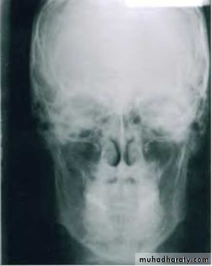
Posteroanterior (p-a) cephalometric radiograph
Image Receptor and Patient Placement:Image receptor is placed in front of the patient
The patient is placed so that the canthomeatal line is perpendicular to the image receptor
Uses :
Provides information related to skull widthSkull symmetry
Vertical proportions of skull, craniofacial complex & oral structuresFor assessing growth abnormalities & trauma
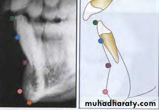
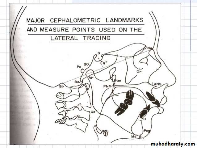
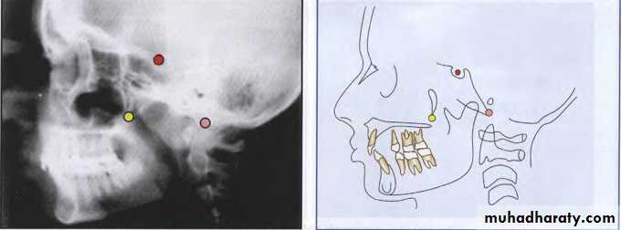
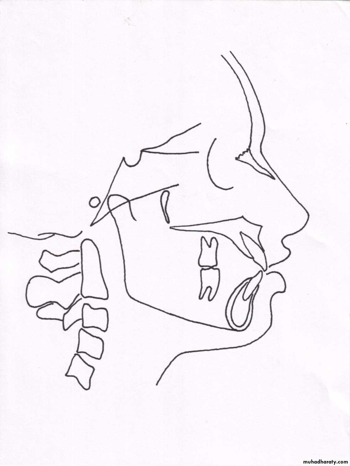
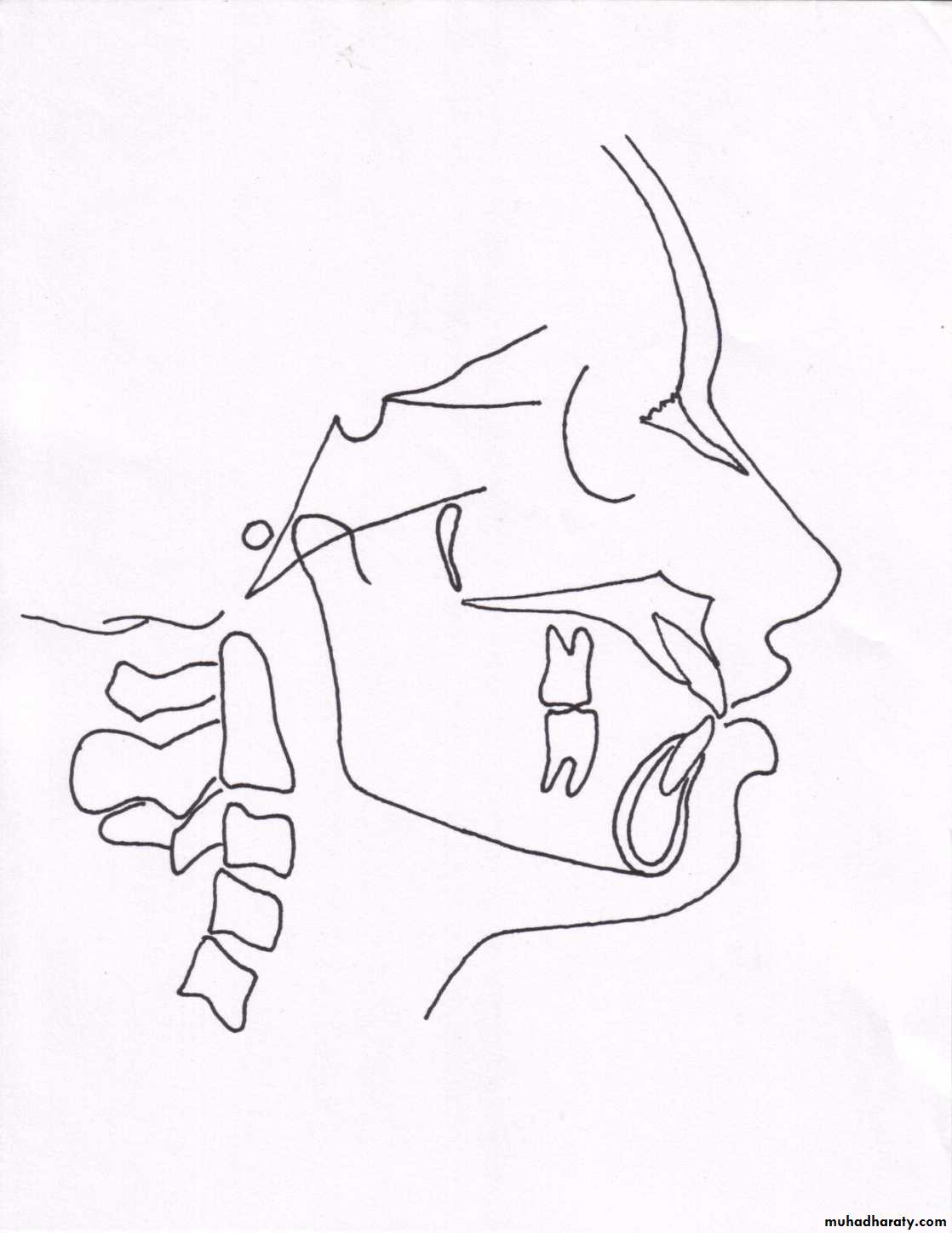
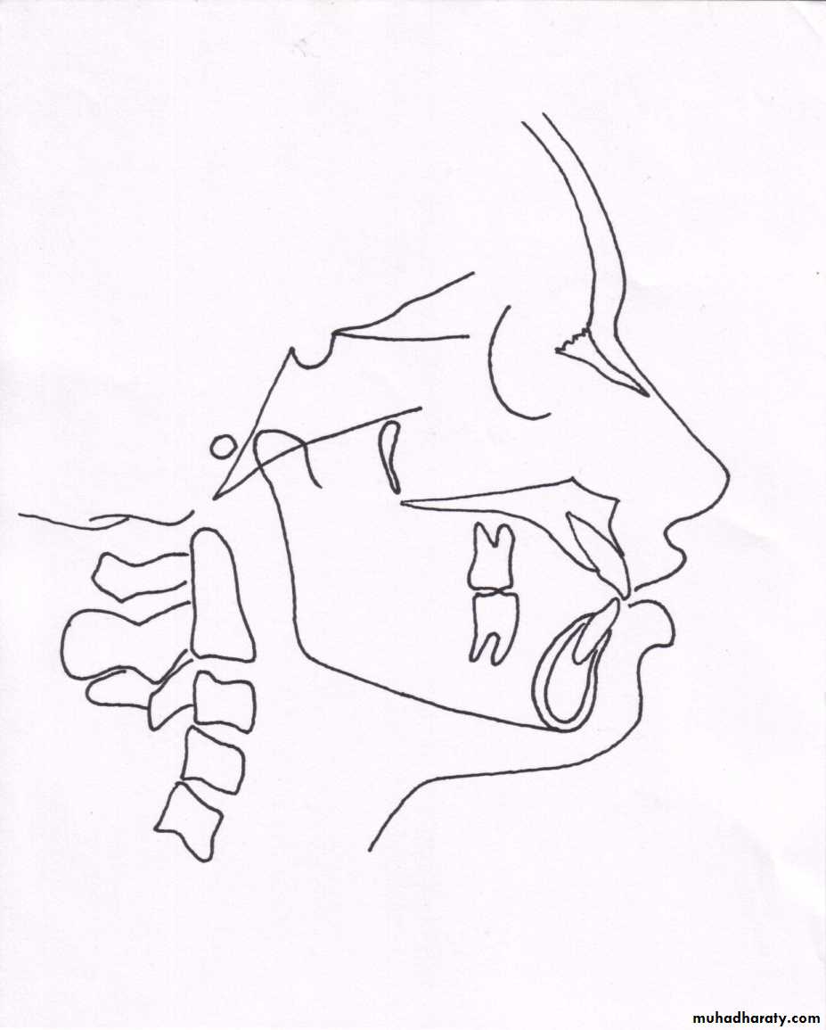
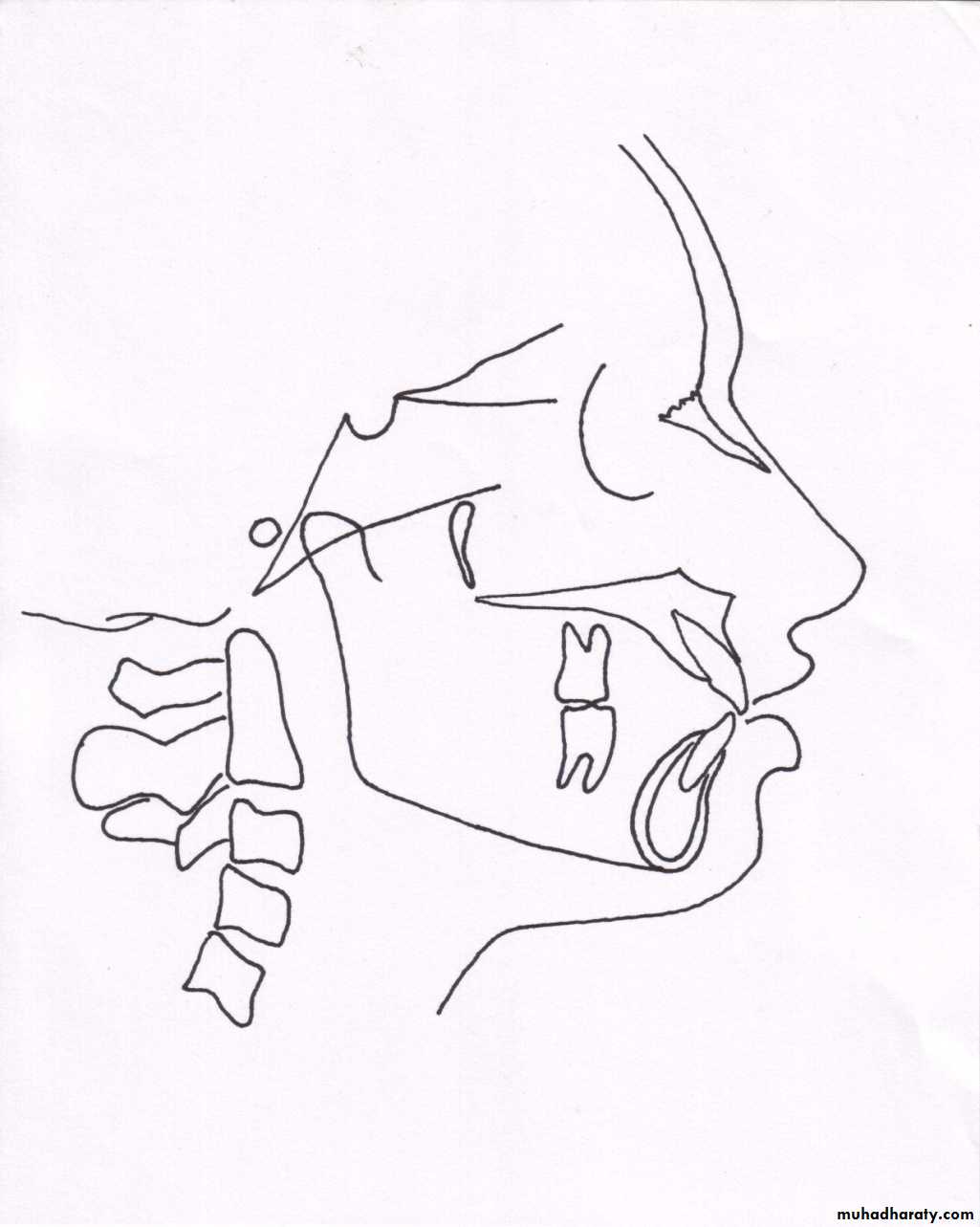
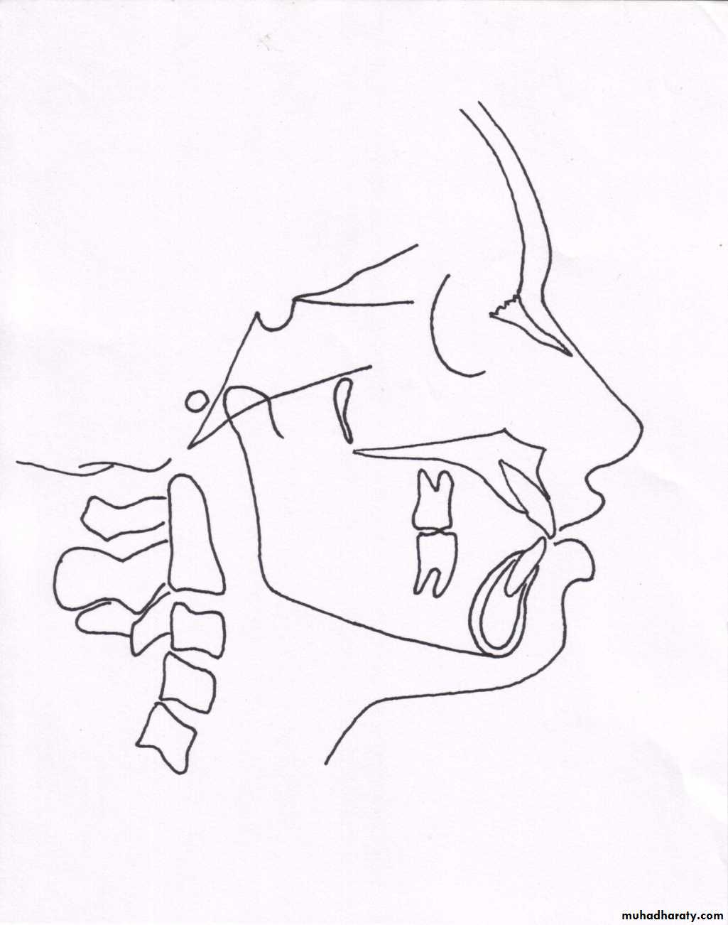
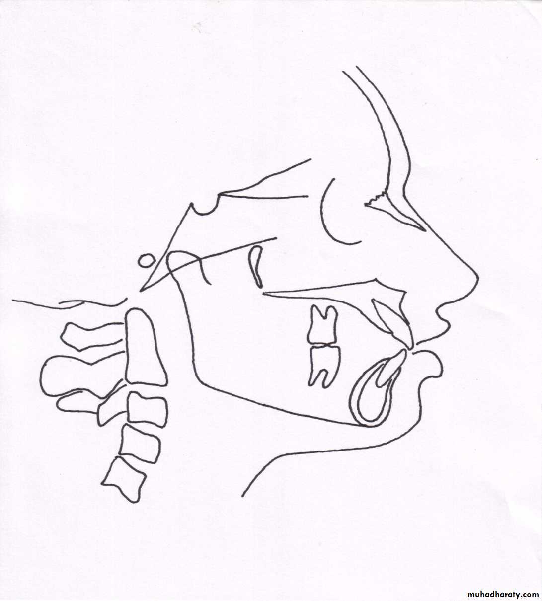
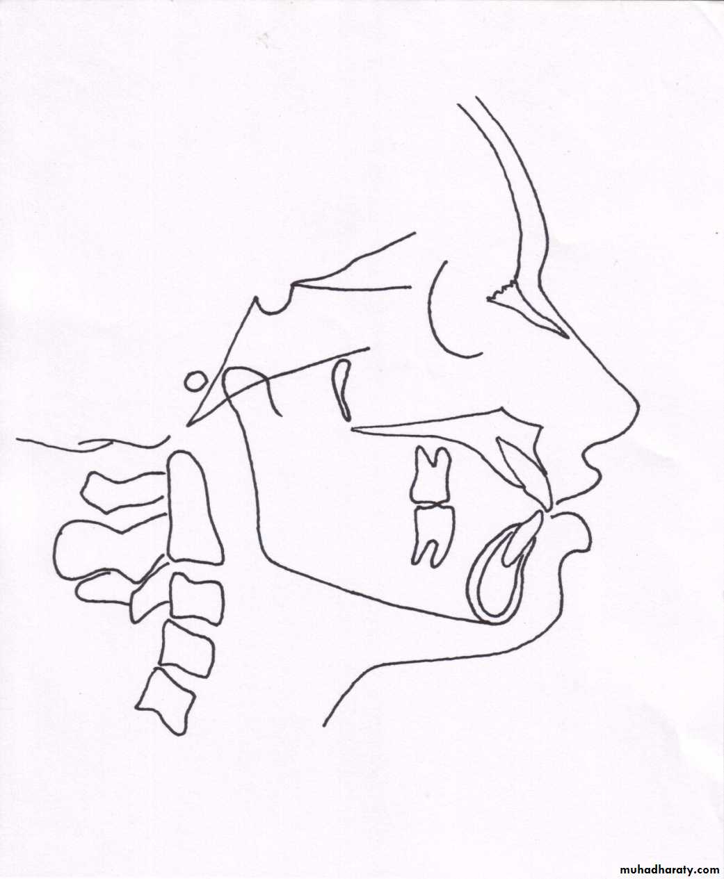
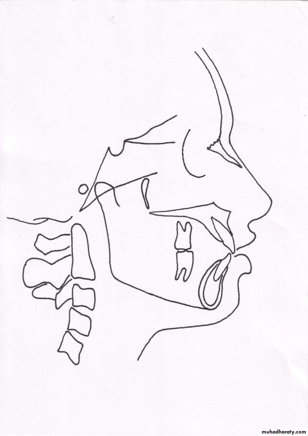
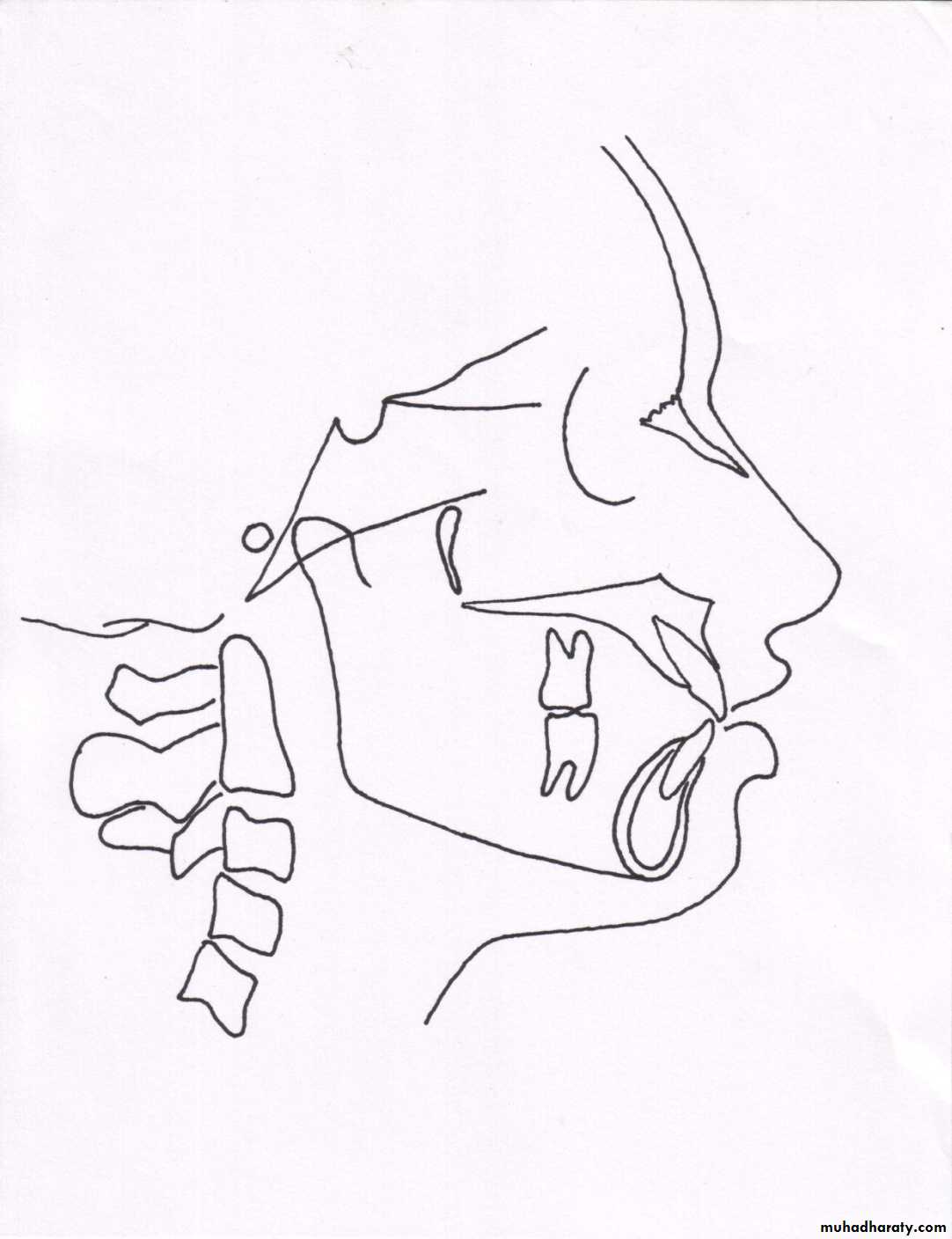
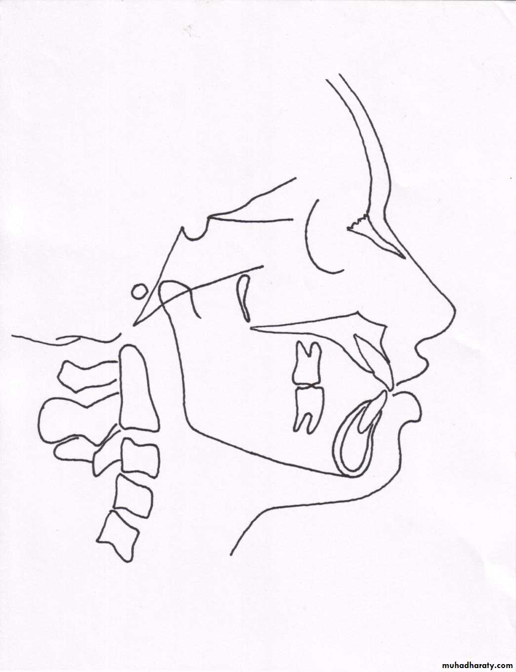
Cephalometric landmarks
A conspicuous point on a cephalogram that serves as a guide for measurement or construction of planes – JacobsonRequisites for a landmark
Should be easily seen on the x ray filmBe uniform in outline
Easily reproducible
Should permit valid quantitative measurement of lines and angles
Lines and planes should have significant relationship to the vectors of growth
Lateral Cephalometric
• Hard tissue landmarks
Nasion (N)The frontonasal suture at its most superior point on
the curve at the bridge of the nose.
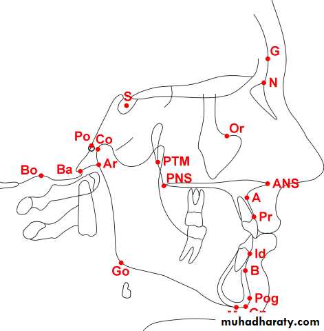
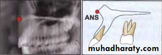
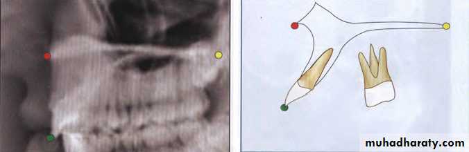
Anterior Nasal Spine (ANS)
The most anterior point on the maxilla at the level ofthe palate.
Superior Prosthion (SPr or PR)
Also termed supradentale. The most anterior inferiorpoint on the maxillary alveolar process.
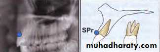
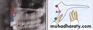
Subspinale ("A" Point)
The most posterior point on the curve between ANSand PR (SPr). "A" point is usually found 2mm anterior
to the apices of the maxillary central incisor root.
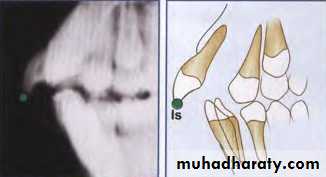
Incision Superius (Is)
The incisal tip of the most anterior maxillary centralincisor.
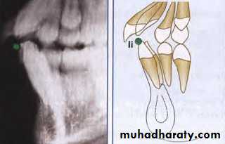
Incision Inferius (Ii)
The incisal tip of the most labial mandibular centralIncisor.
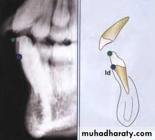
Intradentale (Id)
The most anterosuperior point on the mandibularalveolar process usually found near the CEJ
of the mandibular central incisor. Also
termed inferior prosthion.
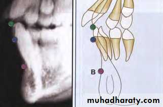
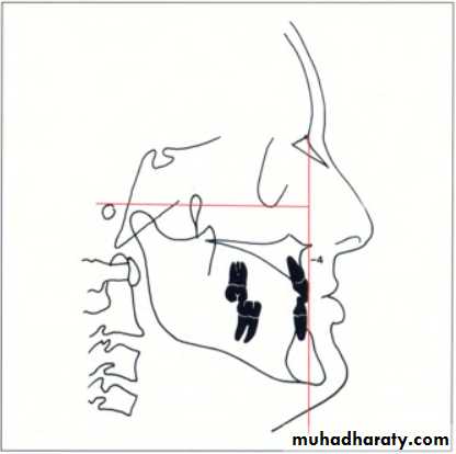
Supramentale (« B » point)
The most posterior point of the bony curvature of themandible below infradentale and above Pogonion. "B"
Point is usually found near the apical third of the roots. of the mandibular incisors and may be obscured
during the eruption of these teeth. When the profile
of the chin is not concave, "B" point cannot be
determined.
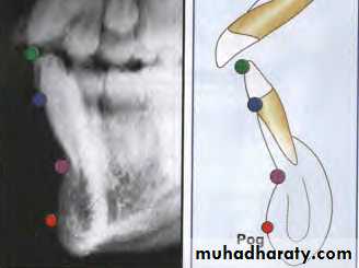
Pogonion (Pog)
Pogonion is the most anterior point on the contour ofthe chin. Pogonion usually is located by a tangent
perpendicular to the mandibular line or a tangent
dropped to the chin from nasion.
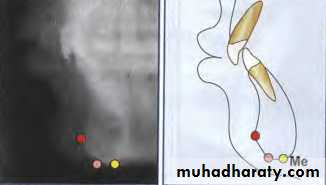
Menton (Me)
Menton is the lowest point on the symphyseal outlineof the chin.
Gnathion (Gn)
The most anteroinferior point on the lateral shadowof the chin. Gnathion may be approximated by the
midpoint between pogonion and menton on the
contour of the chin
Basion (Ba)
The most inferoposterior point in the sagittal planeon the anterior rim of the foramen magnum-the tip
of the posterior cranial base.
Bolton Point (BO)
The highest point in the upward curvature of the
retrocondylar fossa.
Posterior Nasal Spine (PNS)
The most posterior point on the bony hard plate inthe sagittal plane: usually the meeting point of the
inferior and superior surfaces of the hard plate.
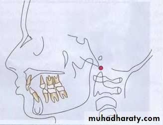
Sella(S)
The center of the hypophyseal fossa (sella turcica). Itis selected by the eye, since that procedure has been
shown to be as reliable as a constructed center.
BILATERAL LANDMARKS
Orbitale (Or)Orbitale has been defined as the lowest point of the
bony orbit. In the PA cephaJogram, each may be
identified but in the lateral cephalograms. the outlines
of the orbital rims overlap. Usually, the lowest point
on the average outline is used to construct the
Frankfort plane.
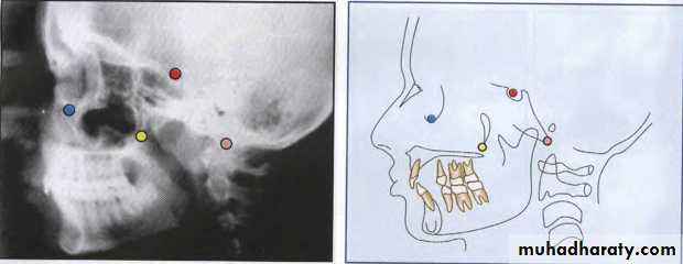
Gonion (Go)
Gonion is the most posteroinferior point at the angleof the mandible. It may be determined by inspection
or by bisecting the angle formed by the junction of the
ramal and mandibular lines, and extending this
bisector through the mandibular border.
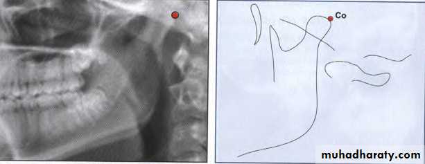
Condylion (Co)
Condylion is the most posterosuperior point on thecondyle of the mandible.
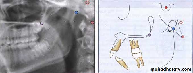
Articulare (Ar)
The intersection of the radiographic shadows:the inferior surface of the cranial base and the posterior
surfaces of the necks of the condyles of the mandible.
Articulare is systematically used for condylion when
the latter is not reliably discernible. Displacement of
the condyle moves the articulare.
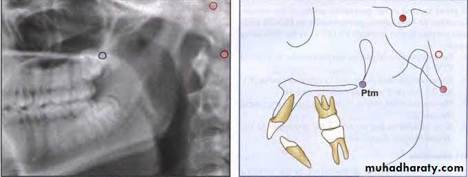
Pterygomaxillary Fissure (Ptm)
A bilateral teardrop-shaped area of radiolucency, theanterior shadow of which is the posterior surfaces of
the tuberosities of the maxilla.
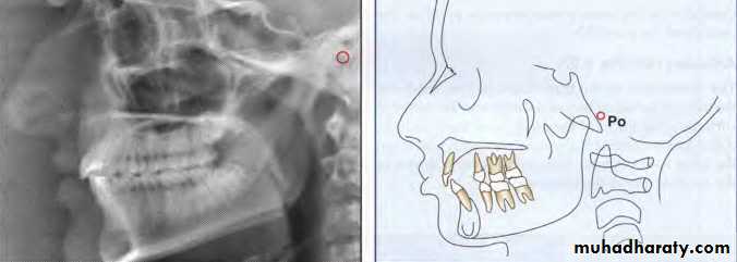
Porion (Po)
The "top" of the external auditory meatus. Sometimes,because porion is quite unreliable, the "top" of the shadow of the ear rods is used, which is known as
“machine porion".
Soft tissue landmarks

Tracing technique
Tracing supplies & equipmentsLateral ceph, usual dimensions of 8 x 10 inches (patients with facial asymmetry requires antero posterior head film)
Acetate matte tracing paper.
A sharp 3H drawing pencil or a very fine felt-tipped pen
Masking tape
A few sheets of cardboard (preferably black), measuring approximately 6 x 12 inches, and a hollow cardboard tube
A protractor and tooth-symbol tracing template for drawing the teeth (optional)
Dental casts trimmed to maximal intercuspation of the teeth in occlusionViewbox (variable rheostat desirable, but not essential)
Pencil sharpener and an eraser
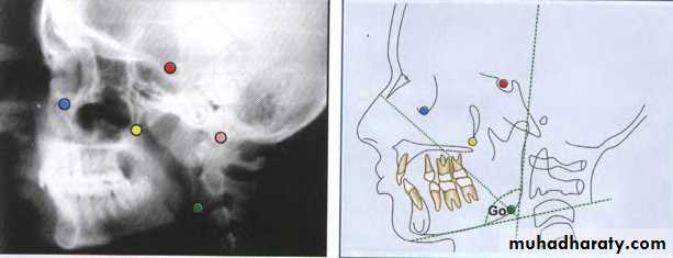
Cephalometric planes
Are derived from at least 2 or 3 landmarksUsed for measurements, separation of anatomic divisions, definition of anatomic structures of relating parts of the face to one another
Classified into horizontal & vertical planes
Horizontal planes
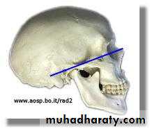
Frankfurt Horizontal plane
Porion-orbitale.
P
O
Sella-Nasion plane
S
N
Basion-Nasion plane:
Palatal plane:
Occlusion plane:
Ba
N
ANS
PNS
Mandibular plane: Different definitions are given in different analysis
1. Tweed- Tangent to lower border of the mandible2. Downs analysis – extends from Go to Me
3. Steiner’s anlysis – extends from Go to Gn
Go
Gn
Me
Vertical planes
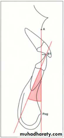
Facial plane
A-Pog line
Facial axis
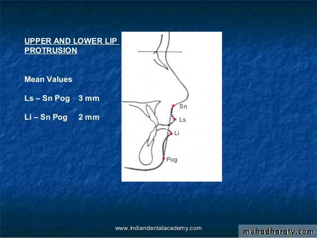
E. plane (Esthetic plane)
Ptm
Gn
N
Pog
A
E plane
Measurement analysis :
Downs analysisSteiner analysis
Tweed analysis
Wits appraisal
Rickets analysis
Mc Namara analysis
Holdaway soft tissue analysis
MEASUREMENT ANALYSIS
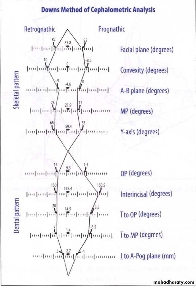
DOWN’S ANALYSIS
Given by WB Downs, 1925
One of the most frequently used cephalometric analysis after St analysis
Based on findings on 20 caucasian individuals of 12-17 yrs age group belonging to both the sexes
Consists of 10 parameters of which 5 are skeletal & 5 are dental
Skeletal parameters :Facial angle
Average value is 87.8°, Range 82-95°
Gives an indication of anteroposterior positioning of mandible in relation to upper faceMagnitude increases in skeletal class 3 cases, decreases in skeletal class 2 cases
FH plane
NPog
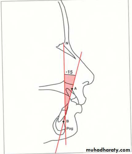
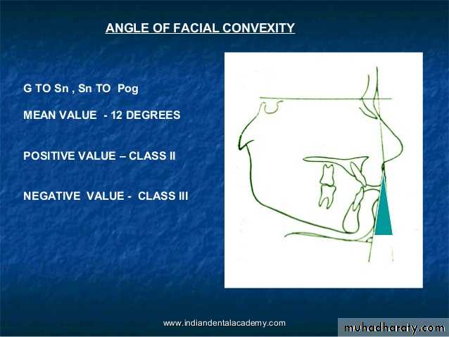
Angle of convexity
Reveals convexity or concavity of skeletal profile
Average value 0°, Range = -8.5 to 10°Positive angle or increased angle – prominent maxillary denture base relative to mandible
Decreased angle , negative angle.prominent of mand.

N
A
Pog
A-B plane angle
Mean value = -4.6°, Range = -9 to 0°Indicative of maxillary mandibular relationship in relation to facial plane
Positive angle in class 3 malocclusion
Mandibular plane angle
Mean value = 21.9°, Range = 17 to 28°
Increased mandibular plane angle suggestive of vertical grower with hyperdivergent facial pattern
FHplane
GoMe
Y- axis (growth axis)
Mean value = 59° , range = 53 to 66°Angle is larger in class 2 facial patterns than in class 3 patterns
Indicates growth pattern of an individual
Angle greater than normal – vertical growth of mandible
Angle smaller than normal – horizontal growth of mandible
S
Gn
FH plane
Dental parameters
Cant of occlusal plane
occlusal plane to FH
Mean value = 9.3° , Range = 1.5 to 14°
Gives a measure of slope of occlusal plane relative to FH plane

FH plane
Inter- incisal angleAverage reading = 135.4° ,
Angle decreased in class 1 bimaxillary protrusion & class 2 div 1 malocculsion
Increased in class II div II case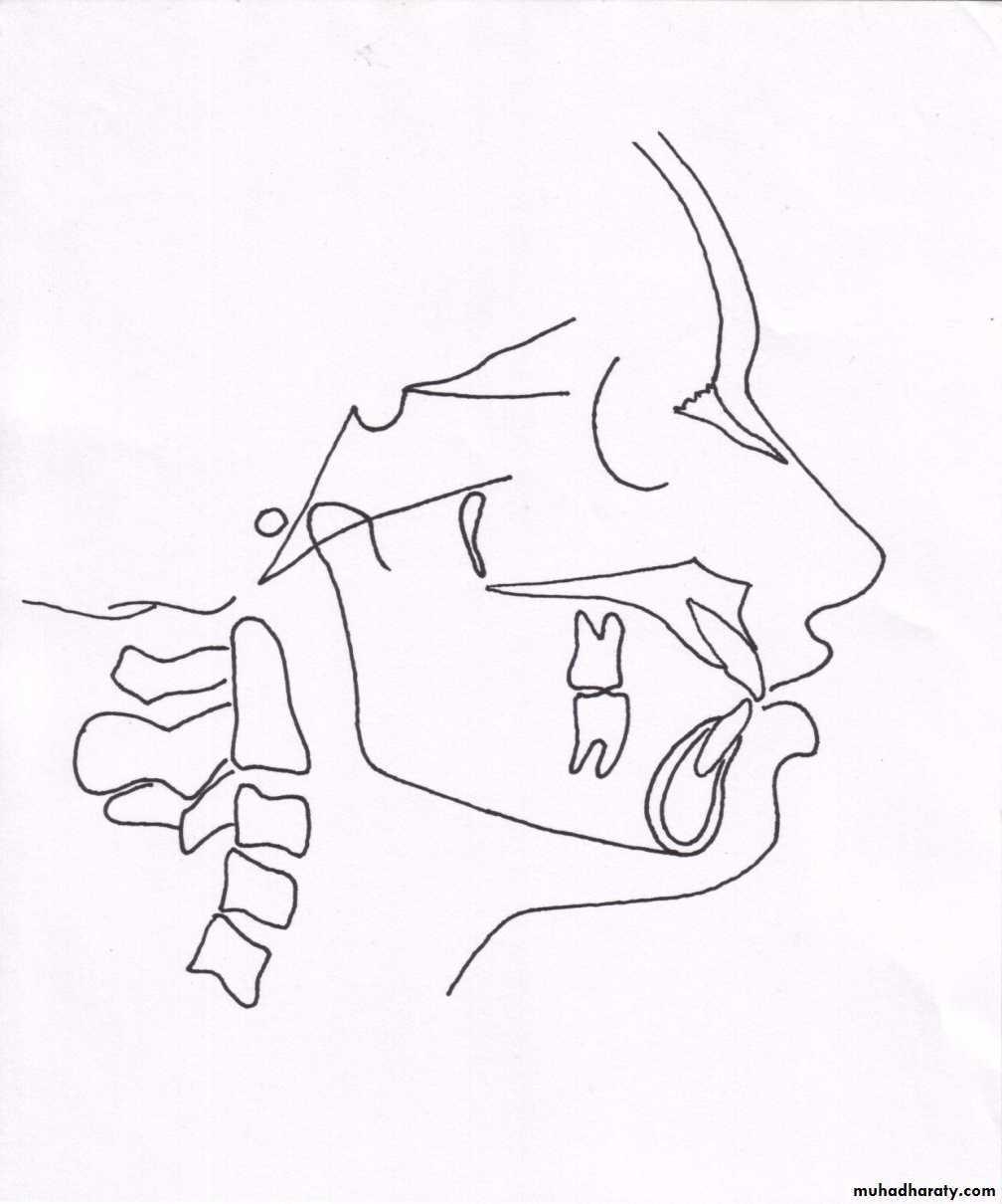
Incisor occlusal plane angle
Average value = 14.5°, range = 3.5 to 20°
Increase in the angle is suggestive of increased lower incisor proclination
Incisor mandibular plane angle
Mean angulation is 1.4, range = -8.5 to 7°Increase in angle is indicative of lower incisor proclination
Upper incisor to A-Pog line
Average distance is 2.7mm (range -1 to 5 mm)Measurement is more in patients with upper incisor proclination
Limitations of Downs analysis
Too many landmarksToo many measurements
Time consuming
-- Jacobson
STEINER ANALYSIS
Developed by Steiner CC in 1930 with an idea of providing maximal information with the least no. of measurementsDivided the analysis into 3 parts
SkeletalDental
Soft tissue
Skeletal analysis
S.N.A angleIndicates the relative antero-posterior positioning of maxilla in relation to cranial base
>82° -- prognathic maxilla (Class 2)
< 82°– retrognathic maxilla (class 3)
S
N
A
Mean value -- 82°
S.N.B angle
Indicates antero-posterior positioning of the mandible in relation to cranial base> 80°-- prognathic mandible
< 80°-- retrusive mandible
S
N
B
Mean value-- 80°
A.N.B angle
Denotes relative position of maxilla & mandible to each other> 2° –- class 2 skeletal tendency
< 2°–- skeletal class 3 tendency
A
N
B
Mean value = 2°
Mandibular plane angle
Gives an indication of growth pattern of an individual< 32° -- horizontal growing face
> 32°– vertical growing individualS
N
Mean value = 32°
Occlusal plane angle
Mean value = 14.5°Indicates relation of occlusal plane to the cranium & face
Indicates growth pattern of an individualS
N
Dental analysis
Upper incisor to N-A(angle)Normal angle = 22°
Angle indicates relative inclination of upper incisorsIncreased angle seen in class 2 div 1 malocclusion
N
A
Upper incisor to N-A ( linear)
Helps in asssessing the upper incisor inclinationNormal value is 4 mm
Increase in measurement – proclined upper incisorsN
A
Inter-incisal angle
< 130 to 131° -- class 2 div 1 malocclusion or a class 1 bimax> 130 to 131° – class 2 div 2 malocclusion
Mean value = 130 to 131°
Lower incisor to N-B (angle)Indicates inclination of lower central incisors
>25 °-- proclination of lower incisors< 25 °– retroclined incisors
N
B
Mean value of 25 °
Lower incisor to N-B (linear)
Helps in assessing lower incisor inclinationIncrease in measurement indicates proclined lower incisors
Normal value– 4mm
N
B
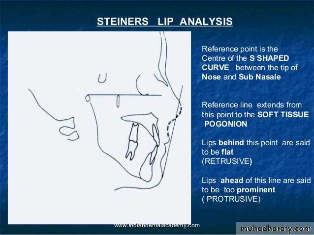
Soft tissue analysis
S line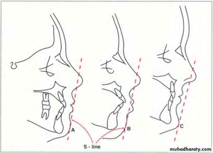


TWEED ANALYSIS
Given by Tweed CH, 1950• Used 3 planes to establish a diagnostic triangle --
• Frankfurt horizontal plane
• Mandibular plane
• Long axis of lower incisor
• Determines position of lower incisor
FMPA = 25 °
IMPA = 90 °FMIA = 65 °
FH planeMand plane
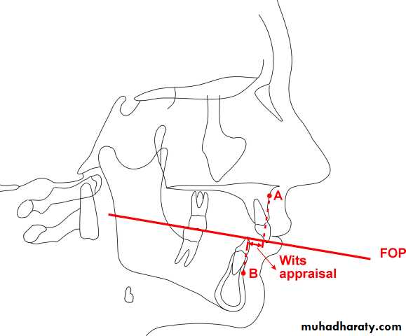
WITS APPRAISAL
It is a measure of the extent to which maxilla & mandible are related to each other in antero-posterior or sagittal planeUsed in cases where ANB angle is considered not so reliable due to factors such as position of nasion & rotation of jaws
In males point BO is ahead of AO by 1mm
In females point AO & BO coincideIn skeletal class 2 tendency BO is usually behind AO( positive reading)
In skeletal class 3 tendency BO is located ahead of AO ( negative reading)
RICKETTS ANALYSIS
Also known as Ricketts’ summary descriptive analysisGiven by RM Ricketts in 1961
The mean measurements given are those of a normal 9 year old child
The growth dependent variables are given a mean change value that is to be expected and adjusted in the analysis.

Dr. RM Ricketts
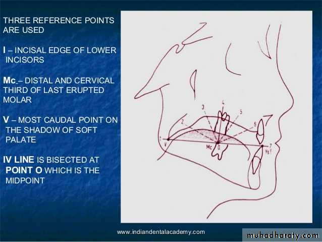
-- JacobsonLandmarks
This is a 11 factor summary analysis that employs specific measurements toLocate the chin in space
Locate the maxilla through the convexity of the faceLocate the denture in the face
Evaluate the profile
This analysis employs somewhat less traditional measurements & reference points
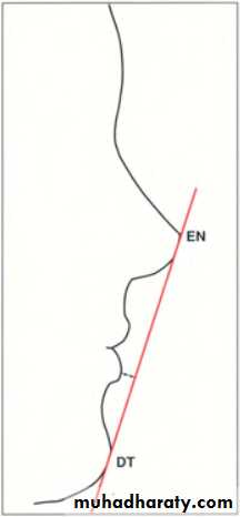
En = nose
DT = soft tissue
Ti = Ti point
Po = Cephalometric
Gn = Gnathion
A6 = upper molar
B6 = Lower molar
Go = gonion
C1 = condyle
DC=articular
CC = Center of cranium
CF = Points from planes at pterygoid
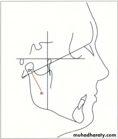
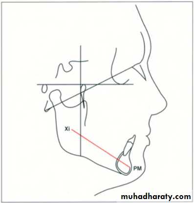
Xi point
A point located at the geometric center of the ramus. Location of Xi is keys of geometrically to Po-Or (FH) and perpendicular through Pt (pterygoid vertical [PtV]; aline perpendicular to FH at the posterior margin of the pterygopalatine fossa) in the following steps:1. Planes perpendicular to FH and PtV are constructed.
2.The constructed planes are tangent to points R1, R2, R3, and R4 on the borders of the ramus.3.The constructed planes form rectangular enclosing the ramus.
4. Xi is located in the center of the rectangle at the intersection of the diagonals.
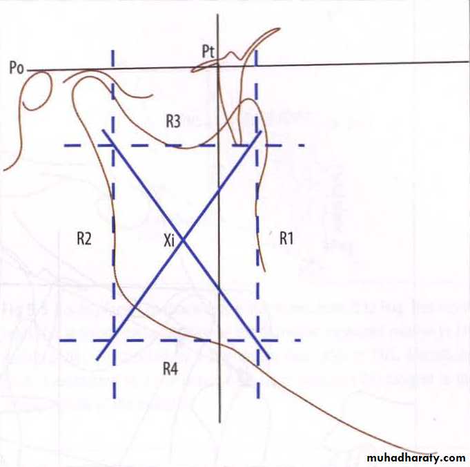
Xi point --

Rl·mand. The deepest point on the curve of the anterior border of the ramus, one half the distance between the inferior and superior curves.
R2- mand. Apoint located on the posterior border of the ramus of the mandible.
R3-mand. Apoint located at the center and most inferior aspect of the sigmoid notch of the ramus of the mandible.R4-mand. Apoint on the lower border of the mandible, directly inferior to the center of the sigmoid notch of the ramus.
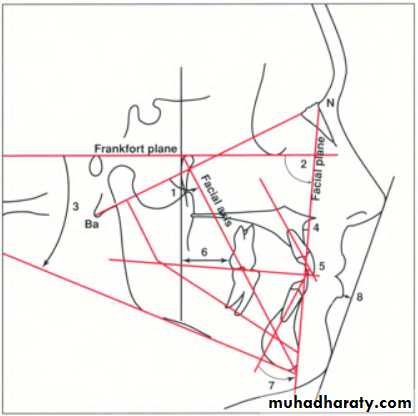
Planes
Frankfurt horizontal -- Extends from porion to orbitaleFacial plane -- Extends from nasion to pogonion
Mandibular plane -- Extends from cephalometric gonion to cephalometric gnathion

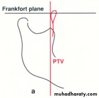
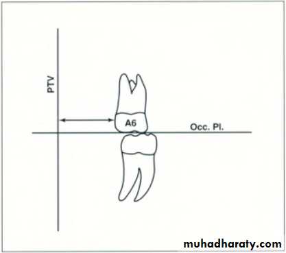
Pterygoid vertical -- A vertical line drawn through the distal radiographic outline of the pterygomax fissure & perpendicular to FHP
Ba-Na plane -- Extends from basion to the nasion. Divides the face and cranium.
Occlusal plane -- Represented by line extending through the first molars & the premolars.
A-pog line -- Also known as the dental plane.
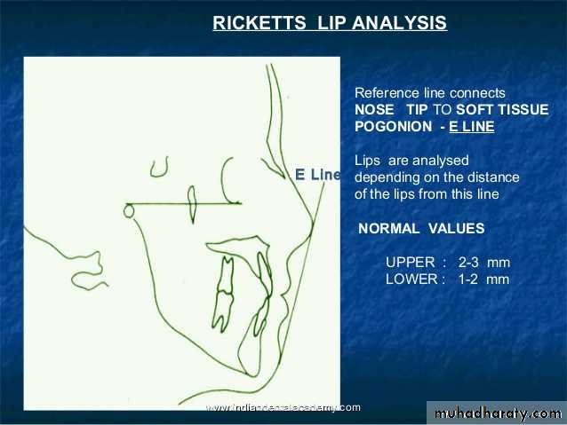
E-line -- Extends from soft tissue tip of nose to the soft tissue chin point.
Axis
Facial axis
PtmGn
Condylar axis
Corpus axis
Interpretation
This consists of analyzing:Chin in space
Convexity at point ATeeth
Profile
Chin in Space
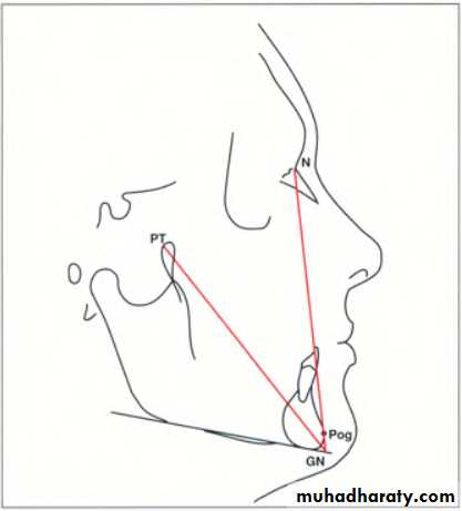
This is determined by :Facial axis angle
Facial (depth) angleMandibular plane angle
Facial axis angle
• Mean value is 90˚ ± 3˚• Does not changes with growth
• Indicates growth pattern of the mandible & also whether the chin is upward & forward or downward & backwards
Facial (depth) angle
• Changes with growth• Mean value is 87˚± 3˚ with an increase of 1˚ every 3 years
• Indicates the horizontal position of the chin & therefore suggests whether cl.II or cl.III pattern is due to the position of the mandible
Facial (depth) angle
Mandibular plane angle• Mean -- 26˚± 4˚at 9 yrs with 1˚decrease every 3 yrs
• High angle -- open bite – vertically growing mandible
• Low angle – deep bite – horizontally growing mandible
• Also gives an indication about ramus height
Po
O
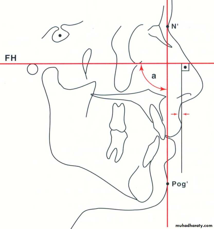
Convexity at point A
This gives an indication about the skeletal profileDirect linear measurement from point A to the facial plane
Normal at 9 yrs of age is 2mm & becomes 1mm at 18 yrs of age, since mandible grows more than maxillaHigh convexity – Cl II pattern
Negative convexity – Cl III pattern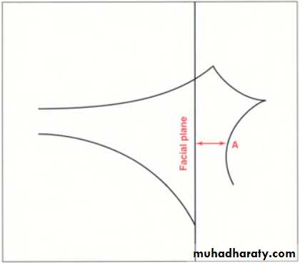
Teeth
Lower incisor to A-PogReferred to as denture plane
Useful reference line to measure position of anterior teeth
Ideally lower incisor should be located 1 mm ahead of A-Pog line
Used to define protrusion of lower arch
Upper molar to PtV
Measurement is the distance between pterygoid vertical to the distal of upper molarMeasurement should equal the age of the patient +3.0mm
Determines whether the malocclusion is due to position of upper or lower molars
Useful in determining whether extractions are necessary
Lower incisor inclinations
Angle between long axis of lower incisors & the A-Pog planeOn average this angle this angle should be 28 degrees
Measurement provides some idea of lower incisor procumbency
Profile
Lower lip to E planeDistance between lower lip & esthetic plane is an indication of soft tissue balance between lips & profile
Average measurement is -2.0mm at 9 yrs of age
Positive values are those ahead of E- line
Mc NAMARA ANALYSIS
Given By Mc Namara JA, 1984In an effort to create a clinically useful analysis, the craniofacial skeletal complex is divided into five major sections.
• Maxilla to cranial base
• Maxilla to mandible
• Mandible to cranial base
• Dentition
• Airway

Dr. Mc Namara JA
-- JacobsonMAXILLA TO CRANIAL BASE
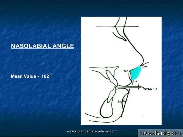
Soft tissue evaluationNasolabial angle
Acute nasolabial angle – dentoalveolar protrusion, but can also occur because of orientataion of base of nose
Cant of upper lip
Line is drawn from nasion perpendicular to upper lip14 degree in females
8 degree in malesHard tissue evaluation
Anterior position of point A = +ve valuePosterior position of point A = -ve value
In well-balanced faces, this measurement is 0 mm in the mixed dentition and 1 mm in adult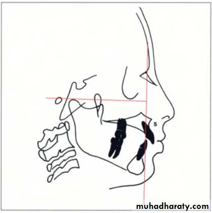
Maxillary skeletal protrusion
Maxillary skeletal retrusion
Maxilla to mandible
Anteroposterior relationshipLinear relationship exists between effective length of midface & that of mandible
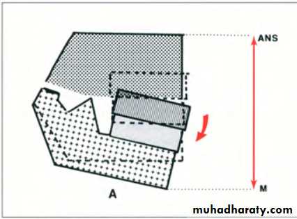
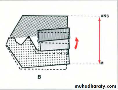
a) Lower Anterior Face Height (LAFH)
LAFH is measured from ANS to MeIn well balanced faces it correlates with the effective length of midface
Vertical relationshipVertical maxillary excess – downward & backward rotation of mandible, increasing lower anterior facial height
Vertical maxillary deficiency – upward & forward rotation of mandible, decreasing lower anterior facial height
b) Mandibular plane angle
On average, the mandibular plane angle is 22 degrees ± 4 degreesA higher value excessive lower facial height
lesser angle Lower facial height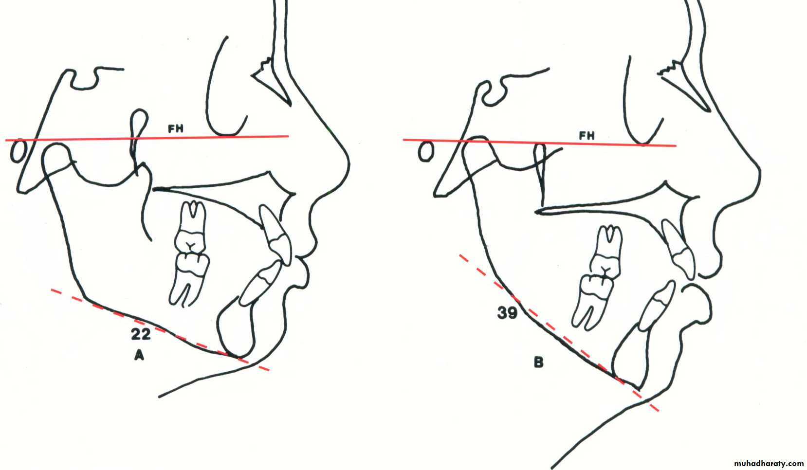
c) The facial axis angle
In a balanced face --90 degrees to the basion-nasion lineA negative value excessive vertical development of the face
Positive values deficient vertical development of the faceDentition
a) Maxillary incisor positionThe distance from the point A to the facial surface of the maxillary incisors is measured
The ideal distance 4 to 6 mm
b) Mandibular incisor positionIn a well-balanced face, this distance should be 1 to 3 mm
AIRWAY ANALYSIS
Upper PharynxWidth measured from posterior outline of the soft palate to a point closest on the pharyngeal wall
The average nasopharynx is approximately 15 to 20mm in width.
A width of 2mm or less in this region may indicate airway impairment
Lower Pharynx
Width – point of intersection of posterior border of tongue & inferior border of mandible to closest point on posterior pharyngeal wallThe average measurement is 11 to 14 mm, independent of age
Greater than average lower pharyngeal width-- possible anterior positioning of the tongueTHE HOLDAWAY SOFT TISSUE ANALYSIS
Given by Dr. Reed Holdaway, 1984Dr. Reed Holdaway in series of two articles outlined the parameter of soft tissue outline
Analysis consists of 11 measurement
Dr. Reed Holdaway
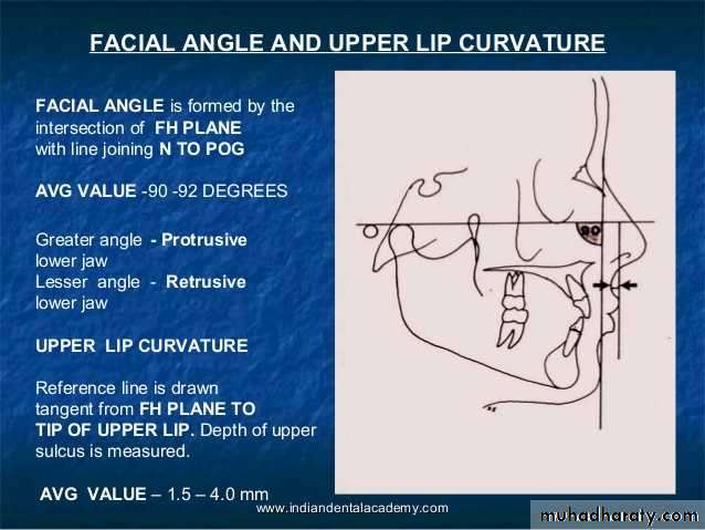
-- Jacobson• Facial Angle (90 degree)
• Ideally the angle should be 90 to 92 degrees>90 degree: mandible too protrusive
<90 degree: recessive lower jaw
•
2. Upper lip curvature (2.5mm)
Depth of sulcus from a line drawn perpendicular to FH & tangent to tip of upper lipLack of upper lip curvature – lip strain
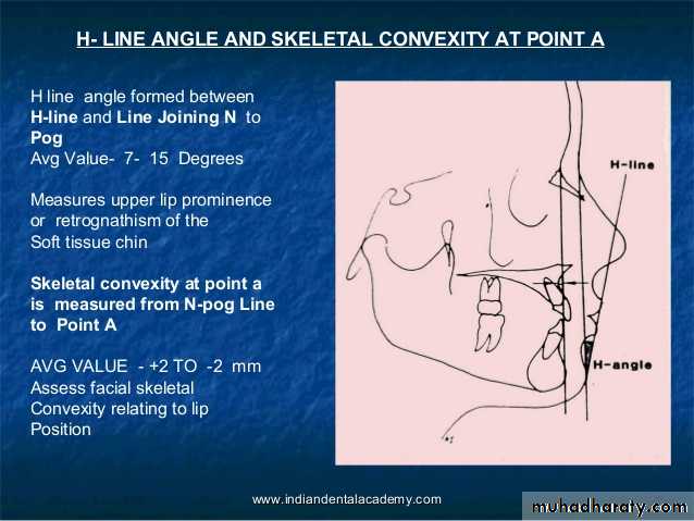
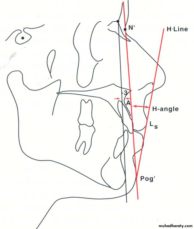
Excessive depths could be caused by jaw overclosure3. Skeletal convexity at point A (-2to 2mm)
Measured from point A to N’-Pog’ lineNot a soft tissue measurement but a good parameter to assess facial skeletal convexity relating to lip position
Dictates dental relationships needed to produce facial harmony
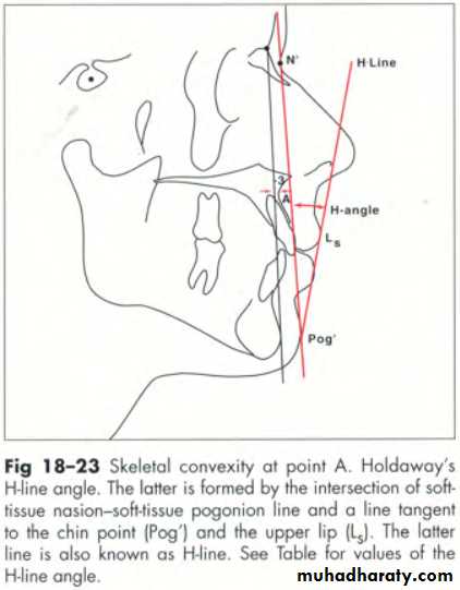
4. H-Line Angle(7-15 degree)
Formed between H-line & N’-Pog’ lineMeasures either degree of upper lip prominence or amount of retrognathism of soft tissue chin
If skeletal convexity & H-line angles donot approximate, facial imbalance may be evident
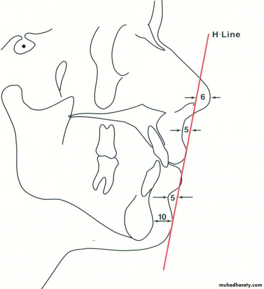
5. Nose tip to H-line (12mm maximum)
Measurement should not exceed 12mm in individuals 14 yrs of age6. Upper sulcus depth (5mm)
Short/thin lips -measurement of 3 mm may be adequateLonger/thicker lips- 7mm may still indicate excellent balance
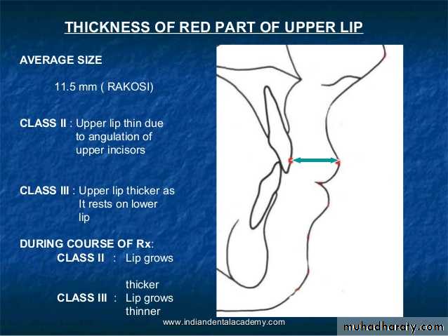
7.Upper lip thickness (15mm)
Measured horizontally from a point on outer alveolar plate 2mm below point A to outer border of upper lip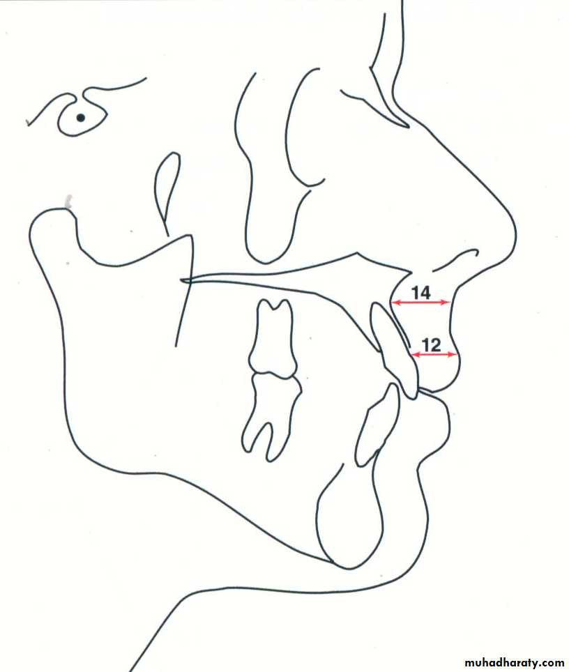
8. Upper lip strain
Measured from vermillion border of upper lip to labial surface of maxillary center inc.Measurement should be approx same as the upper lip thickness (within 1mm)
Measurement less than upper lip thickness – lips are considered to be strained9. Lower lip to H-line(0mm)
Measured from the most prominent outline of the lower lipNegative reading – lips are behind the H line
Positive reading – lips are ahead of H line
Range of -1 to +2mm is regarded normal
10. Lower sulcus depth (5mm)
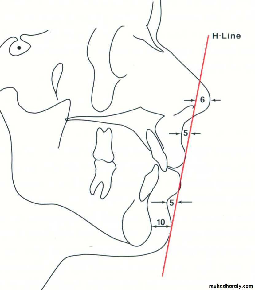
11. Soft tissue-chin thickness (10-12mm)
Measured as distance between bony & soft tissue facial planesIn fleshy chins, lower incisors may be permitted to stay in a more prominent position, allowing for facial harmony