ANATOMY OF ANTERIOR WALL &
INGUINAL REGION
.أ
د
.
عبد الجبار الحبيطي

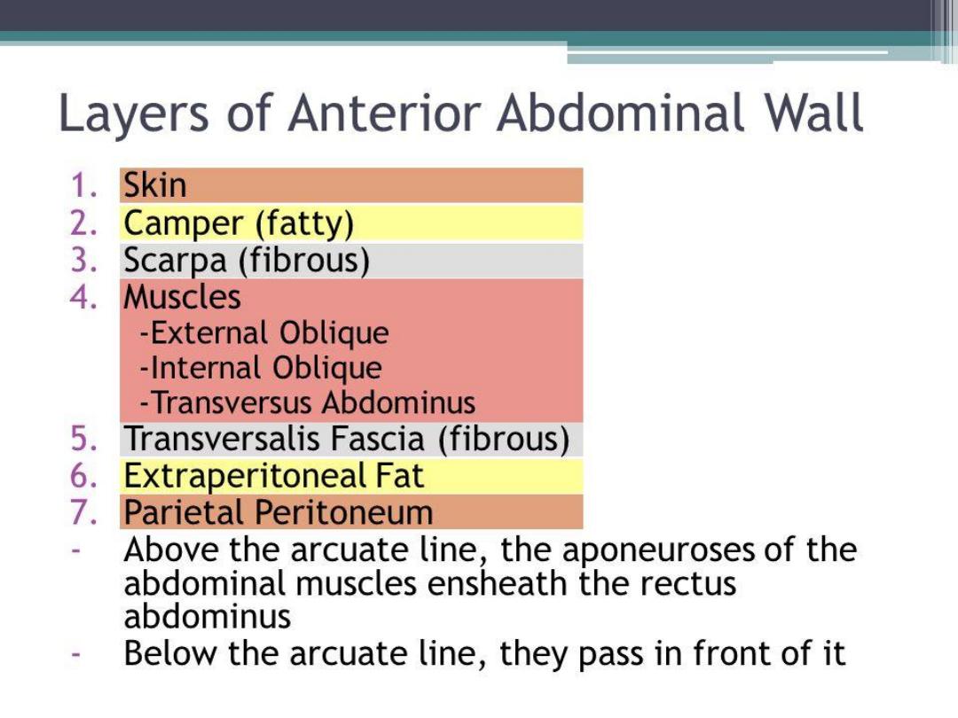
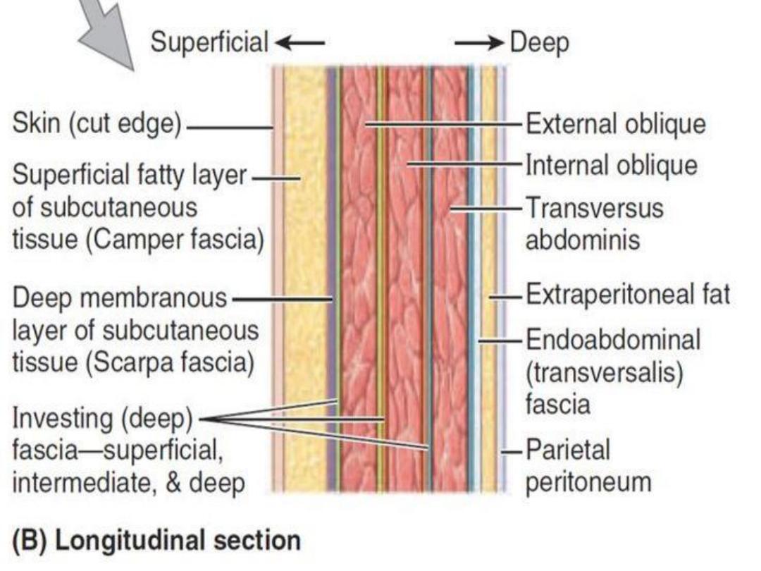
• A mid line canula through anterior abdominal
wall passes through the following layers:
1-Skin
2-Superficial fascia
3-Linea alba
4-Transversalis fascia&extraperitoneal fat
5-The parietal peritoneum.

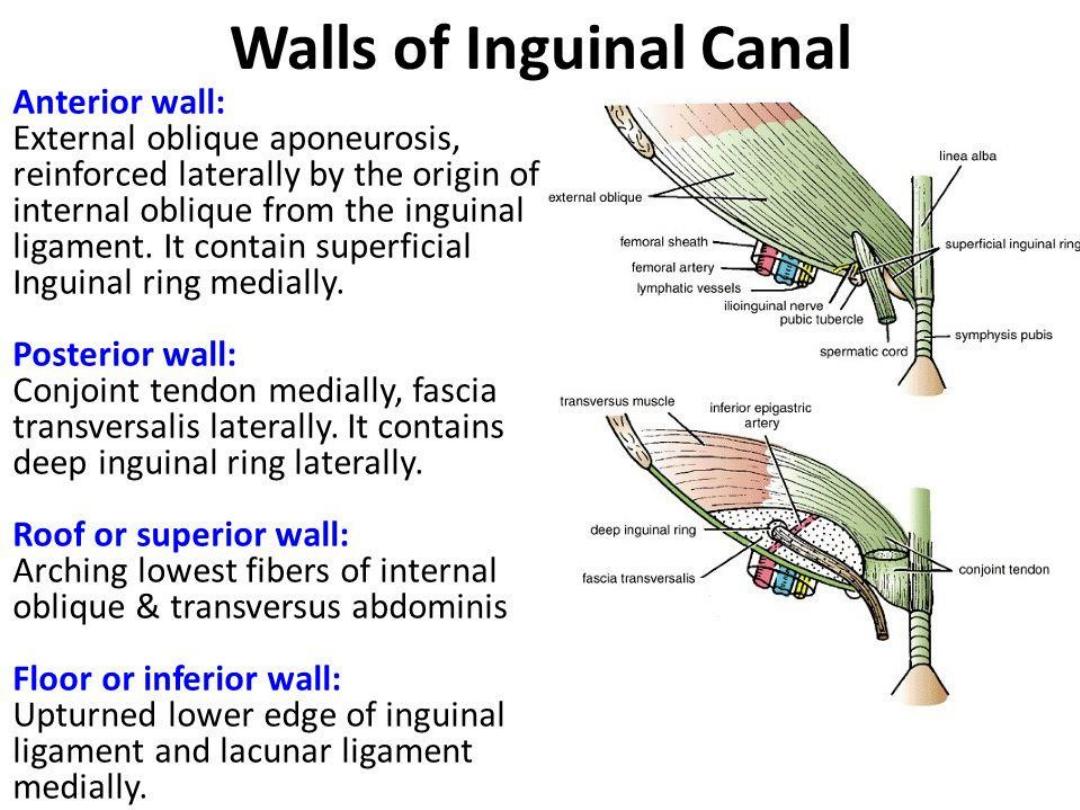
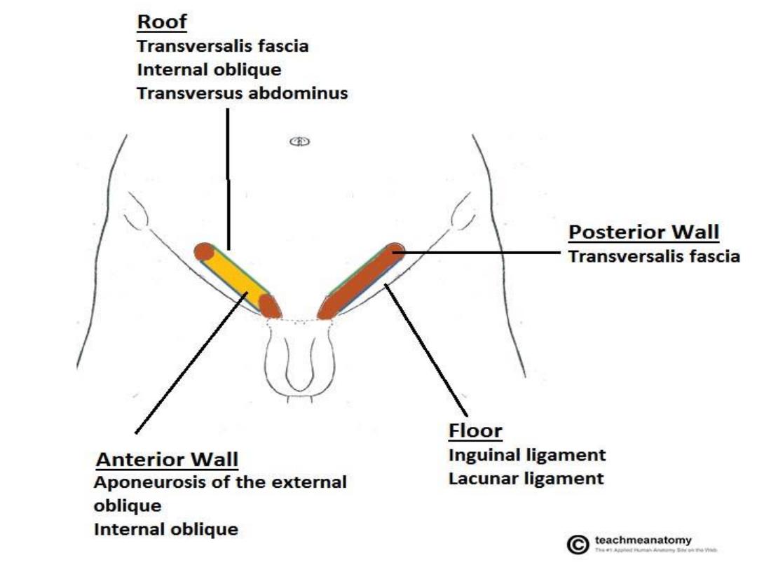
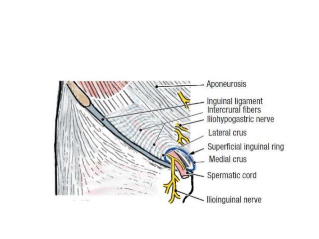
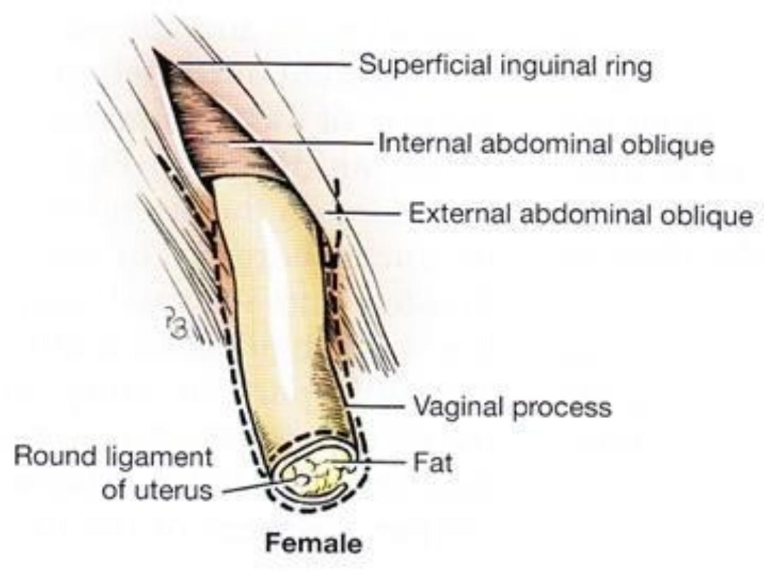
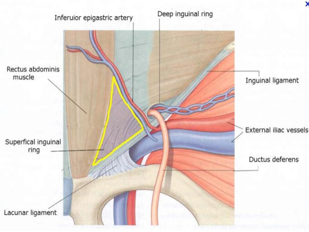
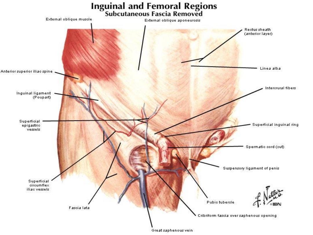
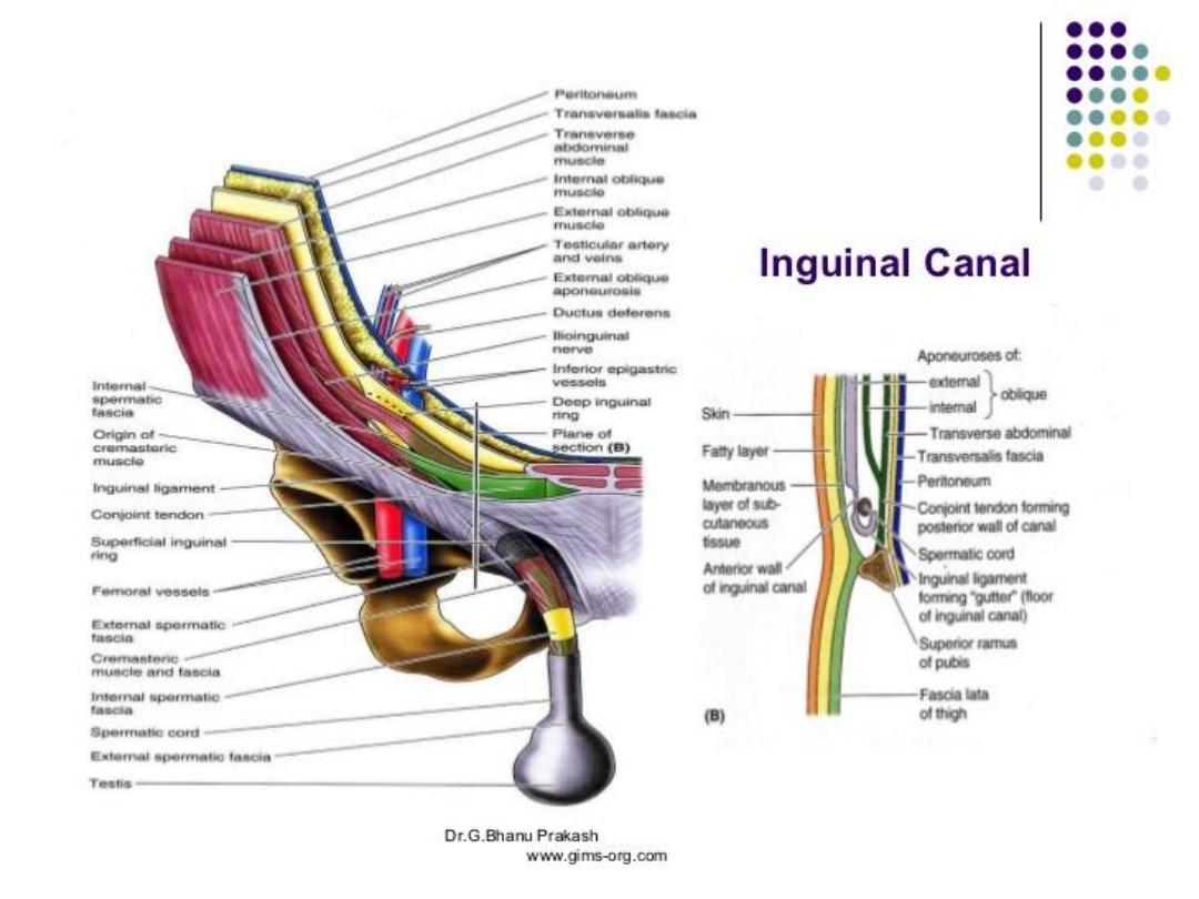
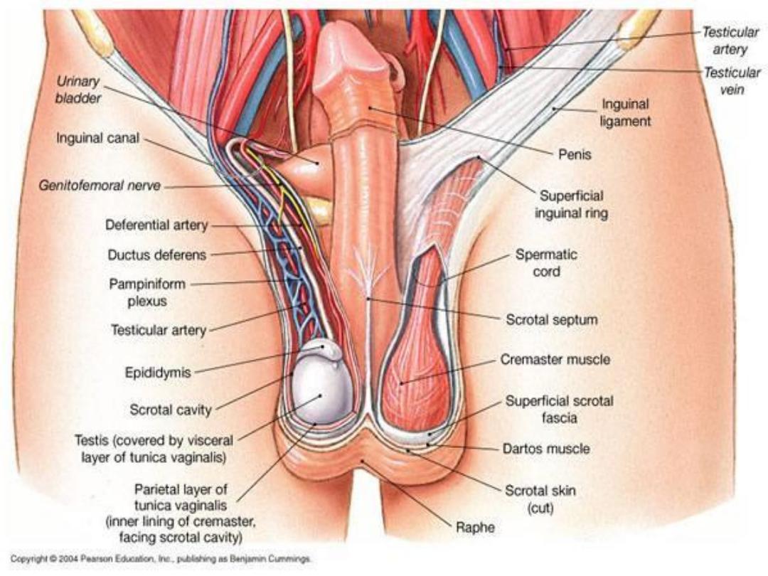
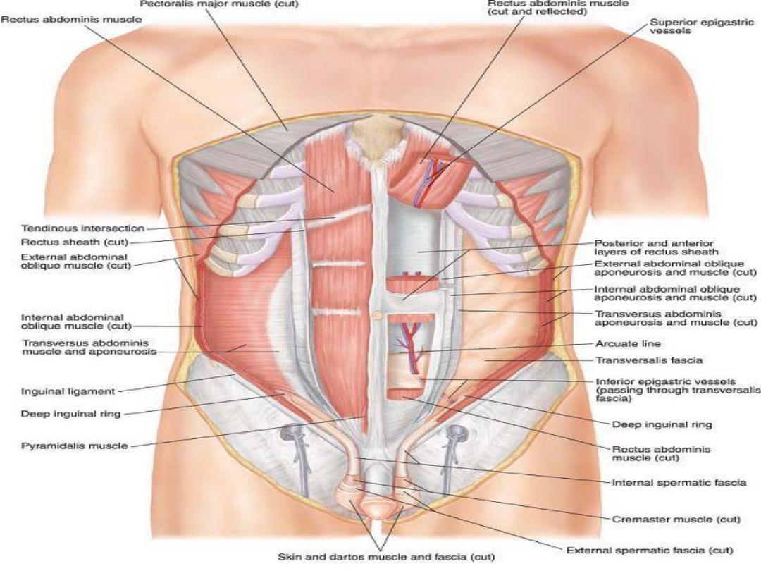
• The superficial inguinal ring : is triangular in
shape located just above the pubic tubercle.It is
related to the Conjoint tendon posteriorly ,which
forms part of the posterior wall of the inguinal
canal (medial part) .The conjoint tendon
reinforces the superficial inguinal ring.
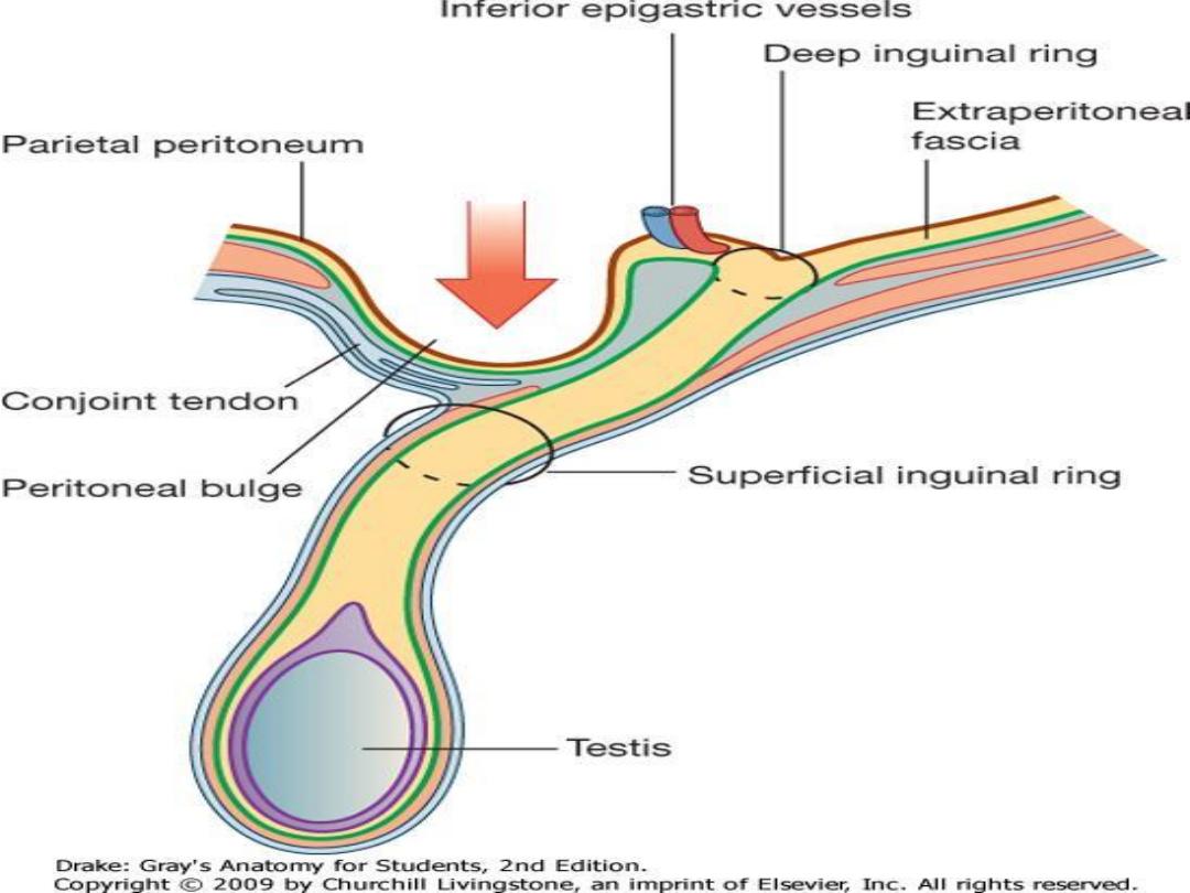
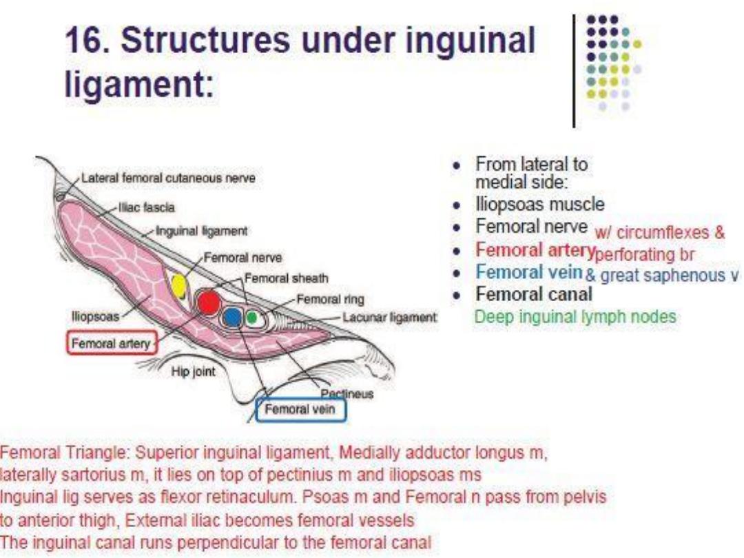
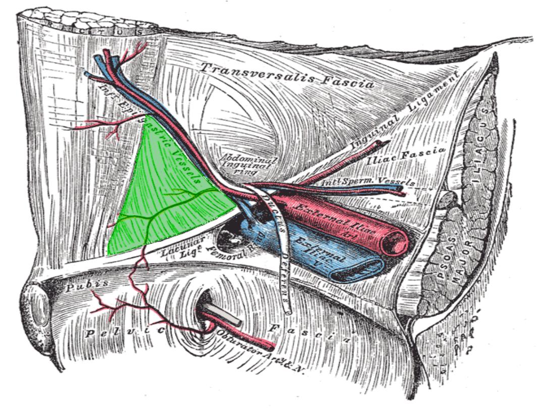
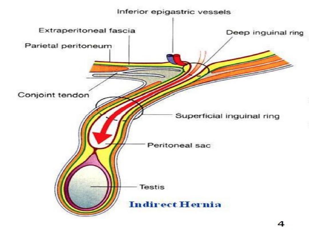
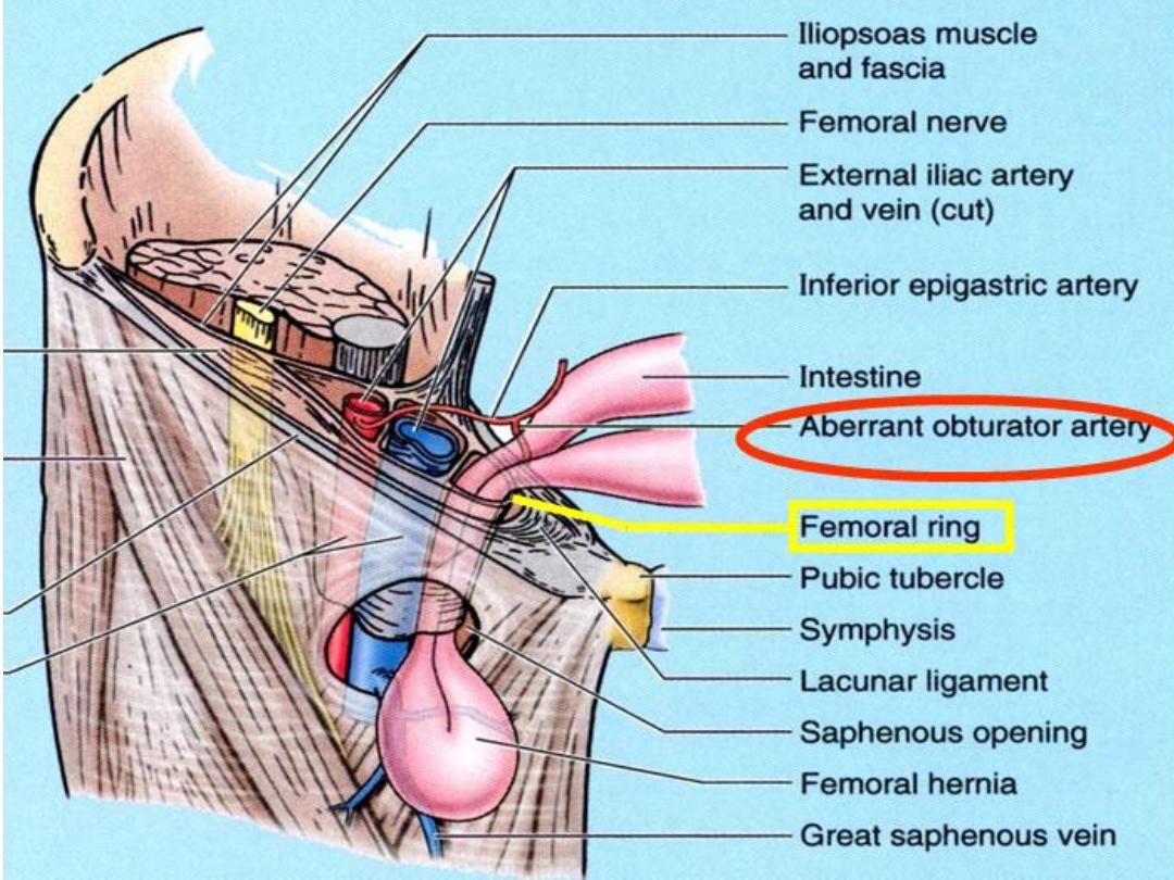
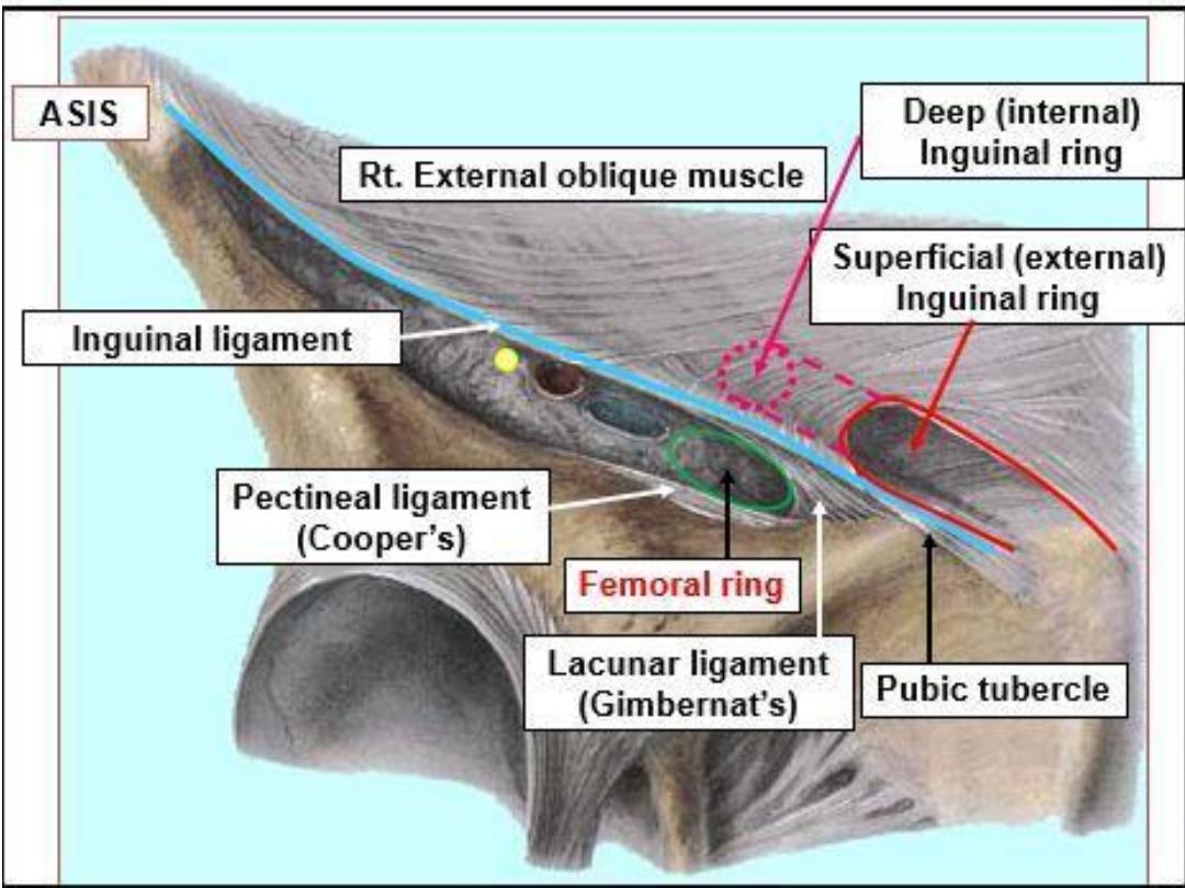
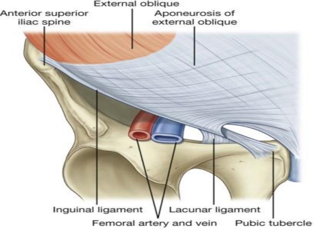
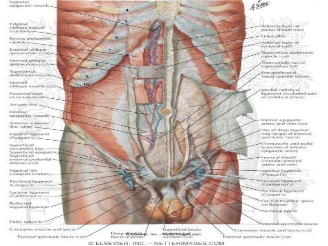
• The Deep Inguinal Ring(D.I.R) is about half an inch
above the mid point of inguinal ligament,it is a
defect through the Transversalis fascia & the
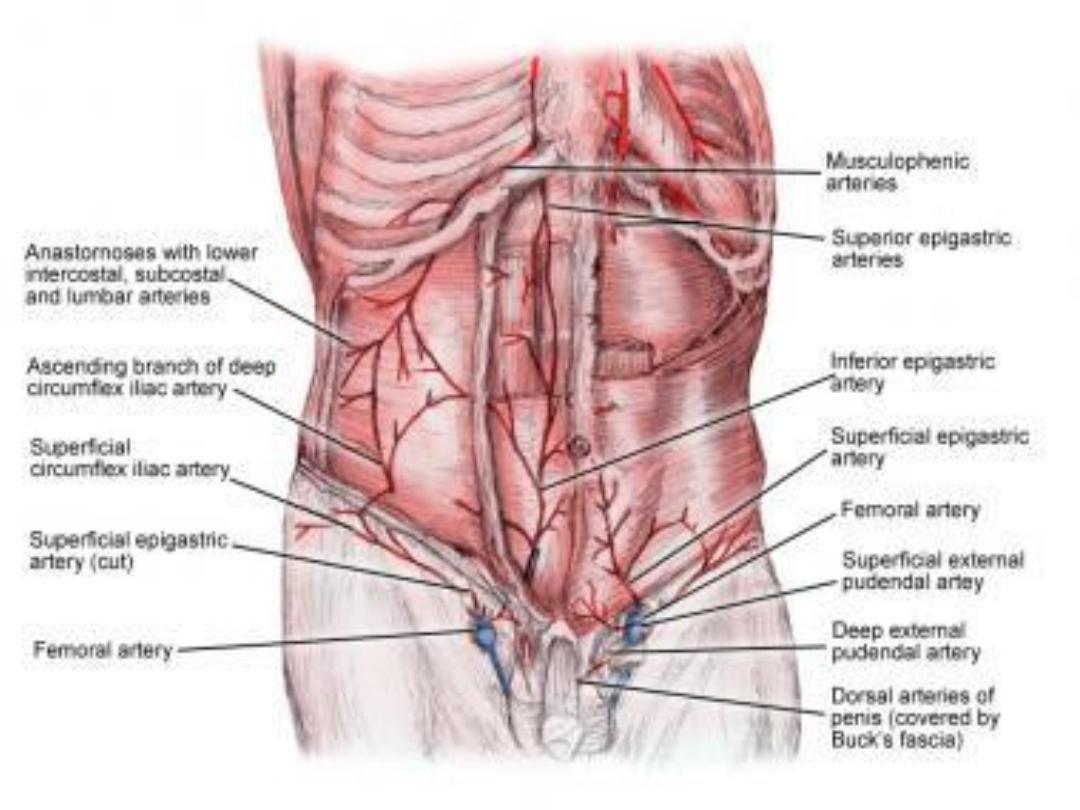
inferior epigastric artery runs just medial to it.

Deep to the inguinal ligament the following
structures pass:
• 1-Iliacus & psoas major Ms
• 2-External iliac vessels (just below lig, become
Femoral)
• 3-The Femoral nerve
• 4- The lateral cutaneous nerve of the thigh.

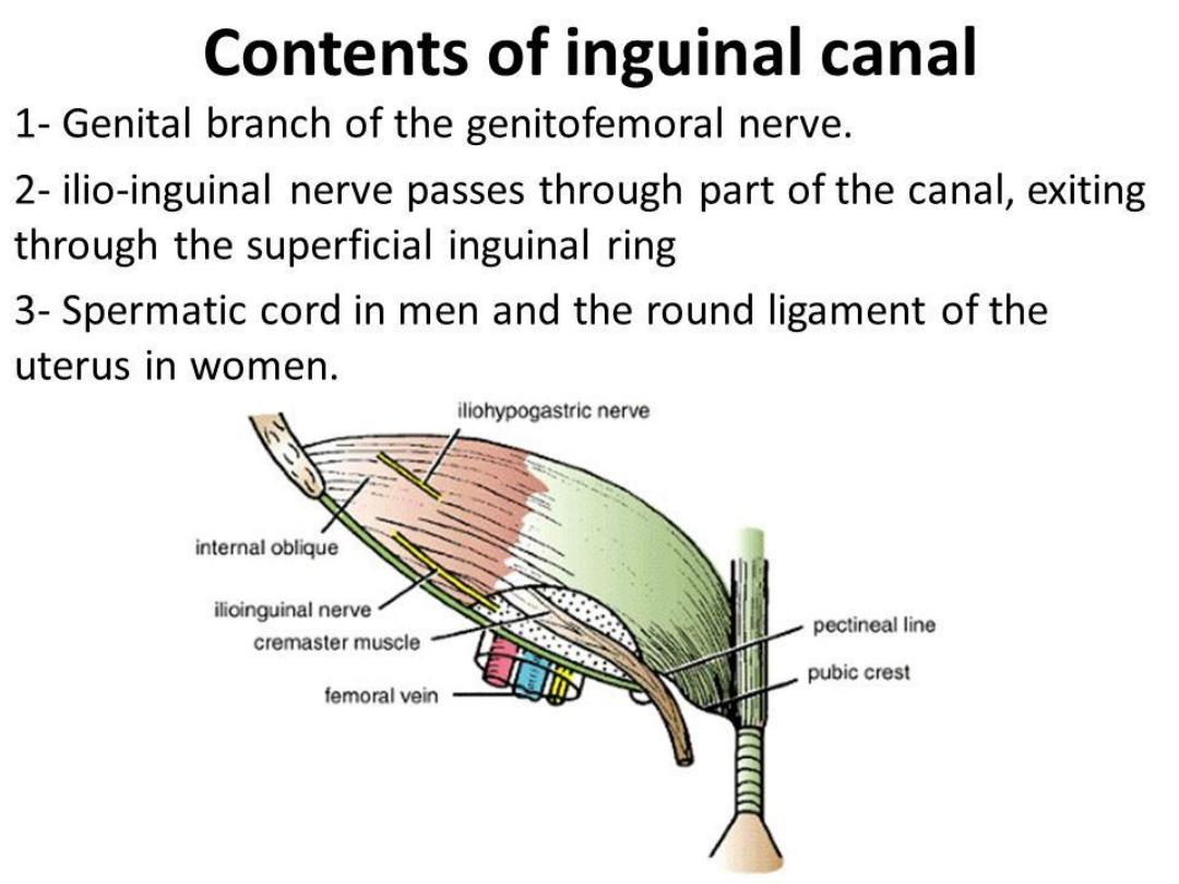
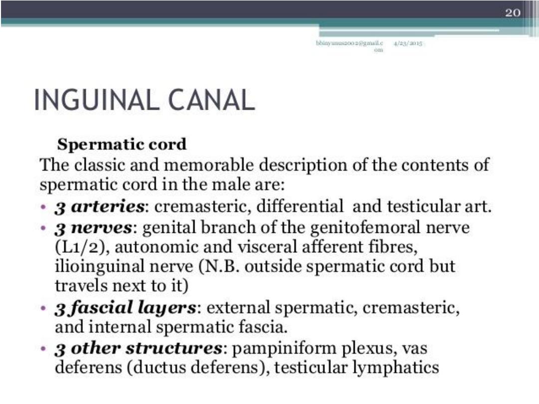
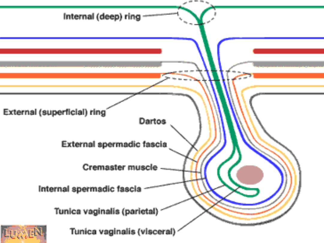
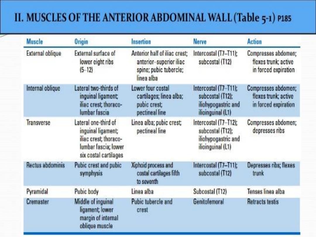
• The Cremaster muscle forms loops on the
spermatic cord,it reaches the base of the
scrotum.It is supplied by genital branch of
genitofemoral nerve( cremasteric nerve ), it
elevates the testes ,while cremasteric reflex is
by ilioinguinal nerve .The cremaster muscle is
absent in the female.

• The Conjoint tendon:It lies behind the S.I.R &
reinforces it .It is formed by contributions
from the aponeurosis of both internal oblique
& transversus abdominis & is attached to the
pubic crest. It forms part of posterior wall of
the inguinal canal& supplied by ilioinguinal
nerve.

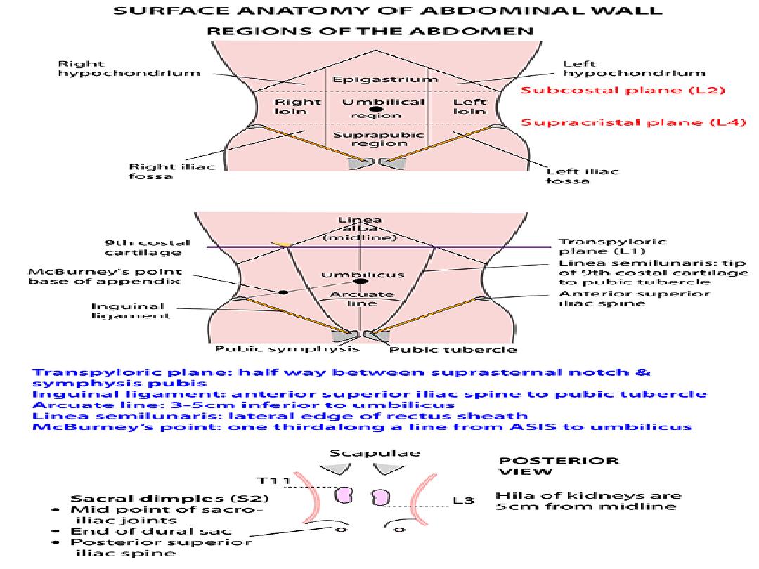
• The following structures are seen at the level
of the transpyloric plane:
• 1-Fundus of the gall bladder.
• 2-the upper limit of the hilum of right kidney.
3-The tip of the 9
th
costal cartilage.
• 4-The pylorus of stomach.
• 5-Body of L1 vertebra.
• 6-Origin of superior mesenteric artery.

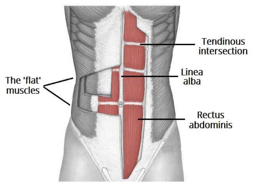
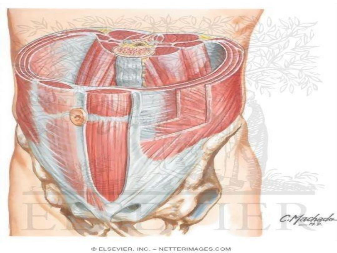
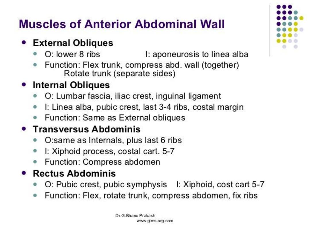
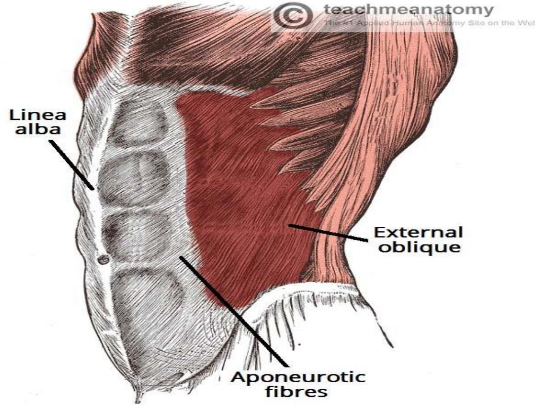
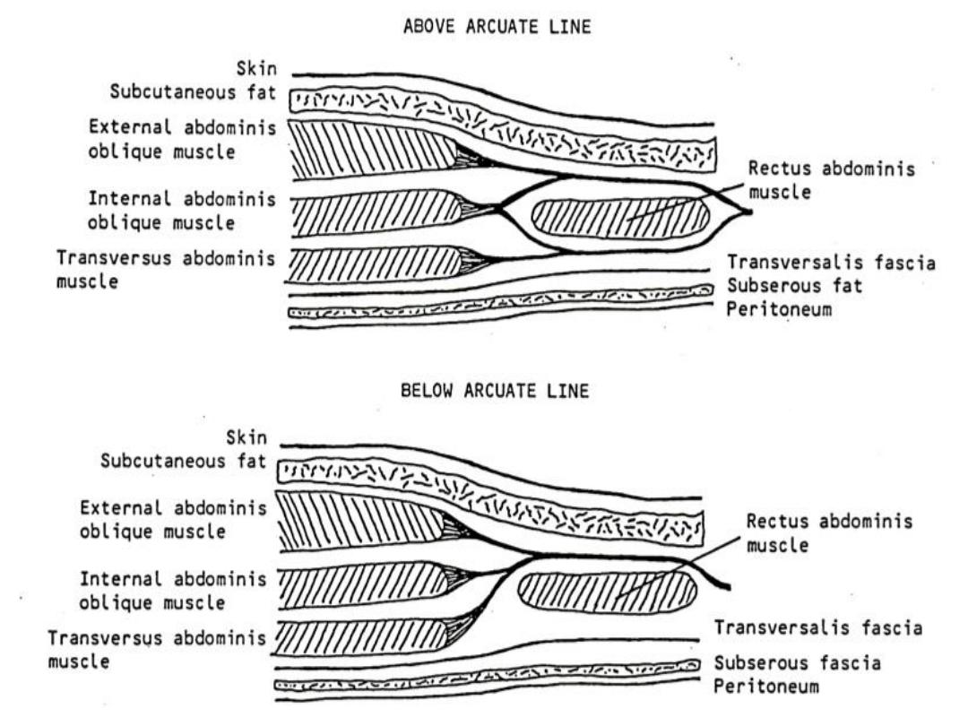
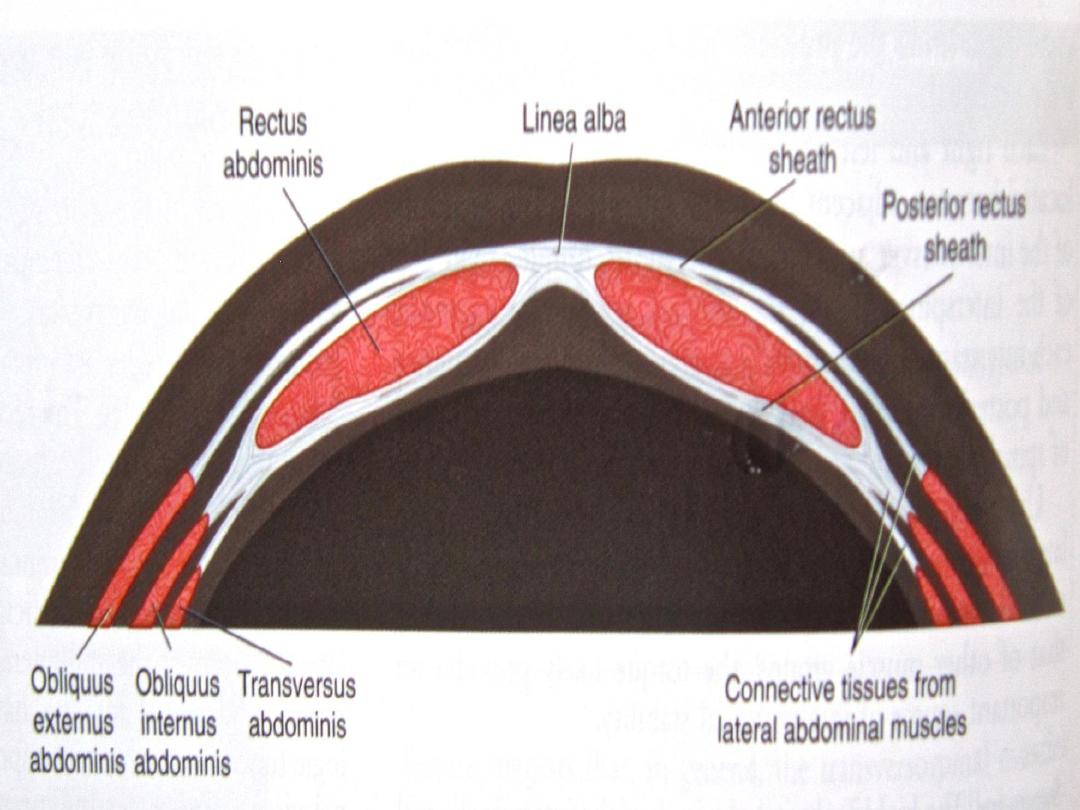
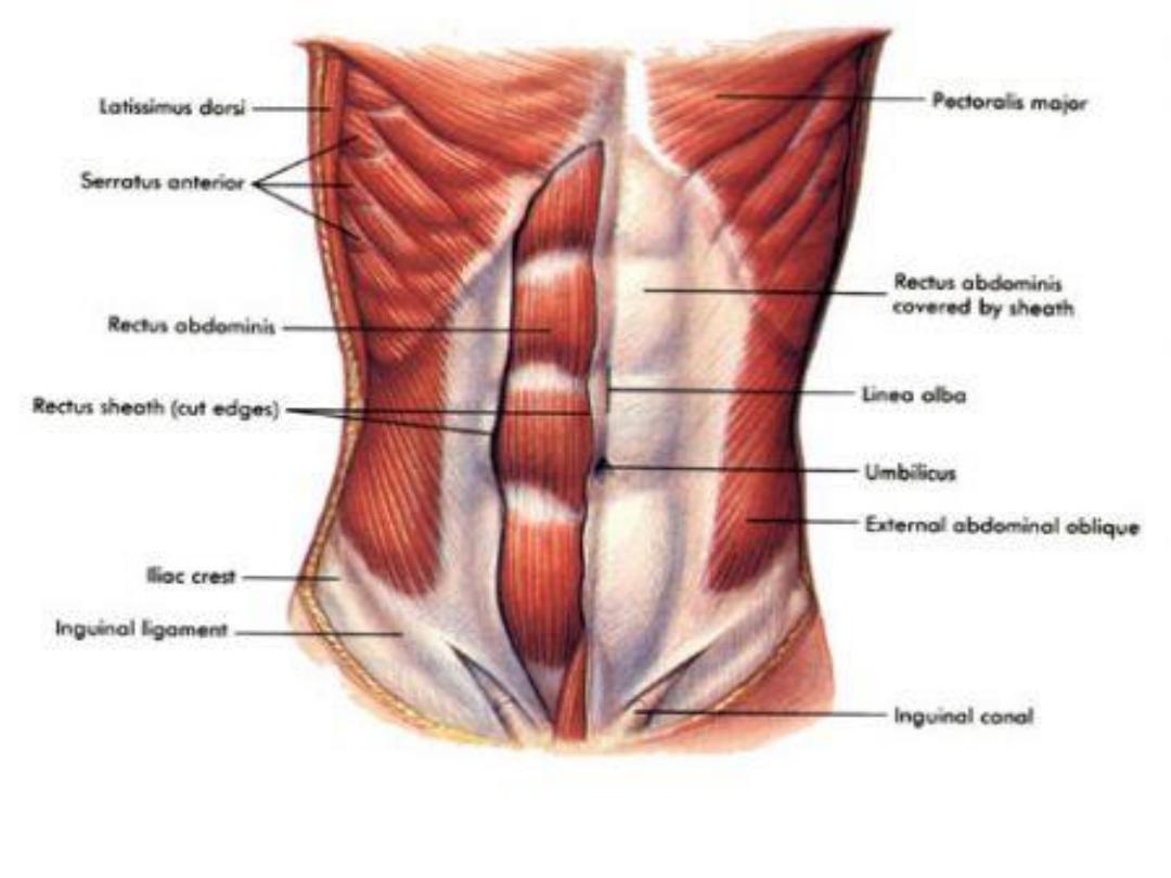
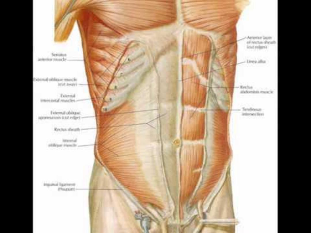
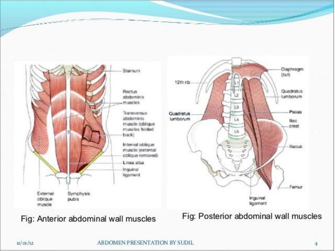
• The Rectus Abdominis Muscle:
• 1- Helps in flexion of the trunk
• 2-Is supplied by lower 5 intercostal nerves
• 3-Shows three tendinous intersections on its
anterior surface.
• 4-The rectus sheath is not attached to its
posterior surface.

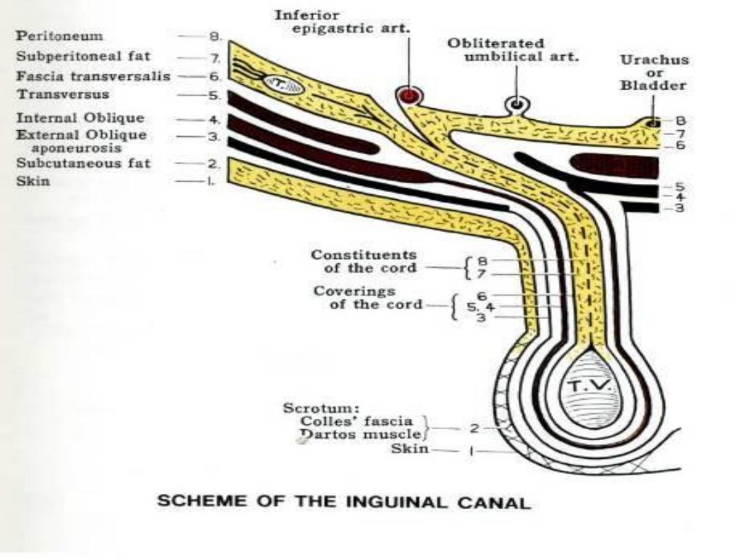
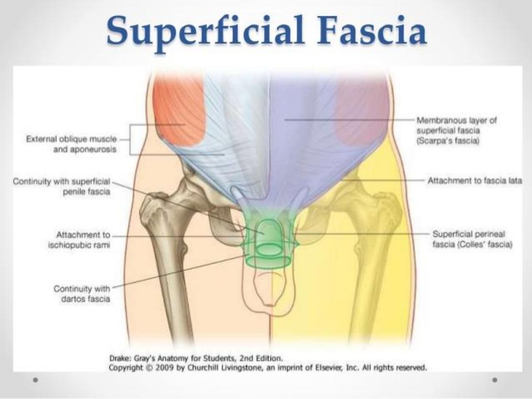
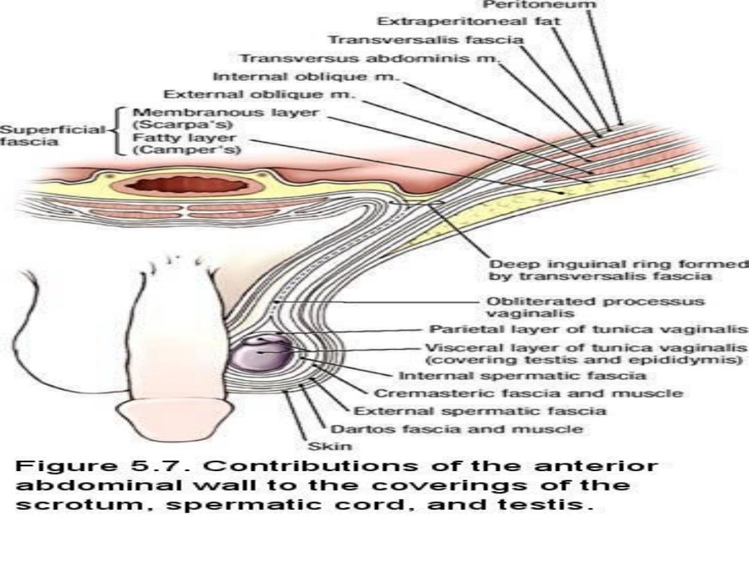
• On tapping Hydrocele (Tunica vaginalis is
distended with fluid) the Canula passes through:
• 1-Skin
• 2- Dartos muscle(smooth m.fs) & membranous
layer of the superficial fascia
• 3-External spermatic fascia
• 4-Cremaster fascia
• 5-Internal spermatic fascia
• 6- The parietal layer of Tunica Vaginalis .

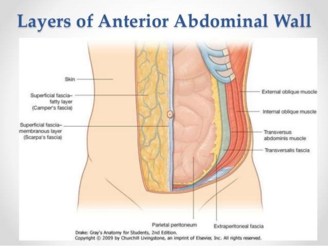
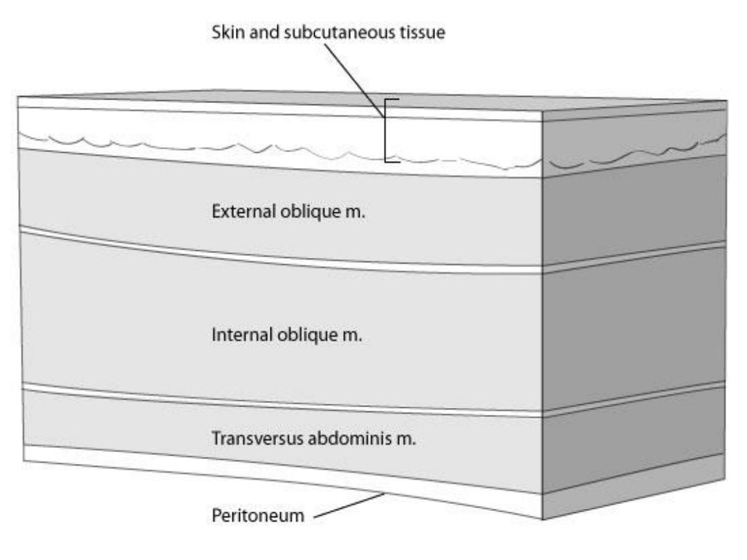
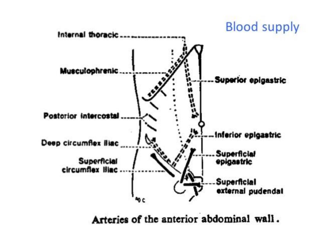
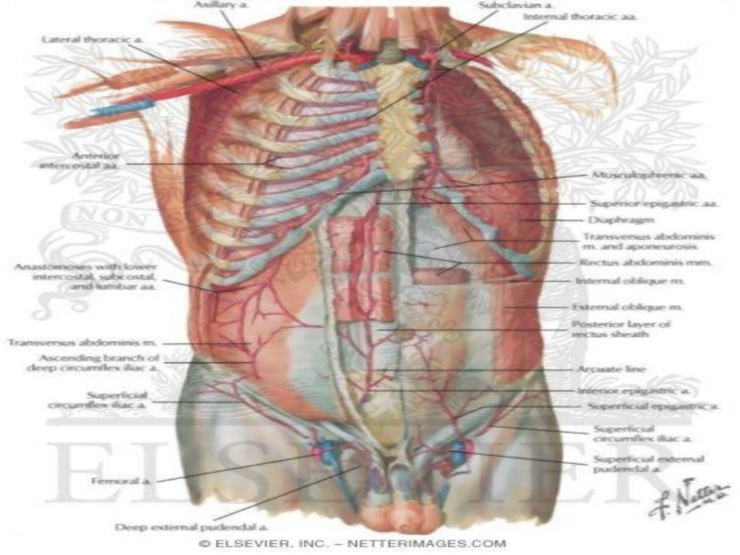
• To withdrow fluid from peritoneal cavity
aneedle(just lateral to the inferior epigastric
artery ) should pass through the following
layers:
• 1-Skin
• 2- Superficial fascia
• 3-External oblique M
• 4-Internal oblique M
• 5-Transversus abdominis M
• 6-Transversalis fascia
• 7-Extraperitoneal fat
• 8-Parietal peritoneum.

•
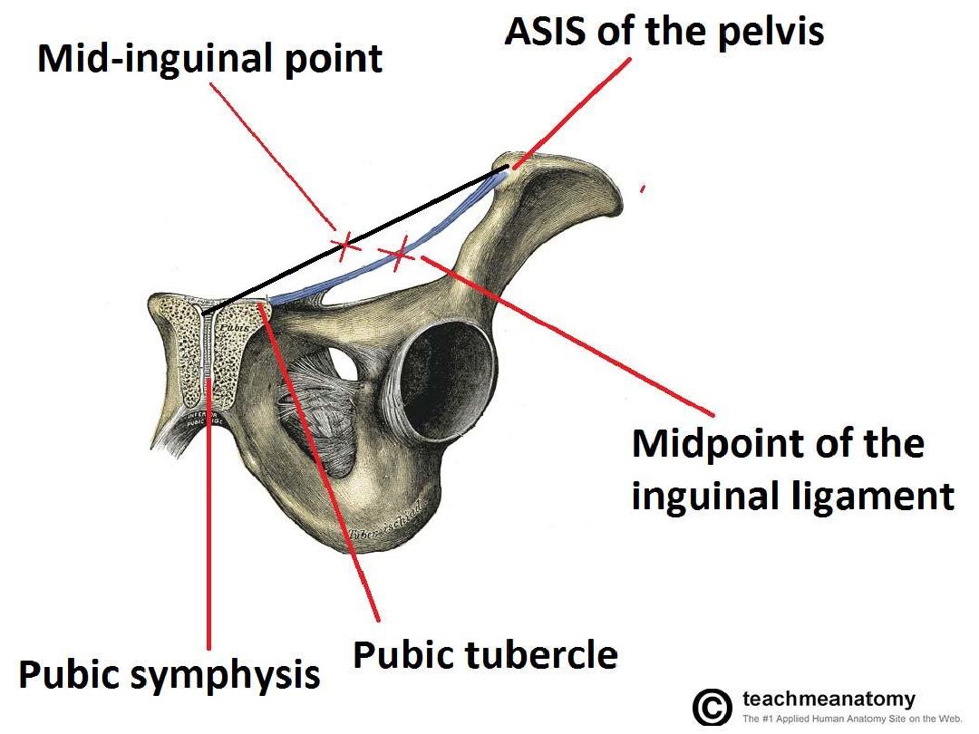
The Mid-inguinal point is the point located at
the middle of the distance between the
Anterior superior iliac spine & Pubic
symphysis.

ANATOMY OF
ANTERIOR ABDOMINAL WALL
.أ
د
.
عبد الجبار الحبيطي


ANATOMY
OF
INGUINAL REGION
.أ
د
.
عبد الجبار الحبيطي