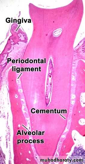
PERIODONTAL LIGAMENT
Periodontium-Is a connective tissue organ, covered by epithelium that attached the teeth to the bones of the jaws and provides a continually adapting apparatus for support of the teeth during function.
-The periodontium comprises four connective tissues:-
-Two mineralized C.T. (cementum and alveolar bone).
-Two fibrous C.T. (periodontal ligament and lamina properia of the gingival).
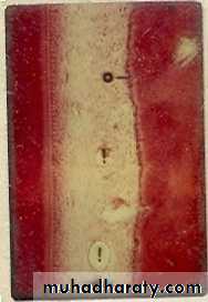
The periodontal ligament is a fibrous C.T ligament located between the alveolar bone proper and cementum. This ligament covers the root of the tooth and connects with the tissue of the gingiva. The p.d.l. occupies the periodontal space and is composed of cell and intercellular substance. It has a thickness of 0.15-0.38 mm, it is thinnest in the mid root zone, and decrease slightly in thickness with aging.
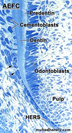
Development of P.d.l.
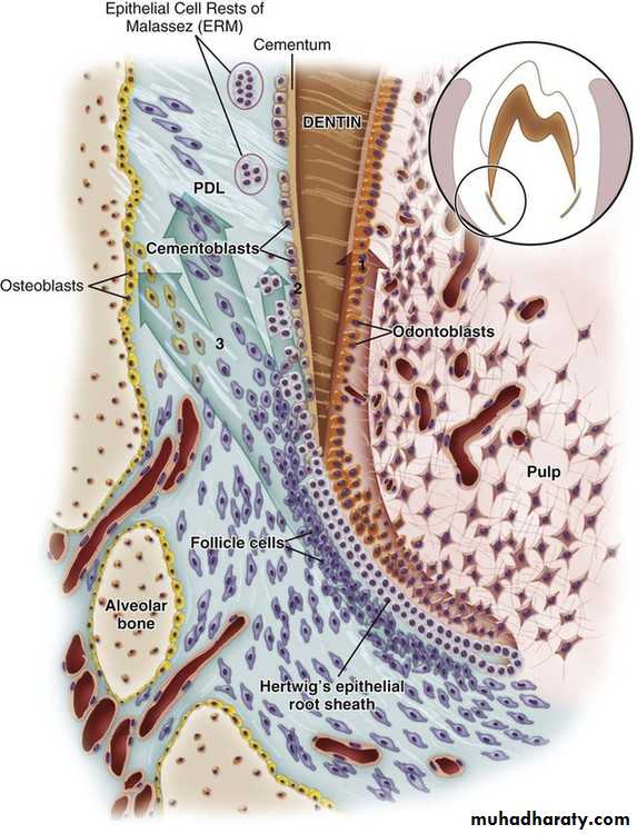
The formation of P.d.l. occurs after the cells of hertwig’s epithelial root sheath have separated, forming the strands known as (the epithelial rests of malassez). This separation permits the cells of the dental follicle to migrate to the exernal surface of the newly formed root dentin. these cells show high degree of mitotic activity, thus differentiating into different types of cells which give rise to cells of periodontal ligament namely: Cementoblast deposit cementum on the surface of dentin, Osteoblast of the developing alveolar bone, and Fibroblast to synthesize the fibers and ground substance of periodontal ligament, The fibers of p.d.l. become embedded in newly developed cementum and alveolar bone, and as the tooth erupt. Other cells of the dental follicle differentiate into fibroblasts, which synthesize the fibers and ground substance of p.d.l
Osteoblast of the developing alveolar bone.
• Cellular Composition
• The cells of periodontal ligament are categorized as:• 1. Synthetic Cells:
• a) Fibroblasts
• b) Osteoblasts
• c) Cementoblasts
• 2. Resorptive Cells:
• a) Fibroclasts
• b) Osteoclasts
• c) Cementoclasts
• 3. Progenitor Cells.
• 4. Epithelial Cell rests of malassez: Apart from these periodontal ligament comprises of (ERM).
• 5. Defence cells derived from hemopoietic line:
• a) Mast cells
• b) Macrophages
Cells of the P.d.l.1-Synthetic cells Fibroblast, Osteoblast, and cementoblast.
These cells are the most numerous and densely packed in periodontal ligament.
They appear as ovoid or flattened with numerous cytoplasmic processes.
• Fibroblast appear to be oriented parallel to the oriented bundles of collagen fibers. Some fibroblast form and destroy collagen as it has ability to simultaneously synthesize and degrade collagen, a process essential to the physiological turnover or remodeling of p.d.l.
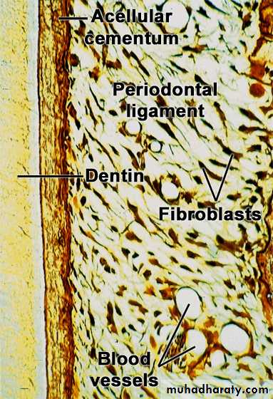
b- Osteoblasts
These are located along the surface of the alveolar bone in various stages of differentiation (developing alveolar bone).c- Cementoblasts
They appear near the cementum of variously differentiated stages and their progenitors,(deposit cementum on the surface of dentin).
2-Resorptive cells a.Osteoclasts
These are the bone resorbing cells and tend to be large and multinucleating but may be small and mononuclear.Derived from circulating monocytes.
Under light microscope these are present in Howship’s lacunae.
Under electron microscope exhibit numerous mitochondria, lysosomes, Golgi apparatus, few ribosomes and few rough endoplasmic reticulum.
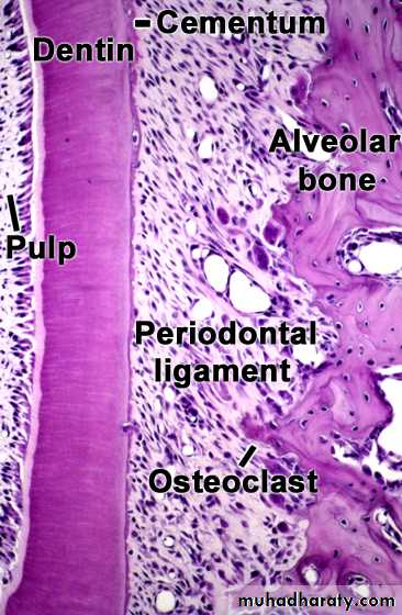
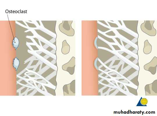
They appeared to occupy bays in bone (howship's lacunae) or surround the end of a bone spicule. They have characteristic folds termed the ruffled or striated border. The ruffled border disappears in inactive osteoclasts. Histochemical tests showed that osteoclasts are rich in acid phosphatase, which is contained in lysosomes, for this osteoclasts in periodontal ligament indicates active resorption , that allows functional changes in the position of teeth that must be accommodated by supporting tissues.
b.Cementoclasts
They resemble osteoclast and occasionally found in the normal functioning, p.d.l. They resorbed cementum under certain circumstances; and in these instances mononuclear or multinucleated giant cells located in Howship's lacunae, are found on the surface of the cementum.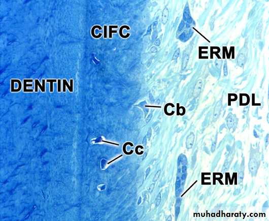
3-Progenitor cells
All C.T., including p.d.l. contain progenitor cells that have the capacity to undergo mitotic division, and replace dying differentiated cells at the end of their life span or as a result of truma.4-Epithelial rests of Malassez
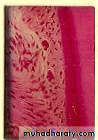
Periodontal ligament contains epithelial cells that lie close to cementum.Described by malassez in 1884.
They are remnants of Hertwig’s epithelial rest sheath that are embedded in periodontal ligament during deviation.
they persist as a network, strand, or tubule-like structures and parallel to the surface of the root.
When certain pathological conditions are present, cells of epithelial rests can undergo proliferation and can produce a variety of cysts and tumor that unique to the jaws.
5-Defence cells :--Mast cells These are small round / oval in shape about 12-15 µm in dia. Contain numerous cytoplasmic granules such as heparin, serotonin, histamine. These granules play a role in inflammation reaction especially histamine. Cells degenerate in response to Ag-Ab reaction.-Macrophages They are important defense cells because of their phagocytic activity and mobility; they take up bacteria, dead cells and foreign bodies.lymphocytes, leukocytes, and plasma cells also appear when p.d.l. is stressed by disease.
Extracellular substance
The extracellular substance of the p.d.L. comprises the following:-Ground substance
*Proteoglycans
*Glycoproteins
-Fibers
*collagen
*oxytalan
Fibers: The fibers of the p.d.l. are made up of collagen and oxytalan. The majority of fibers in the p.d.l. are collagen.
Oxytalan fiber:- these are
immature elastic fibre.
small in diameter.
appear to interlace with the collagen bundles.
supporting the collagen fibers and restricted to walls of blood vessels.
When viewed through a light microscope they appear to be almost longitudinal in the ligament.
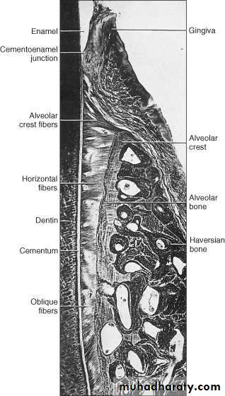
Collagen fibers is a specific, high molecular weight protein to which are attached a small number of sugars. The p.d.l. appears to be made up predominantly of type I collagen and type III collagen are also present .
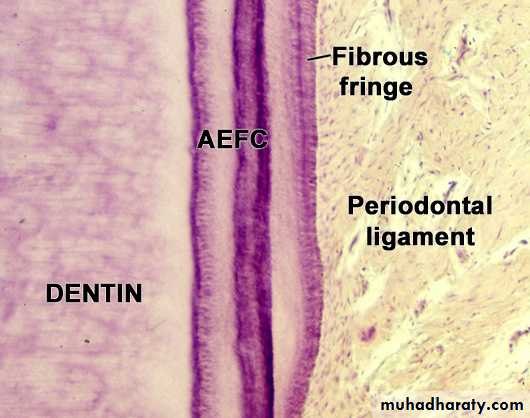
Collagen macromolecules are arranged to form fibrils, these fibrils are packed to form bundles or fibers that oriented relative to periodontal space and these are termed principal fibers.
Groups of principal fibers are named according to their location with respect to the teeth, there are two groups: the gingival group, located around the necks of teeth, and the dentoalveolar group ,which surrounds the roots.
Gingival group
The fibers of this represent four groups of principal fibers, each having a different, orientation and function in support of gingival tissues.1-Free gingival fibers.
2-Attached gingival fibers.
3-Circumferential fibers.
4-Transseptal fibers.
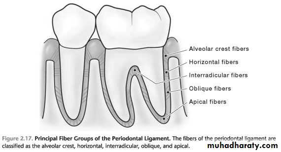
Dentoalveolar group
This group consists of five differently oriented types of principal fiber groups named according to their origin and insertion in the dentoalveolar process.1. Alveolar crest group
2. Horizontal group
3. Interradicular group
4. Oblique group
5. Apical group
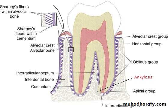
The principal fibers run a wavy course from cementum to bone.
Microscopical examination showed that fibers arising from cementum and bone are joined in the mid-region of periodontal space, giving rise to a zone of distinct appearance, called intermediate plexus, this region provides a site where rapid remodeling of fibers occurs, allowing adjustments in the ligament to be made to accommodate small movements of tooth.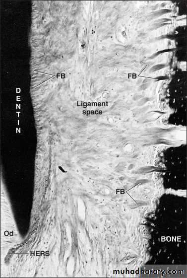
Intercellular tissue
Surrounds and protects the cells of p.d.l. and also the product of these cells, composed of ground substance (glycoprotein, and proteoglycans) that surround the collagen fibers, these protein and polysaccharide provide the cells with vital substance that arise from the blood capillaries and return catabolic from these cells to the vessels.Interstitial tissue
Some of blood vessels, lymphatic and nerves of p.d.l. are surrounded by loose C.T., these area termed interstitial tissue, they occupy spaces between bundles of principal fibers called Interstitial space, these spaces are designed to withstand the impact of force.
Structures present in p.d.l.
-Blood vessels-Lymphatics
-Nerves
-Cementicles
Blood vessels
The arterial vessels of the p.d.l. are derived from three sources1-Branches in the p.d.l. from apical vessels that supply the dental pulp
2-Branches from intra-alveolar vessels, these branches run horizontally, penetrating the alveolar bone to enter the p.d.l.
3-Branches from gingival vessels enter p.d.l. from the coronal direction.
There is rich vascular plexus at the apex and in the cervical part of the ligament. The venous vessels tend to run axially to drain to the apex.
• Lymphatics
• A network of lymphatic vessels following the path of the blood vessels. It drains lymph from p.d.l. into the adjacent alveolar bone.
Nerves
The larger nerve trunks traverse the p.d.l. in the central zone of the tooth's long axis. The nerve fibers are either of large diameter and myelinated concerned with the sensation of touch or be small diameter may or may not be myelinated concerned with pain. Pressure receptors are located among principal fibers of the ligament. Autonomic nerve fibers are associated with blood vessels.Cementicles
These are calcified bodies found in p.d.l. seen in older individual, they may remain free in C.T. or they may fuse into large calcified masses joined with cementum.
Functions of the periodontal ligament
1. supportive• Attachment of teeth to bone.
• Maintenance of gingival tissue in the proper relationship to the teeth.
• Resistance to impact of occlusal forces.
• Provision of a soft tissue cassing to protect the vessels and nerves from injury by mechanical forces.
2.Sensory
The p.d.l. is supplied with abundant receptors and nerves that sense any movement in function when the receptors sense pressure the nerve send signals to the brain to inform the masticatory apparatus, including the temporomandibular joint and muscles of mastication.3.Nutritive
The blood vessels of the ligament provide the essential nutrient for the ligaments' vitality and the heart tissue of the cementum and bone. All cells require nutrition which is carried by the blood vessels to the ligament and also the blood vessels are concerned with the removal of catabolite.4.Homeostatic
It is evident that the cells of the p.d.l. have the capacity to resorb and synthesize the extracellular substance of the C.T. of the ligament, alveolar bone and cementum. Alveolar bone appears to be resorbed and replaced (remodeled) at a rate higher than other bone tissue in the jaws. Furthermore, the collagen of the p.d.l. is turned over at a rate that may be fastest of all C.T in the body.