To define the main functions of lower limbs
To list the bony skeletal parts & describe them
To describe the way of attachment of lower limbs to the trunk
To describe various movements & innervations
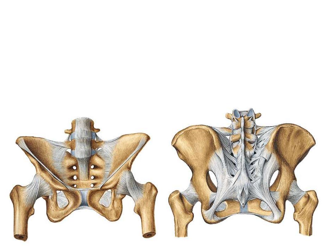
-Lower limbs are the weight bearing & locomotor organs
-The sacrum is the site where the whole weight of the upper part of the
body is supported through its articulation with L5
-The sacroiliac joint transmits this weight to the hip bones
-The hip joint will carry most of this weight to the lower limb bones, the to
the ground
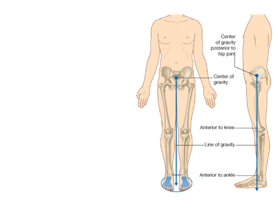
Functions:
1- Weight support:
- When standing erect, a vertical line
through the center of gravity is
slightly posterior to the hip joints,
anterior to the knee and ankle joints,
and directly over the almost circular
support base formed by the feet.
- The
organization
of
ligaments
together with the shape of the
articular surfaces facilitates 'locking'
of these joints into position when
standing, so reducing the muscular
energy
required
to
maintain
a
standing position.
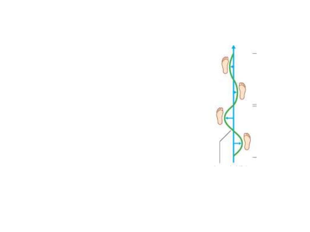
2- Locomotion:
- During
walking,
many
anatomical
features of the lower limbs contribute to
minimizing fluctuations in the body's
center of gravity and so reduce the
amount of energy needed to maintain
locomotion
- They include pelvic tilt in the coronal
plane, pelvic rotation in the transverse
plane, movement of the knees toward the
midline,
and
complex
interactions
between the hip, knee, and ankle
L4
L5
S1
Parts:
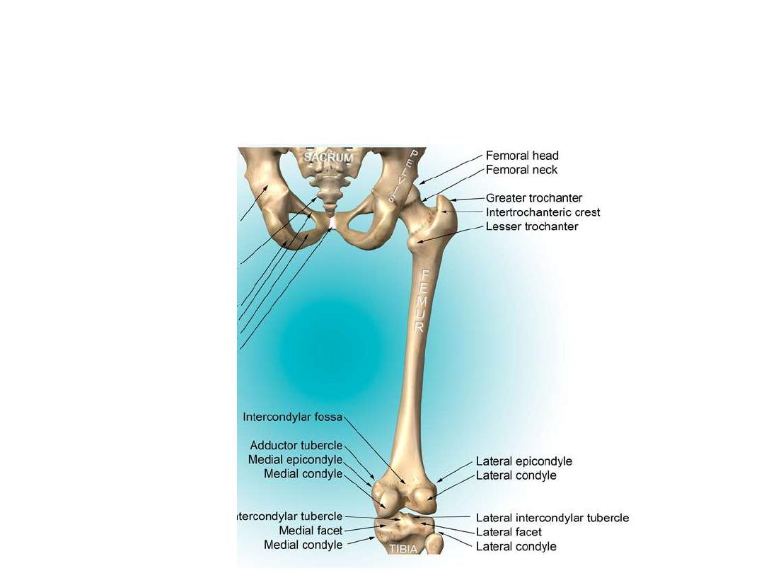
The femur:
– Largest, longest, strongest bone in the body!!
– Receives a lot of stress
– Courses medially (more in women)
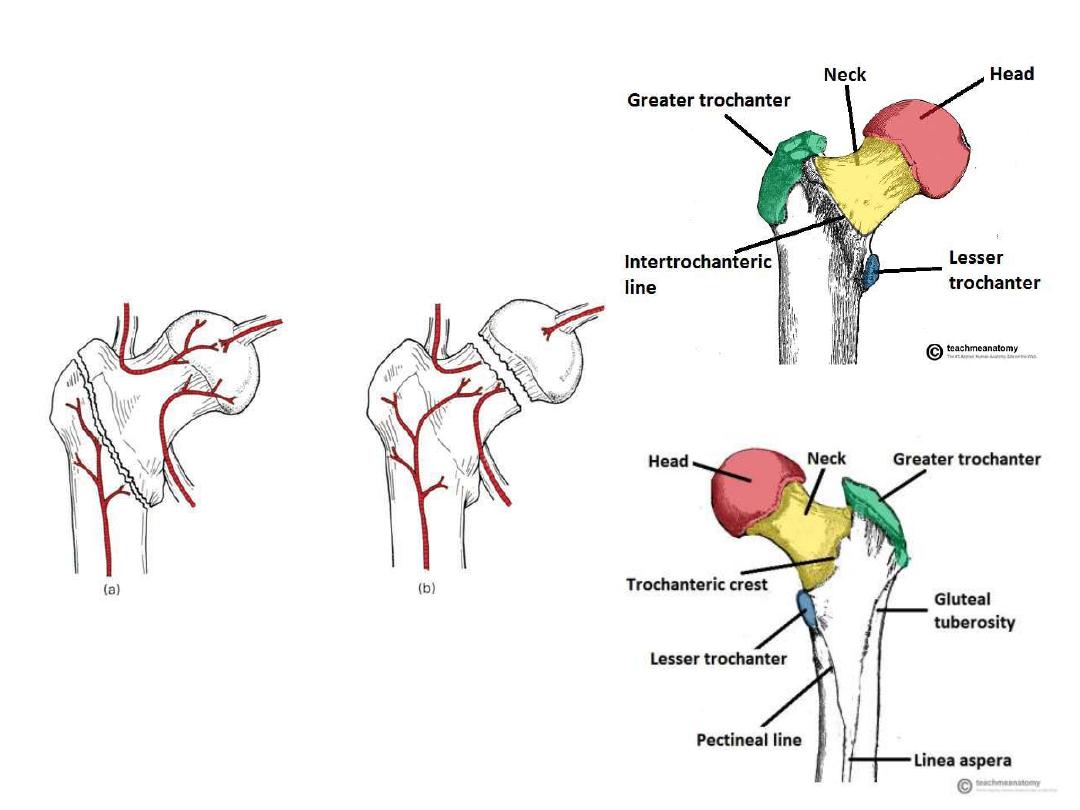
Upper end & blood supply:
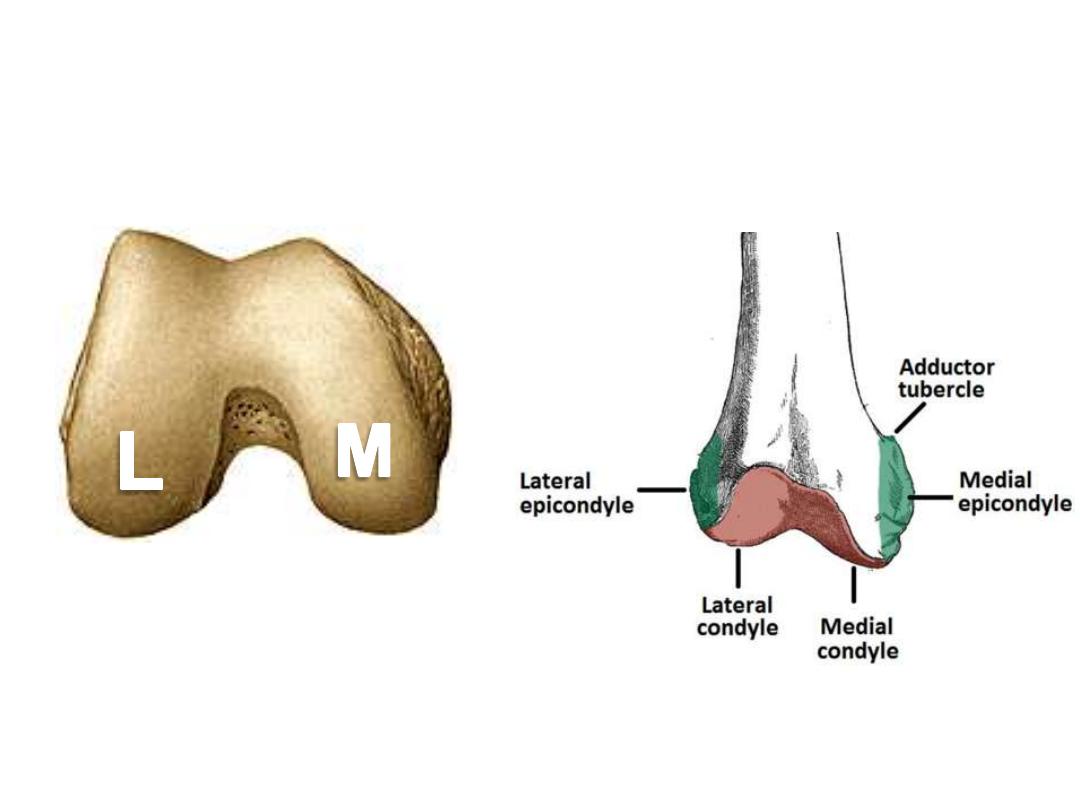
Lower end & patellofemoral articulation:
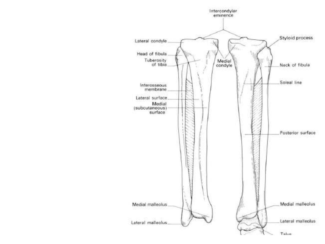
The leg bones:
• Tibia
– Receives the weight of
body from femur and
transmits to foot
– Second to femur in size
and weight
– Articulates with fibula
proximally and distally
• Fibula
– Does NOT bear weight
– Muscle attachment
– Not part of knee joint
– Stabilize ankle joint
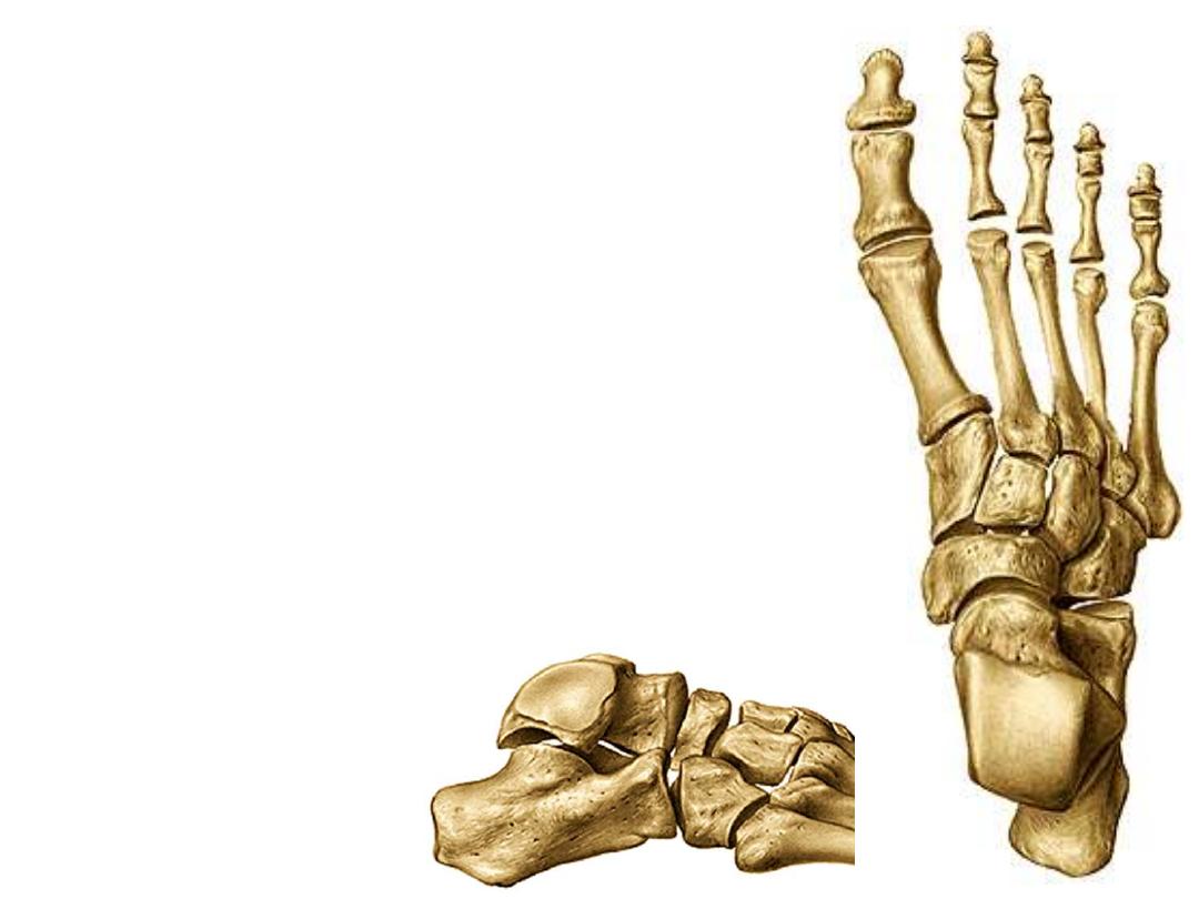
The foot:
• Function:
– Supports the weight of the body
– Act as a lever to propel the body forward
• Parts:
– Tarsals
• Talus = ankle
– Between tibia and fibula
– Articulates with both
• Calcaneus = heel
– Attachment for Calcaneal tendon
– Carries talus
• Navicular
• Cuboid
• Medial, lateral and intermediate
cuneiforms
– Metatarsals
– Phalanges
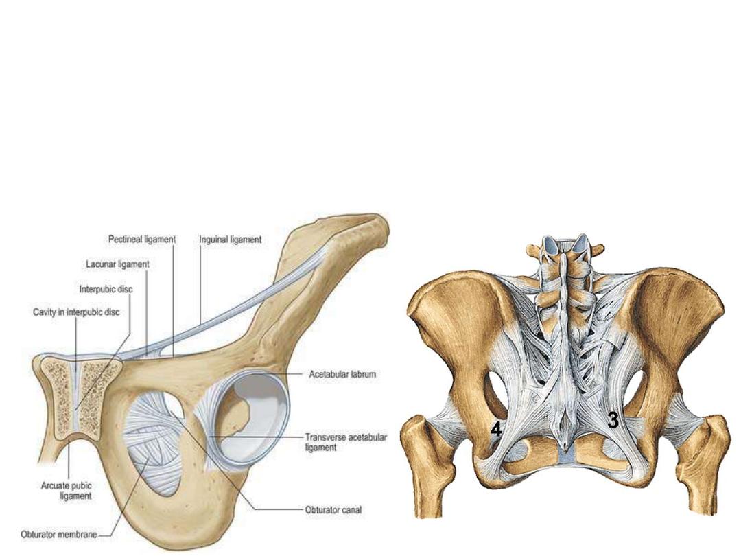
Important ligaments:
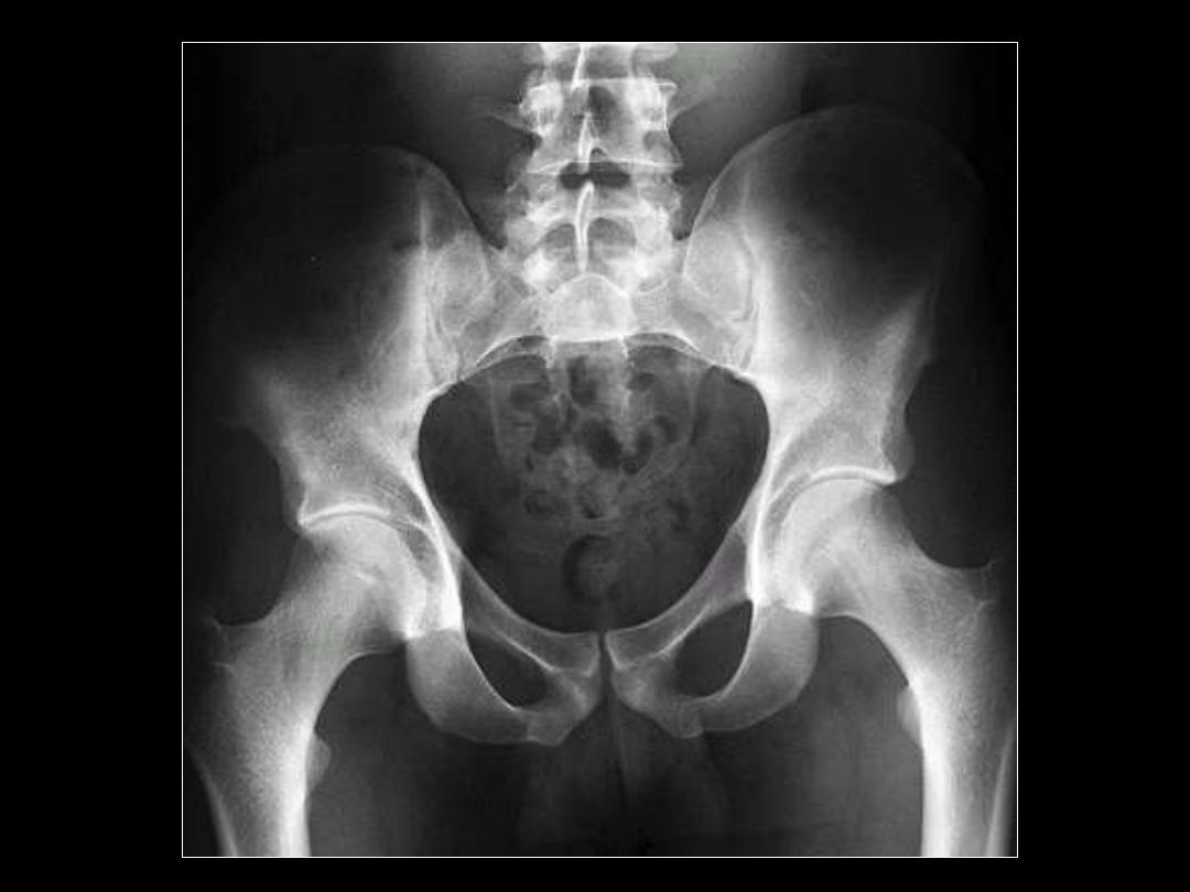
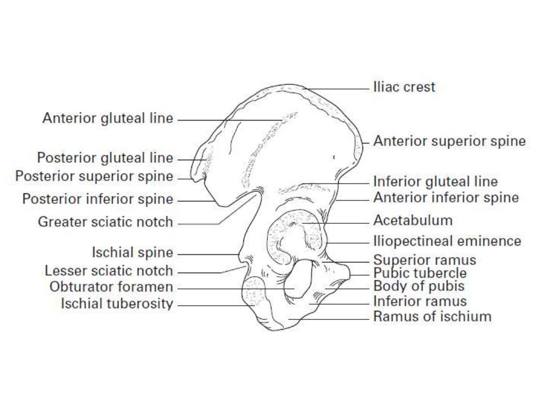
1- The inguinal ligament:
extends between the anterior superior iliac spine &
pubic tubercle.
2- Lacunar ligament:
Lies behind the medial end of IL
3- Sactotuberous ligament:
between the sacrum & ischial tuberosity
4- Sacrospinous ligament:
between the sacrum & ischial spine
1
2
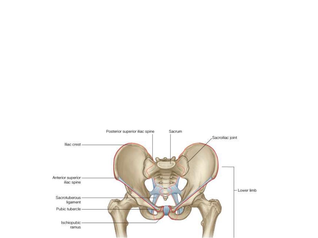
Lines of demarcation:
1- Between the pubic tubercle with the anterior superior iliac spine (position of
the inguinal ligament) & extends to the PSIS to separate the lower limb from
the anterior and lateral abdominal walls
2- Between PSIS & the coccyx to separate the lower limb from the muscles of
the back
3- Between the medial margin of the sacrotuberous ligament, the ischial
tuberosity, the ischiopubic ramus, and the pubic symphysis to separate the
lower limb from the perineum
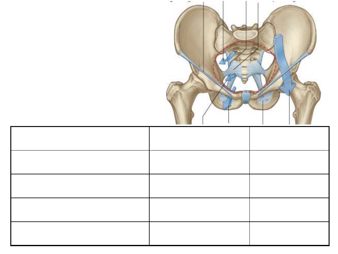
Pathway
Lower limb part
Body part
Gap between inguinal L & hip
Femoral triangle
Iliac fossa
Greater sciatic foramen
Gluteal region
Pelvis
Obturator canal
Medial compartment
Pelvis
Lesser sciatic foramen
Gluteal region
Perineum
Unlike in the upper limb where
most structures pass between the
neck and limb through a single
axillary inlet, in the lower limb,
there are four major entry and exit
points:
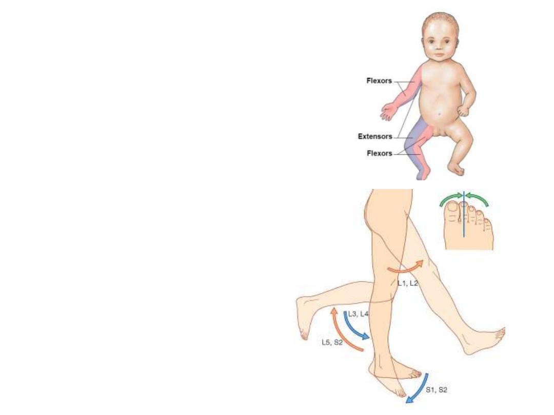
Embryological rotation:
The LL bud had rotated in utero so that:
-Flexion of the hip remained the ventral
movement of the thigh against anterior
abdominal wall, while hip extension is the
reverse
-Knee flexion is the posterior movement
of the leg while extension is the ventral
movement (straightening the leg)
-Dorsiflexion of the ankle is to move toes
off the ground while plantar flexion is to
press the ground by toes
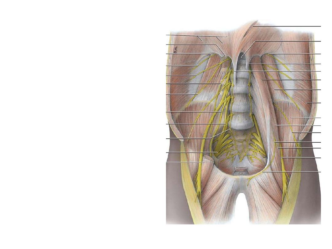
Lumbar plexus:
-The anterior primary rami of L1-L5
once leave the vertebral canal lie
within psoas major substance
-They
supply
it
&
quadratus
lumborum segmentally
-They divide into anterior & posterior
divisions then re-unite within the
muscle to give the branches
-All branches leave through psoas
major in the posterior abdominal wall
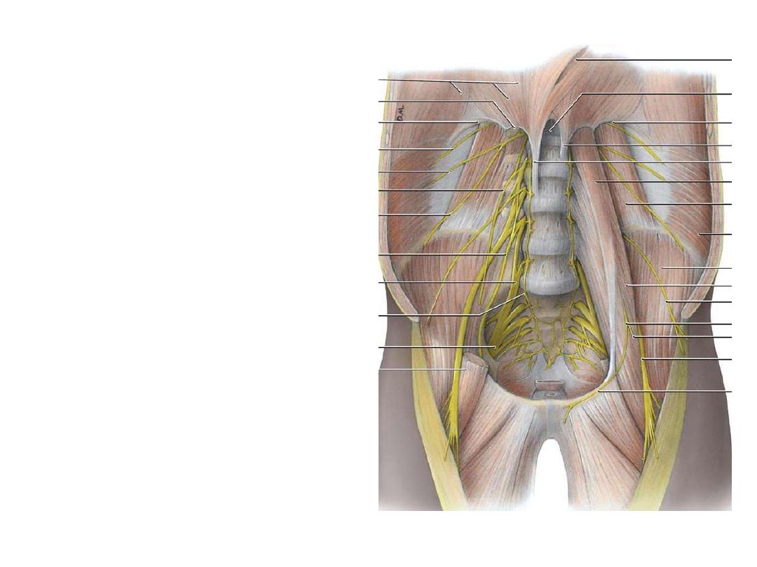
-L1 goes to the abdomen & external
genitalia
-L2-4 give the thigh nerves
-The reminder of L4 + L5 enter the
pelvis as the lumbosacral trunk
-LS trunk shares with the sacral
nerves in the formation of sacral
plexus which will supply the rest of
the lower limb & the pelvis
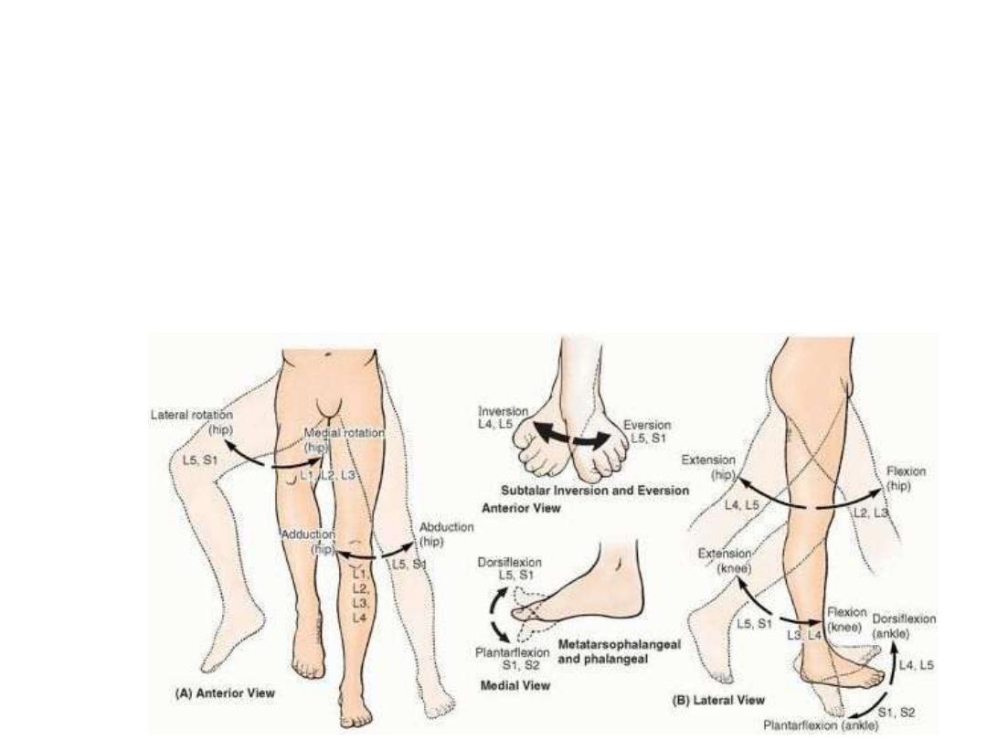
Rules of innervation in the limbs:
-Plexus roots are the anterior primary rami of corresponding spinal nerves (not the
roots of spinal nerve)
-Most proximal muscles are supplied by highest roots of the plexus
-Flexor, adductor & medial rotator muscles are supplied by anterior plexus
divisions
-Extensor, abductor & lateral rotator muscles are supplied by posterior plexus
divisions
