
To classify the joints relative to structure & shape
To describe the anatomy of the hip joint
To describe the ankle joint
To memorize their blood & nerve supply
JOINTS:
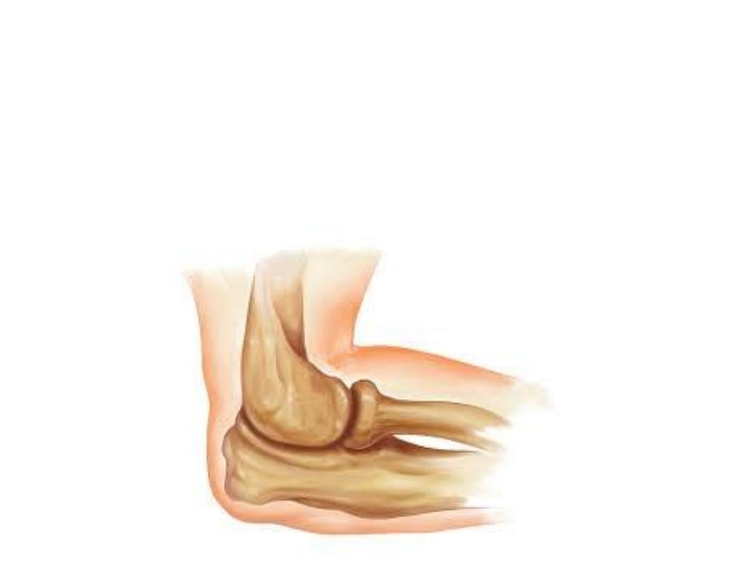
Joints are sites where skeletal structures (bones &/or cartilages) are
connected to each other
Joints are designed for movements though some are immobile
Joints can form between two bones (most joints), two cartilages (like
laryngeal joints), or bone & cartilage (like costo-chondral junctions)
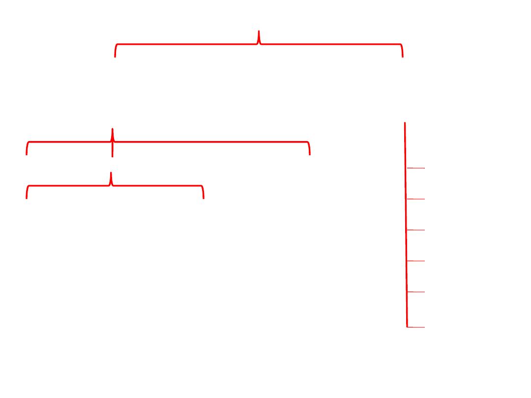
Classification
Structural
(according to the material which
holds the joint)
Morphological
(according to the shape of articulating
surfaces)
Fibrous
Cartilagenous
Synovial
Primary
Secondary
Ball & socket
Condyloid
Gliding
Hinge
Pivot
Saddle
Structural types:
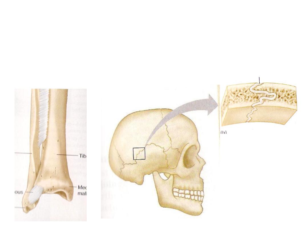
Fibrous joints:
-Articulating surfaces are fastened together by dense connective tissue
containing collagen
-Most of them are immobile
-Examples; skull sutures, tibiofibular joints
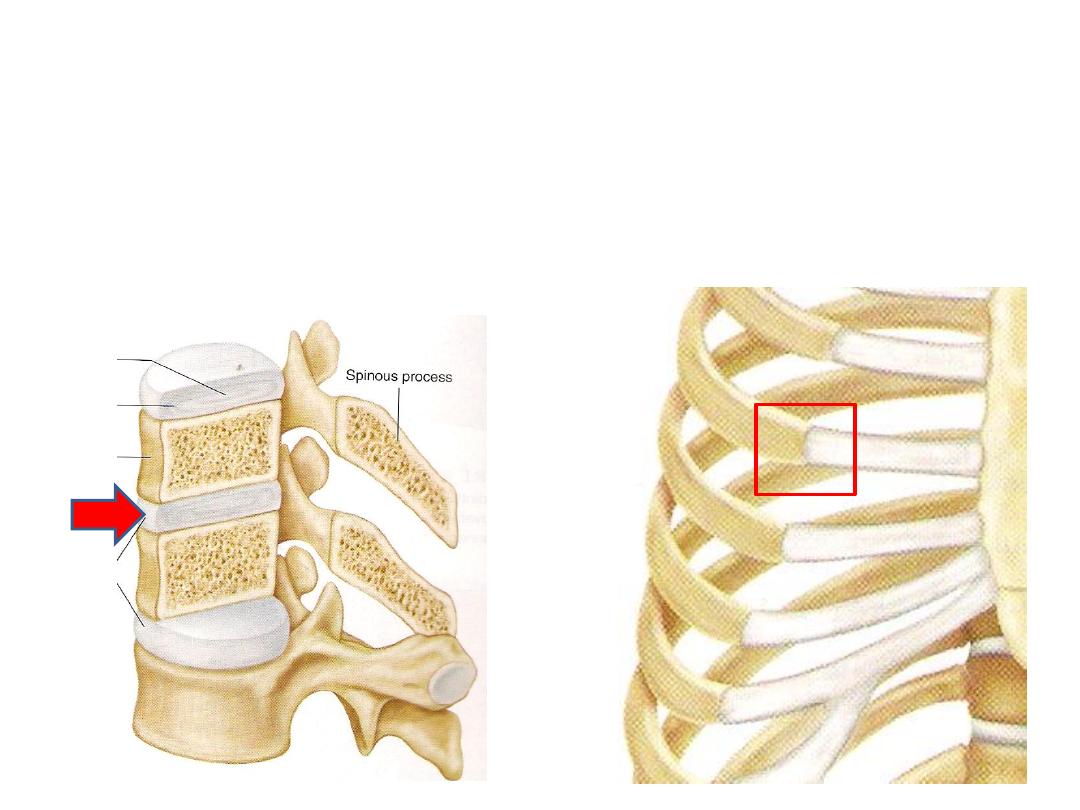
Cartilagenous joints:
-Articulating surfaces are connected by hyaline cartilage or fibrocartilage
-They provide little or no movement
1- Primary cartilagenous joints;
bone meets hyaline cartilage, like the costo-chondral
joint
2- Secondary cartilagenous joints (symphesis);
Bones covered by a hyaline cartilage &
held by fibrocartilage, like intervertebral discs
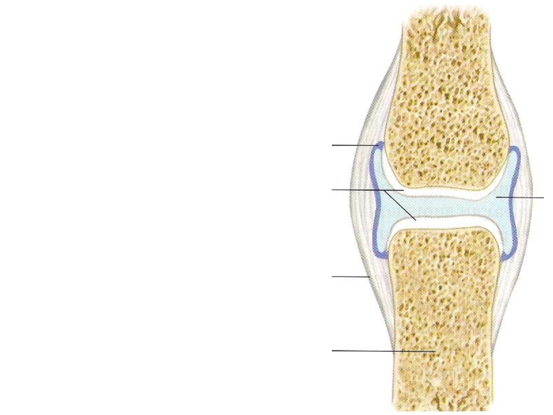
Synovial joints:
Characterized by:
1- Surrounded by a joint capsule
2- Supported by ligaments
3- Lined with synovial membrane
4- Covered by hyaline cartilage
5- Contains synovial fluid
1
4
5
3
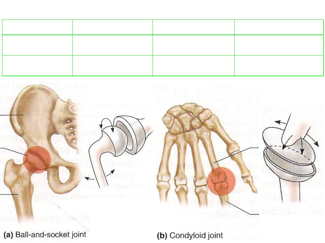
example
Possible movement
Description
Joint type
Hip, shoulder
Free + rotation
Ball-shaped head +
cup-shaped socket
1- Ball & socket
Metacarpo-phalyngeal
Free without rotation
Oval condyle +
elliptical cavity
2- Condyloid
Morphological types:
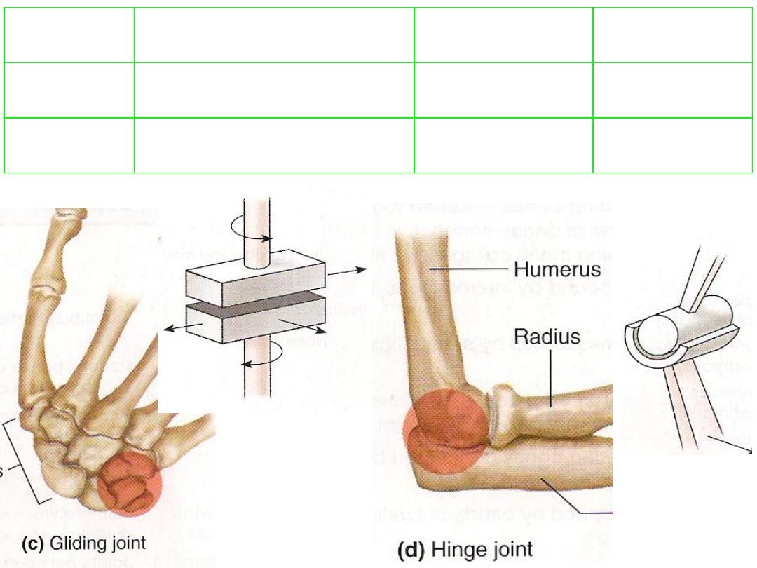
example
Possible movement
Description
Joint type
Intermetacarpal
Sliding or twisting
Articulating surfaces are flat or
slightly curved
3- Gliding
Elbow
Flexion-extension
Convex surface + concave one
4- Hinge
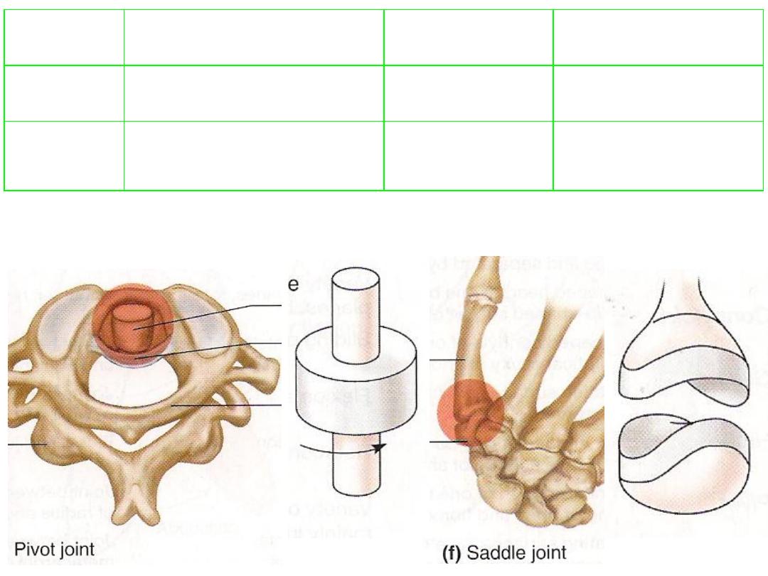
example
Possible
movement
Description
Joint type
Atlantoaxial
Rotation
Cylindrical surface + ring
surface
5- Pivot
Carpometacarpal joint
of thumb
Mainly movements
in two planes
Articulating surface is both
convex & concave against
complementary surface
6- Saddle
Joint stability:
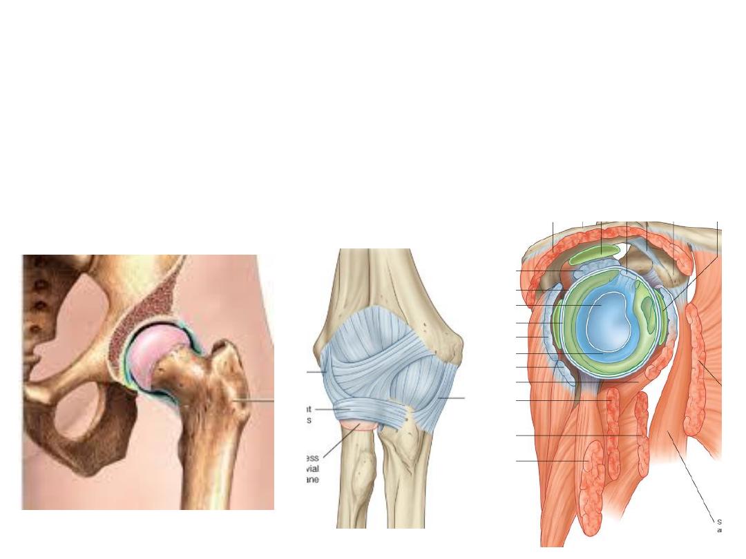
The stability of joint is maintained by three main factors, morphological,
ligamentous & muscular
All these factors usually share in joint stability, although one of them will
prdominates in specific joints
Examples:
1- The shape of the joint is the main stabilizing factor in the hip joint
2- Ligaments are the main stabilizing factor in the elbow joint
3- Muscular factor is the main stabilizing factor in the shoulder joint
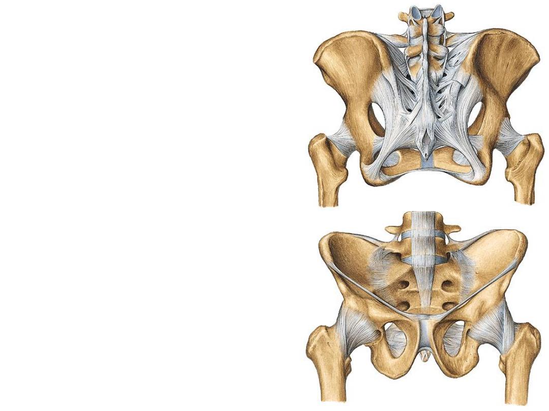
Sacro-iliac joints:
-The SIJ transmit forces from the lower
limbs to the vertebral column.
-They are synovial joints between the
articular facets on the lateral surfaces of
the sacrum and similar facets on the iliac
parts of the pelvis
-The joint surfaces have an irregular
contour and interlock to resist movement.
Ligaments:
1- Anterior sacro-iliac ligament
2- Interosseous sacro-iliac ligament
3- Posterior sacro-iliac ligament
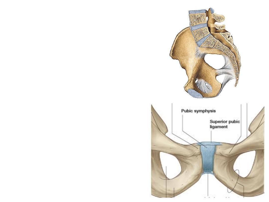
Pubic symphysis:
-A secondary cartilagenous joint
between the adjacent surfaces of the
pubic bones
-Joint surfaces are linked across the
midline by fibrocartilage
Ligaments:
1- The superior pubic ligament
2- The inferior pubic ligament
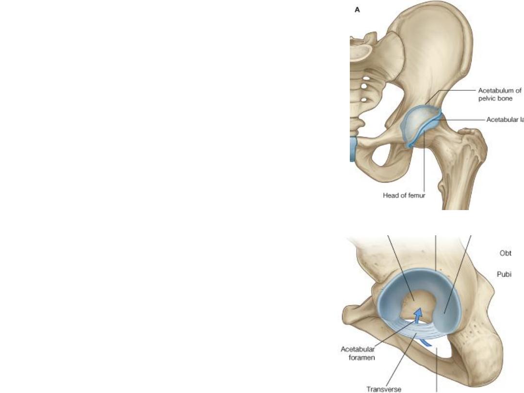
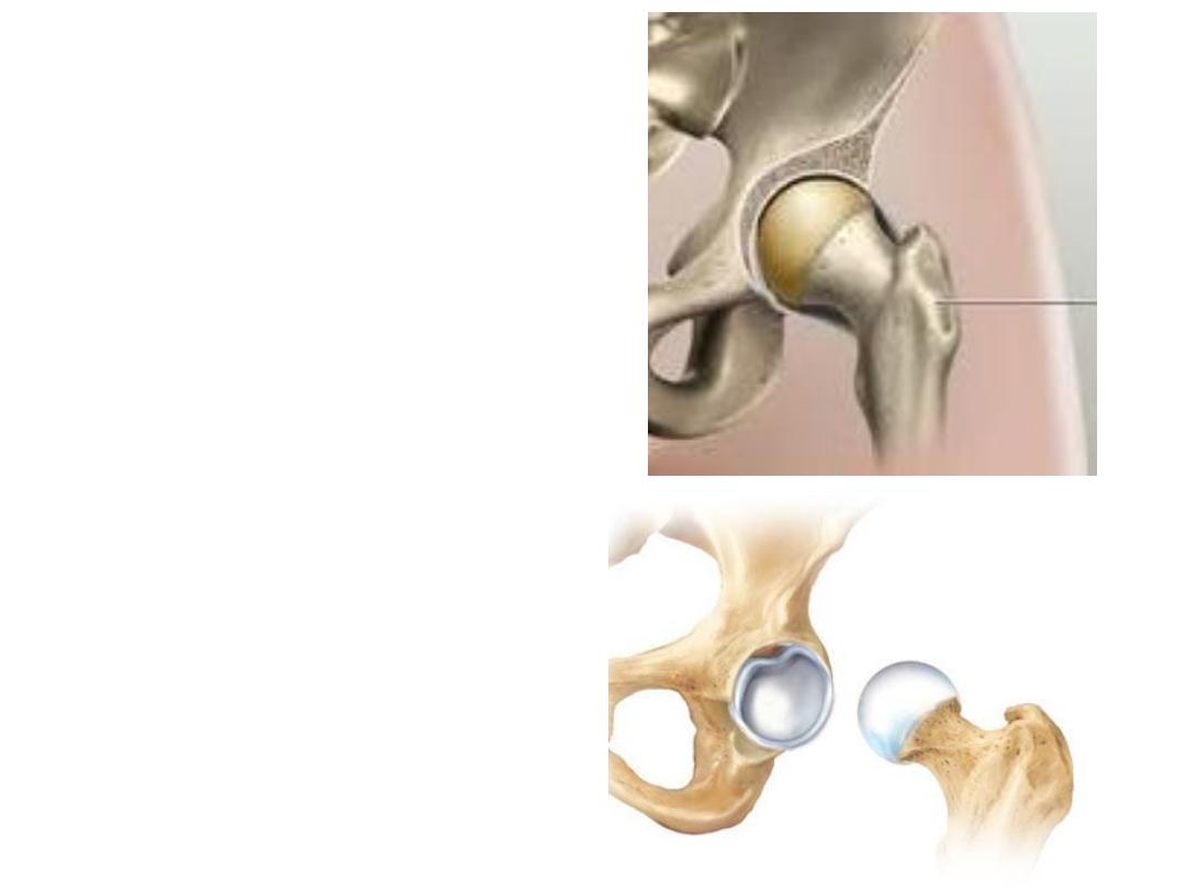
The hip joint:
-Is a synovial articulation between the
head of the femur & the lunate surface of
the acetabulum of the pelvic bone
-The joint is a multiaxial ball and socket
joint designed for stability and weight
bearing at the expense of mobility.
-Movements at the joint include flexion,
extension, abduction, adduction, medial
and lateral rotation, and circumduction.
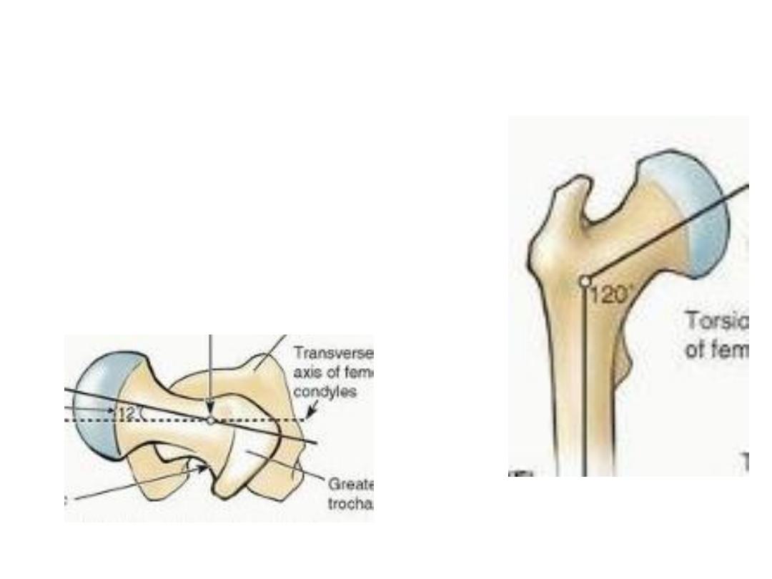
Angles of the femur:
Angle of inclination;
The femur is
“bent” so that
the long axis of the head and neck lies at an angle
of about 125
O
to that of the shaft.
Angle of torsion;
when looking at the femur from
above, it can be seen that the axis of the head and
neck forms a 12
° angle with the transverse axis of
the femoral condyles
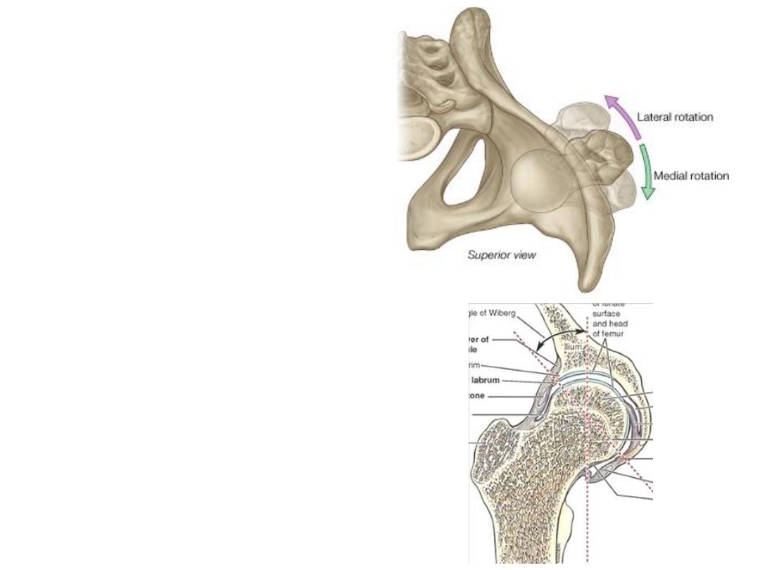
Mechanics:
When considering the effects of
muscle action on the hip joint, these
angles must be borne in mind.
For example, medial and lateral
rotation of the femur involves
muscles that move the greater
trochanter forward and backward,
respectively,
relative
to
the
acetabulum
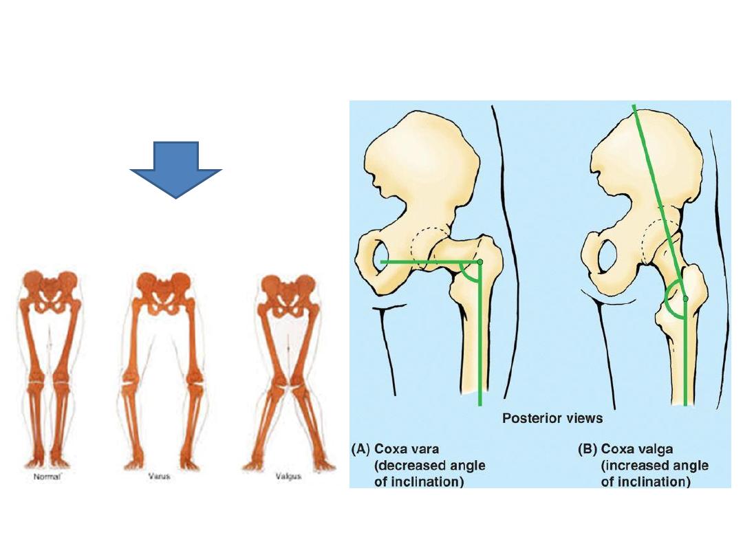
Abnormal inclination angle
Limits certain movements
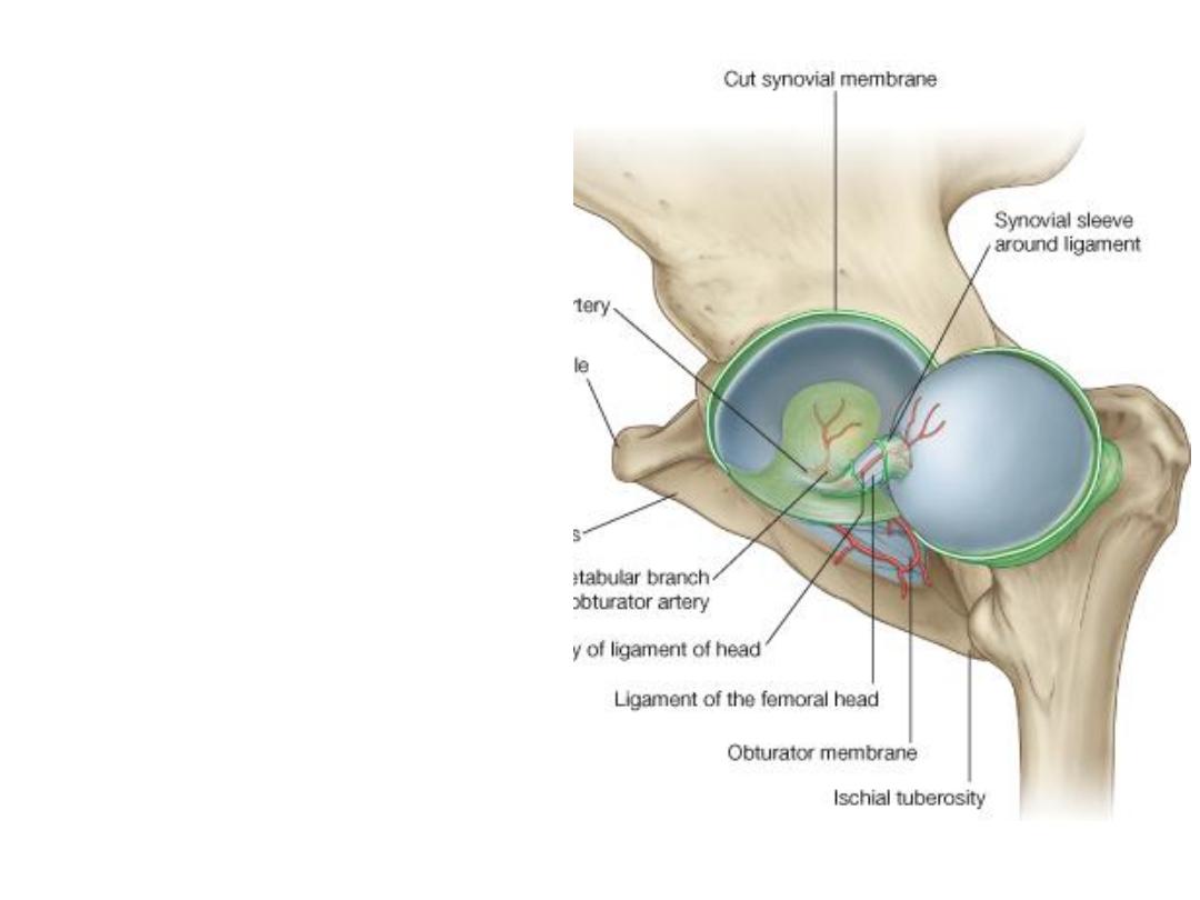
The ball & socket property:
Complete typical ball-and-socket
design of the hip is the major factor
which predispose to its stability
To increase this property, a rim of
the acetabulum is raised slightly by
an acetabular labrum
Acetabular notch is bridged by the
labrum & transverse acetabular
ligament
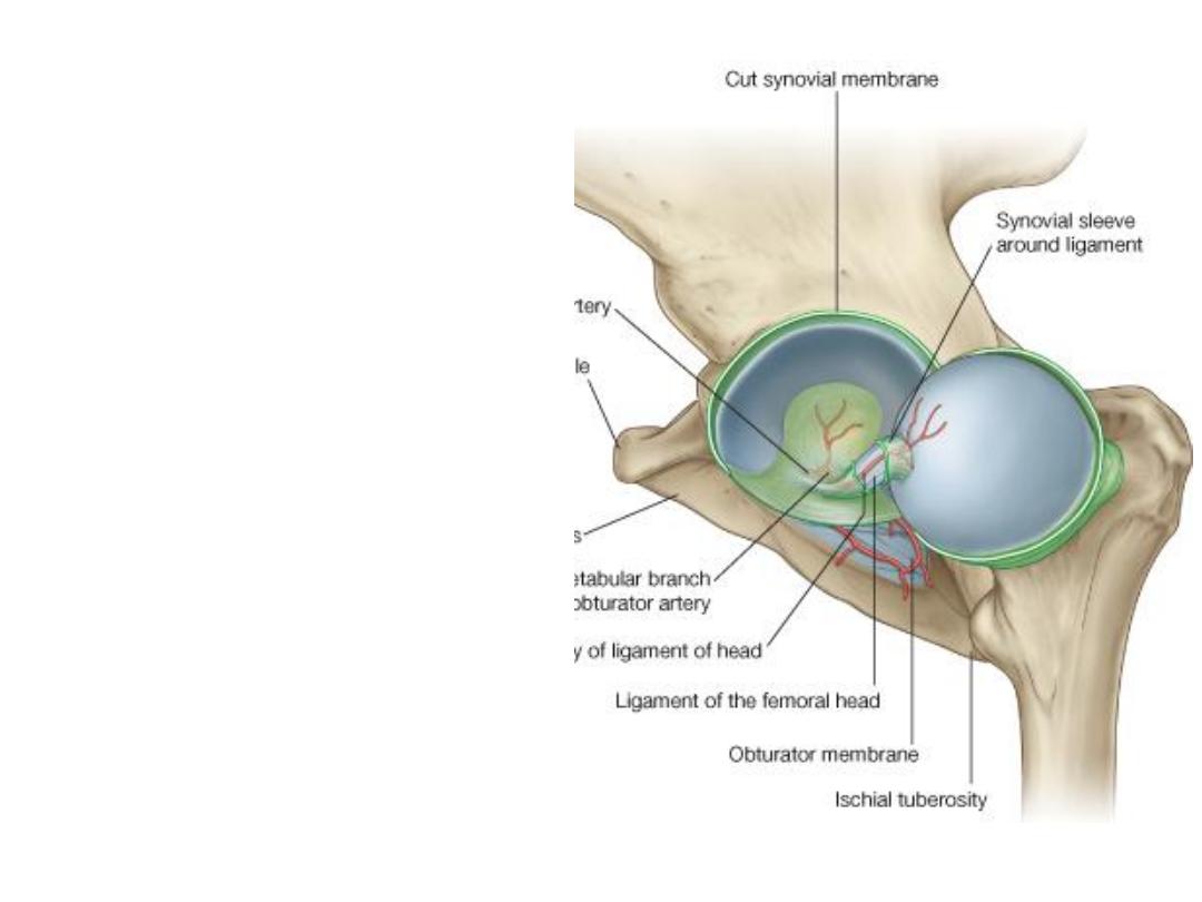
Articular surfaces:
Except for the fovea, the head of
the femur is covered by hyaline
cartilage as well as the lunate
surface of the acetabulum
The non articular acetabular fossa
contains loose connective tissue.
Ligament of head of femur extends
between these two non articular
surfaces
The synovium:
-The synovial membrane attaches to
the margins of the articular surfaces
of the femur and acetabulum
-From its attachment to the margin
of the femoral head, the membrane
covers the neck of the femur before
reflecting onto the fibrous capsule
The capsule:
-Medially, it is attached to the margin of the acetabulum, the transverse
acetabular ligament
- Laterally, it is attached to the intertrochanteric line anteriorly & to the neck
posteriorly
-From the distal attachment, some of the capsule fibers return back on the
neck of femur binding arterial branches to the femoral head
(retinacular fibers)
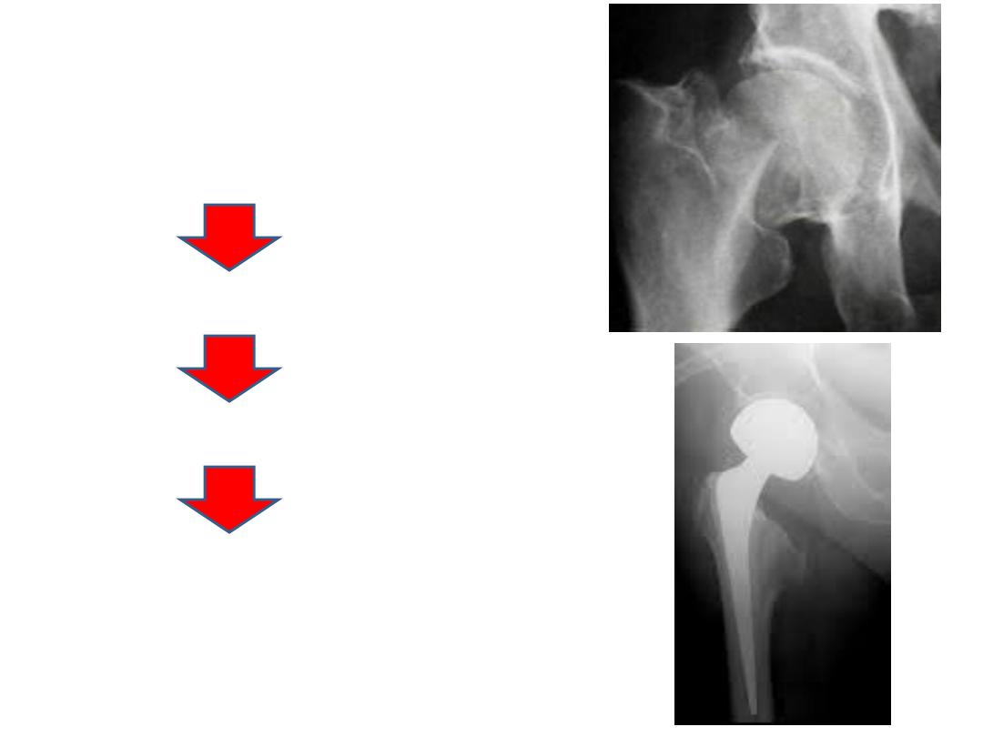
Austin Moore
prosthesis
Partial hip
replacement
Fracture neck of femur
Damage to retinacular fibers
Avascular necrosis of the head
Often needs surgery
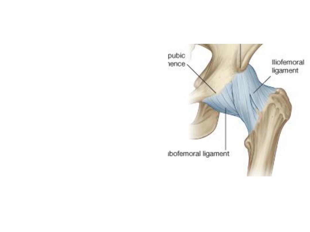
Ligaments:
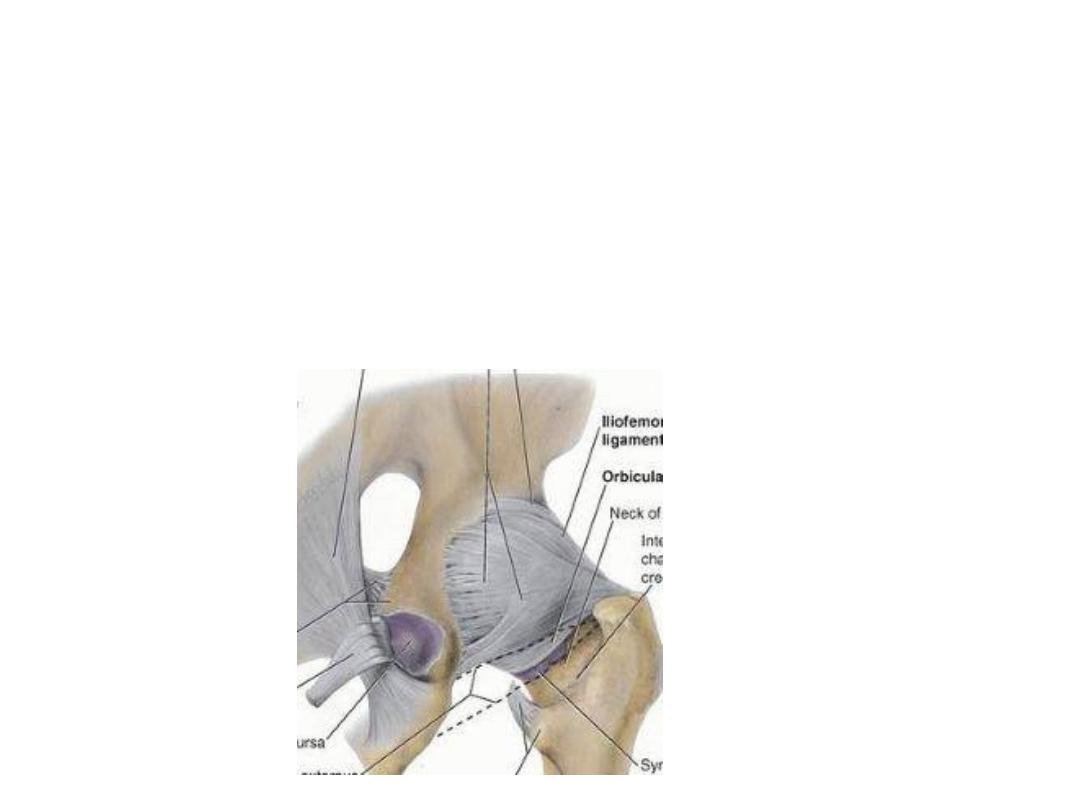
The iliofemoral ligament:
-Y- shaped ligament, lies anterior to the
hip joint
-The stem is attached between the
anterior inferior iliac spine & acetabulum
while its base is attached between the
ends of the intertrochanteric line.
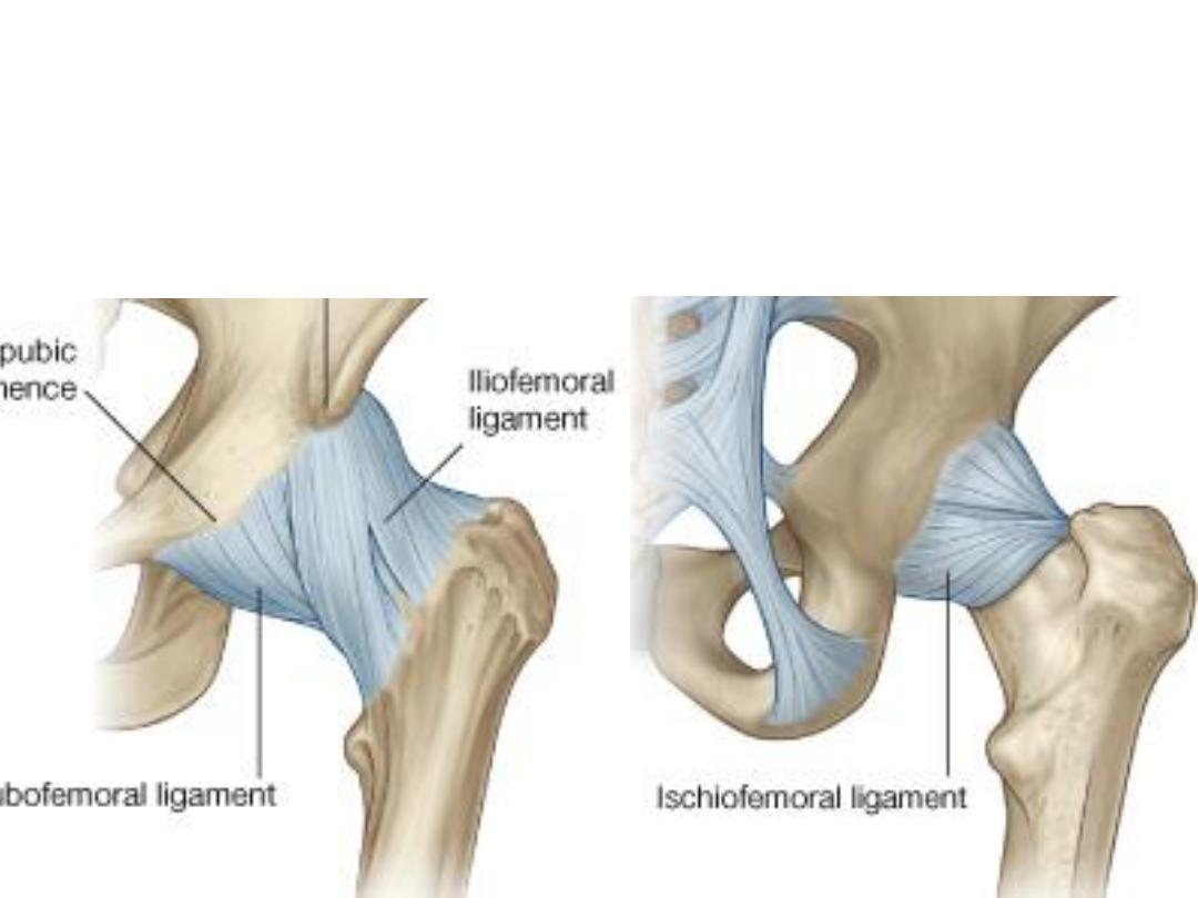
The pubofemoral ligament:
Lies on the anterior surface of the joint deep to the iliofemoral one
The ischiofemoral ligament:
Reinforces the posterior aspect of the fibrous membrane
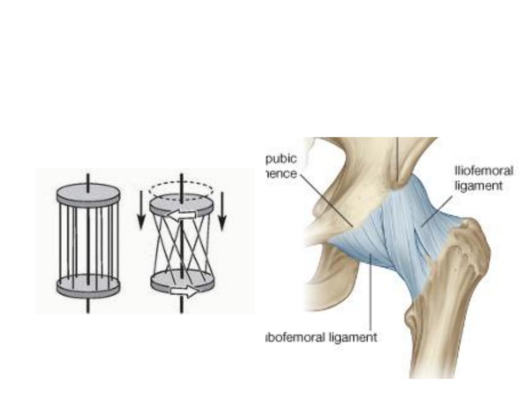
The fibers of all three ligaments are oriented in a spiral fashion around the hip
joint so that they become taut when the joint is extended.
This stabilizes the joint and reduces the amount of muscle energy required to
maintain a standing position.
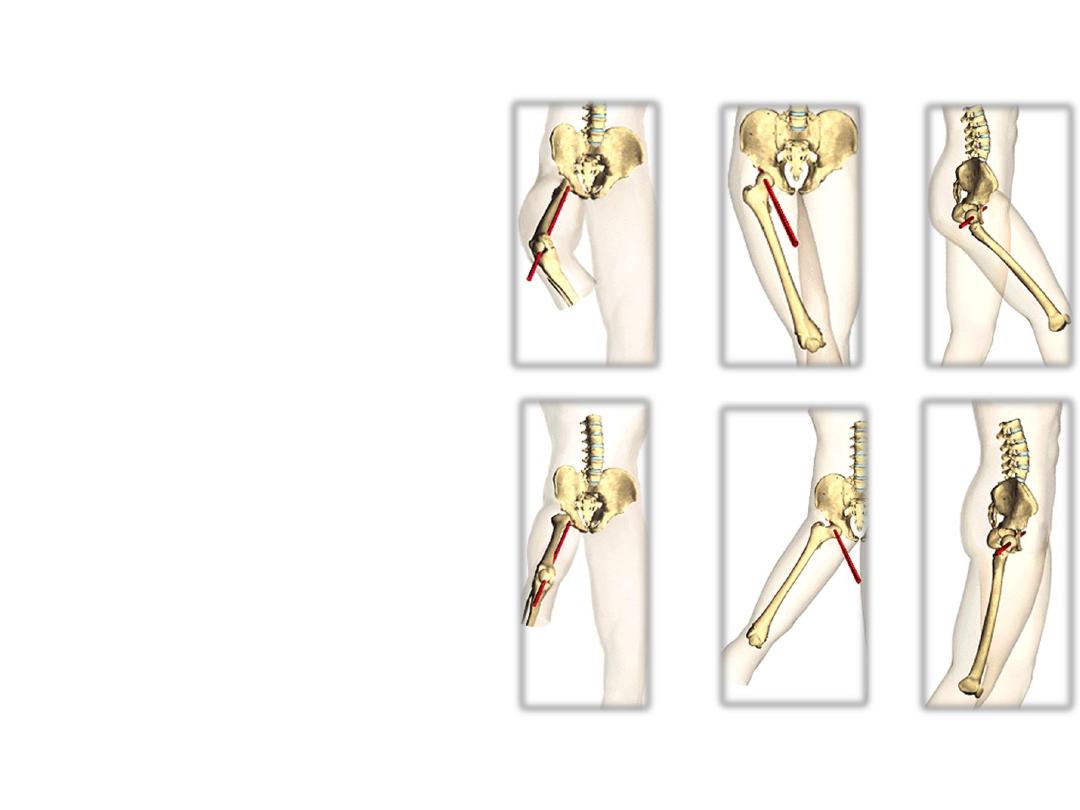
Hip movements:
-
Movements of the trunk at the hip
joints are also important, such as
those occurring when a person lifts
the trunk from the supine position
-
The degree of flexion and extension
possible at the hip joint depends on
the position of the knee. Flexed knee
with
relaxed
hamstrings
permits
active flexion
-
Iliofemoral
ligament
limits
hip
extension
-
Abduction & lateral rotation have
more
range
of
movement
than
adduction & medial rotation because
of the shape of joint & contact of
lower limbs with each other
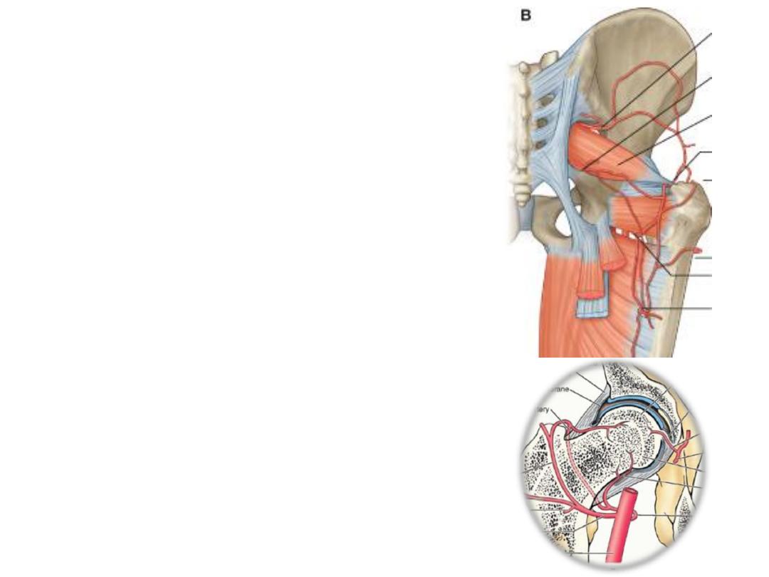
Vascular supply:
1- Head of femur; trochanteric anastomosis
via retinacular fibers passing across the
neck, the artery of head of femur is
insufficient for the supply.
Fracture of femoral neck leads to death of
femoral head!
2- The obturator, circumflex, gluteal & the
first perforating branch of the deep artery of
the thigh form a vascular network which
supplies the joint.
Innervation:
The hip joint is innervated by articular
branches from the femoral, obturator,
superior gluteal nerves and nerve to the
quadratus femoris
(Hilton’s law).
