Fifth stage
PediatricLec-2
د.خليل
19/10/2016
The cardiovascular systemAcyanotic Congenital Heart Disease
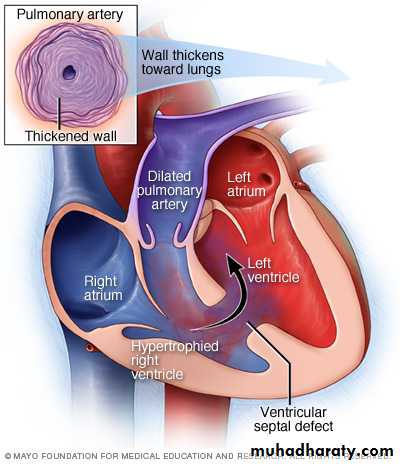
VENTRICULAR SEPTAL DEFECT
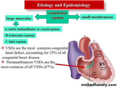
Pathophysiology:-
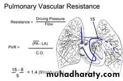
The amount of flow crossing a VSD depends on the size of the defect and the pulmonary vascular resistance.
Even large VSDs are not symptomatic at birth because the pulmonary vascular resistance is normally elevated at this time.
As the pulmonary vascular resistance normally decreases over the first 6to 8 Weeks of life, however,
the amount of shunt increases, and symptoms may develop.
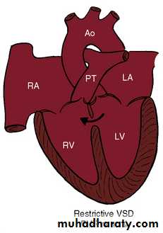
When a small communication is present (usually <0.5 cm2), the VSD is called restrictive and right ventricularpressure is normal.
In large nonrestrictive VSDs (usually >1.0 cm2), right and
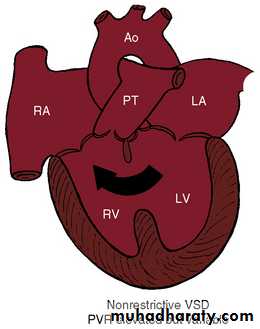
left ventricular pressure is equalized. In these defects, the direction of shunting and shunt magnitude are determined by the ratio of pulmonary to systemic vascular resistance
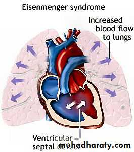
Chronic left-to-right shunt causes gradual ↑pulmonary vascular pressure, ↓gradient between ventricles, and ↓
shunt volume, and finally pulmonary hypertension and right to left shunt, a condition called Eisenmengers’s syndrome
Clinical manifestations
Small VSDs, with little shunt, are often asymptomatic, other than a loud murmur.Moderate to large VSDs result in pulmonary overcirculation and CHF, presenting as fatigue, diaphoresis with feedings, and poor growth.
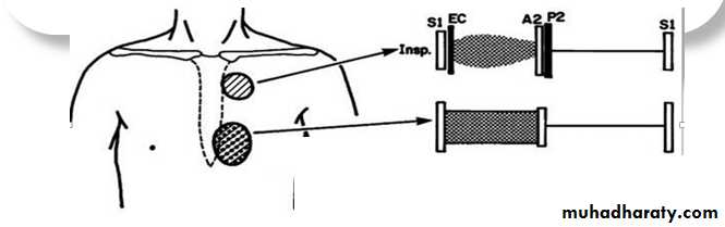
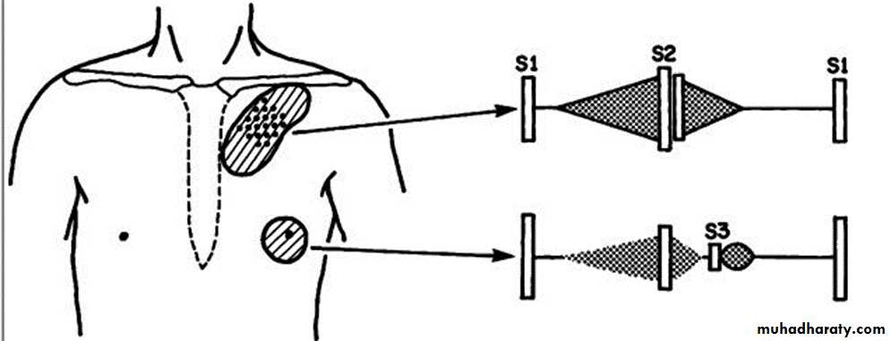
The typical physical finding with a VSD is a pansystolic murmur usually heard best at the lower left sternal border. There may be a thrill in the same region.
Investigations
ElectrocardiographyWith a small VSD, the ECG is normal.
With a moderate VSD, left ventricular hypertrophy (LVH) and occasional left atrial hypertrophy (LAH) may be seen.
With a large defect, the ECG shows biventricular hypertrophy (BVH) with or without LAH .
If pulmonary vascular obstructive disease develops, the ECG shows RVH only.
X-ray Studies
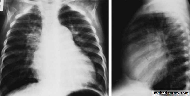
With large VSD cardiomegaly of varying degrees is present and involves the LA, left ventricle (LV), and sometimes RV. Pulmonary vascular markings increase. The degree of cardiomegaly and the increase in pulmonary vascular markings directly relate to the magnitude of the Lt to Rt shunt.CXR of 6 years old child PA and lateral views showing cardiac enlargement and increased pulmonary markings
CXR of 6 years old child PA and lateral views showing cardiac enlargement and increased pulmonary markings
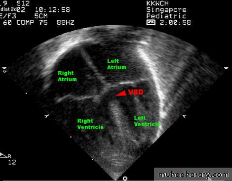
Echocardiography.
Two-dimensional and Doppler echo studies can identify the number, size, and exact location of the defect; estimate PA pressure; identify other associated defects; and estimate the magnitude of the shunt.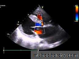
NATURAL HISTORY
Spontaneous closure occurs in 30% to 40% of patients with membranous VSDs and muscular VSDs during the first 6 months of life. It occurs more frequently in small defects.The vast majority of defects that close do so before the age of 4 yrCHF develops in infants with large VSDs but usually not until 6 to 8 weeks of age.
Pulmonary vascular obstructive disease may begin to develop as early as 6 to 12 months of age in patients with large VSDs, but the resulting right-to-left shunt usually does not develop until the teenage years.
Repeated chest infections and arrhythmias.
Infective endocarditis rarely occurs.
Treatment
Small VSDs usually close spontaneously; if they do not close, surgical closure may not be required.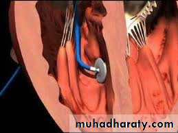
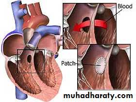
Initial treatment for moderate to large VSDs includes diuretics and digoxin. Continued poor growth or pulmonary hypertension despite therapy requires closure of the defect. Most VSDs are closed in surgery, but some VSDs, especially muscular defects, can be closed with devices placed at cardiac catheterization
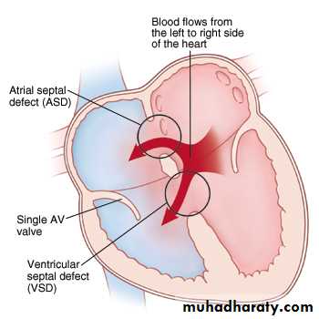
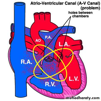
ENDOCARDIAL CUSHION DEFECT (atrioventricular septal defect ) (AV canal defect)
Etiology and EpidemiologyThe defect occurs as the result of abnormal development of the endocardial cushion tissue, resulting in failure of the septum to fuse with the endo-cardial cushion; this results in abnormal AV valves as well.
The complete defect results in a primum ASD, a posterior or inlet VSD, and clefts in the anterior leaflet of the mitral and septal leaflet of the tricuspid valves. In addition to left-to-right shunting at both levels, there may be atrioventricular valvular insufficiency.
The partial defect is presented as ASD primum only.
Clinical manifestation:
The symptoms of CHF usually develop as the pulmonary vascular resistance decreases over the first 6 to 8 weeks of life.Growth is usually poor.
Many children with Down’s syndrome have complete endocardial cushion defects.
Pulmonary hypertension resulting from increased pulmonary circulation often develops early; this results in a prominent S2.

Investigation :
An ECG reveals left axis deviation and combined ventricular hypertrophy and may show combined atrial enlargemen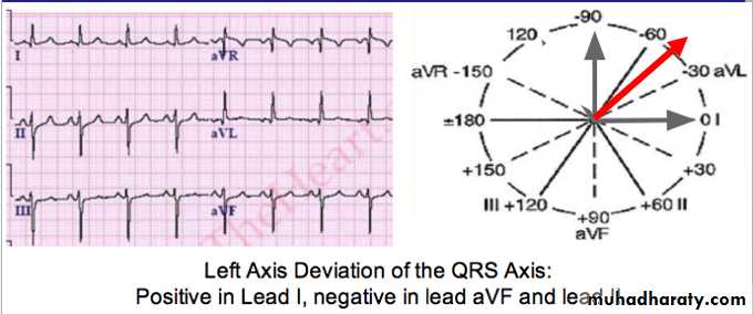
CXR shows cardiomegaly and increased pulmonary vascular markings
Echocaediography is diagnostic and shows the details of the defect.Treatment
Initial management includes digoxin and diuretics for treatment of CHF.Surgical repair of the entire defect ultimately is required, however.
Patent ductus arteriosus
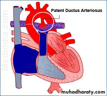
During fetal life, most of the pulmonary arterial blood is shunted through the ductus arteriosus into the aorta .
Functional closure of the ductus normally occurs soon after birth, but if the ductus remains patent when pulmonary vascular resistance falls, aortic blood is shunted into the pulmonary artery.
The aortic end of the ductus is just distal to the origin of the left subclavian artery, and the ductus enters the pulmonary artery at its bifurcation.
Female patients with PDA outnumber males 2 : 1.
Epidemiology
PDA is more common in premature babies and is associated with maternal rubell infection
PDA is seen in 10% of patients with other congenital heart lesions and often plays a critical role in providing pulmonary blood flow when the right ventricular outflow tract is stenotic or atretic or in providing systemic blood flow in the presence of aortic coarctation or interruption
Pathophysiology
As a result of the higher aortic pressure, blood shunts left to right through the ductus, from the aorta to the pulmonary artery.If the PDA is small, pressure within the pulmonary artery, the right ventricle, and the right atrium is normal.
If the PDA is large pulmonary artery pressure may be elevated to systemic levels during both systole and diastole. Patients with a large PDA are at extremely high risk for the development of pulmonary vascular disease if left unoperated.
Pulse pressure is wide because of runoff of blood into the pulmonary artery during diastole.
Clinical manifestation:
SymptomsThey are more less similar to VSD
Patients with small PDAs are asymptomatic.
Moderate to larger shunts produce the symptoms of CHF as the pulmonary vascular resistance decreases over the first 6 to 8 weeks of life.

Physical examination
A widened pulse pressure .A thrill may be palpable with hyperdynamic precordium.
A continuous machine-like murmur can be heard at the left infraclavicular area, and the murmur radiates along the pulmonary arteries and is often well heard over the left back.
Investigations
The chest X-ray and ECG are usually normal with small PDA, but if the PDA is large and symptomatic the features on chest X-ray and ECG are indistinguishable from those seen in a patient with a large VSD.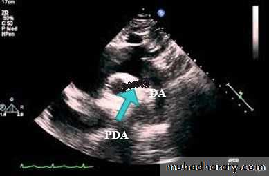
Echocardiography show the PDA and the cardiac chambers.
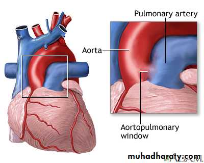
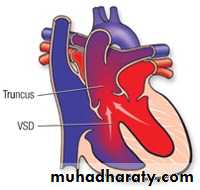
Differential Diagnosis
Aortopulmonary window,
truncus arteriosus,
ventricular septal defect with aortic regurgitation
, or arteriovenous fistula.
Natural HX;
Unlike that in premature infants, spontaneous closure of a PDA does not usually occur in full-term infants and children.CHF or recurrent pneumonia or both develop if the shunt is large.
Pulmonary vascular obstructive disease may develop if a large PDA with pulmonary hypertension is left untreated.
Infective endocarditis may occur.
.Although rare, an aneurysm of PDA may develop and possibly rupture in adult life.
TREATMENT
Irrespective of age, patients with PDA require surgical or transcatheter closure.

HYPERLINK "http://bit.ly/2enArpR" http://bit.ly/2enArpR