Knee joint .anatomy
Jabir ibn HayyanMEDICAL UNIVERSITY


• Dr.Muayad jawad
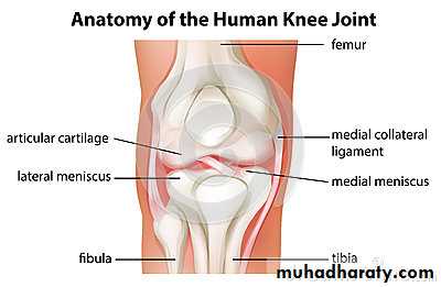
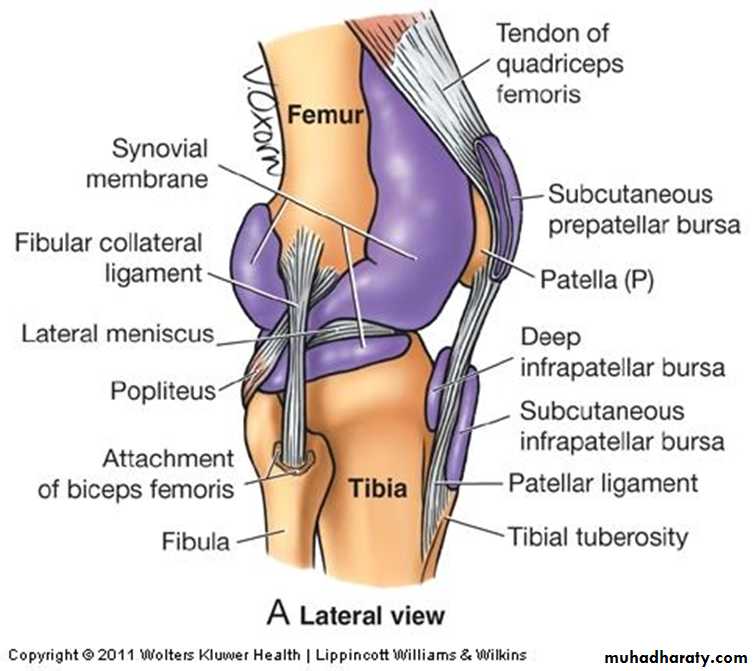
The knee joint is a synovial joint, the largest and most complicated in the body .It is a modified hinge joint; in addition to flexion and extension a small amount of rotation of the leg is possible in the flexed position of the knee.
Three bones take part in forming the knee joint :
1. Lower end of the femur2. Upper end of tibia
3. Patella (knee cup).
Jabir ibn Hayyan
MEDICAL UNIVERSITY
patella
Also known as the knee cap or kneepan is a thick , circular-triangular bone which articulates with the femur (only ) and covers and protects the knee joint.
It is the largest sesamoid bone in the human body.
It have apex (downward) and base (upward) .
It is attached to the tendon of the quadriceps femoris muscle, which contracts to extend/straighten the knee.
◊ The vastus intermedialis muscle is attached to the base of patella.
◊ The vastus lateralis and vastus medialis are attached to lateral and medial borders of patella respectively.
The primary functional role of the patella is knee extension , The patella increases the leverage that the tendon can exerts on the femur by increasing the angle at which it acts
Jabir ibn Hayyan
MEDICAL UNIVERSITY
Articulation
The lower end of femur has a curved articular surface, covered by hyaline articular cartilages ; extends backward and called medial and lateral condyles. The upper end of tibia has oval shaped articular surfaces, medial and lateral condyles.
The femoral and tibial condyles articulates forming femorotibial joint.
This joint flexes and extends the knee.
Jabir ibn Hayyan
MEDICAL UNIVERSITY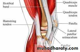
Patella articulates with femur to form patellofemoral joint
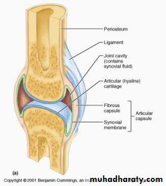
is plane gliding joint , This joint allows the patella to glide over the surfaces of the femur as the knee flexes and extends.The articular surfaces of femur, tibia, and patella are covered with hyaline cartilage.
upper end of fibula (head) does not adjoin the femur .it articulates with lateral side of tibia, just below the lateral tibial condyles .this joint allow slight movement.Jabir ibn Hayyan
MEDICAL UNIVERSITY
Capsule
Femoral attachmentsposteriorly: to the proximal margins of the femoral condyles and the intercondylar fossa.
Medially: the capsule is attached to the articular margin of the femur,
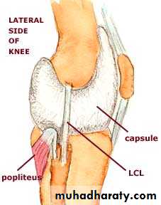
laterally: the attachment is proximal to the groove for the popliteus tendon.
Anteriorly the capsule blends with the patellar retinacula and is attached to the sides of the patellar ligament (tendon) and patella. The capsule is deficient above the level of the patella, permitting the suprapatellar bursa to be in full communication with the joint.
Jabir ibn Hayyan
MEDICAL UNIVERSITY
Tibial attachments
Posteriorly :on the tibia the capsule is attached to the margins of the tibial condyles, and in the intercondylar area to the distal edge of the groove for the posterior cruciate ligament; the attachment to the lateral condyle is interrupted by an aperture for the passage of the popliteus tendon.On the sides: the capsule is attached to the margins of the tibial condyles, and laterally to the head of the fibula as well. A thickening of the capsule on the medial side is a deep component of the tibial collateral ligament and is firmly attached to the medial meniscus. On its deep aspect the capsule has weak attachments to the rims of both menisci, the coronary ligaments, which connect them to the tibia.
Anteriorly the line of attachment on the tibia of the fused capsule and patellar retinacula inclines distally from the medial and lateral condyles to the tibial tuberosity
Jabir ibn Hayyan
MEDICAL UNIVERSITY
LigamentsExtracpsular ligaments
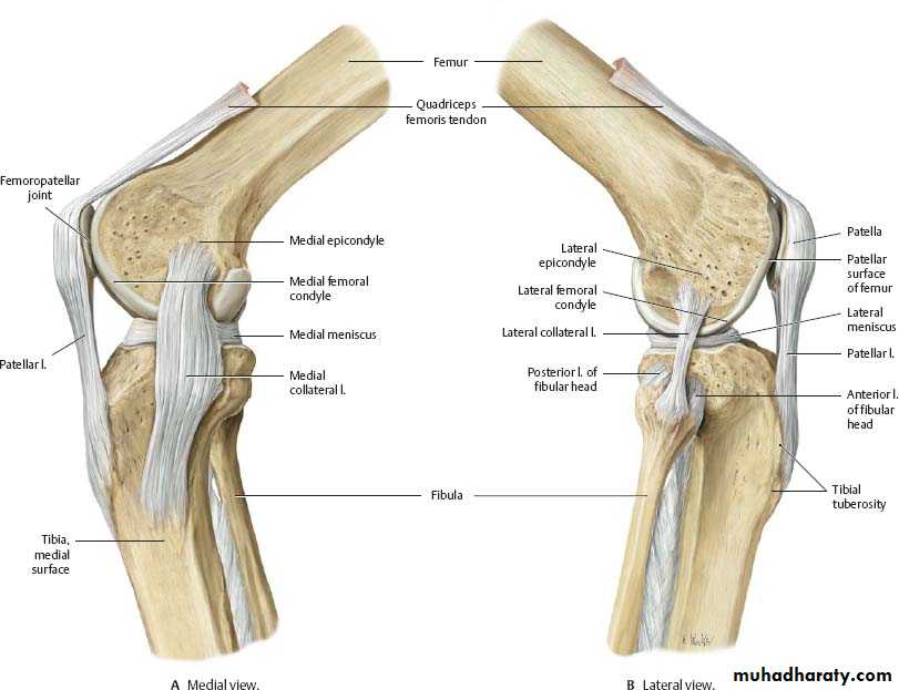
The tibial collateral ligament (medial ligament) is a flat, triangular band attached above to the medial femoral epicondyle, just distal to the adductor tubercle, and attached below to the upper part of the medial surface of the tibia. Its anterior margin, which forms the vertical base of the triangle, is free except at its attached extremities. The posterior apex of the triangular ligament blends with the capsule and is thereby attached to the medial meniscus. Above its distal attachment the ligament is crossed by the tendons of sartorius, gracilis and semitendinosus with a bursa interposed. The medial inferior genicular vessels and nerve and the anterior expansion of the semimembranosus tendon are deep to the distal part of the ligament with another bursa intervening.Jabir ibn Hayyan
MEDICAL UNIVERSITY
The fibular collateral ligament (lateral ligament) is cord-like and is attached proximally to the lateral epicondyle, below the attachment of the lateral head of gastrocnemius and above that of the tendon of popliteus. Its distal attachment is to the head of the fibula overlapped by the tendon of biceps femoris, a bursa intervening between them. The ligament is not attached to the capsule and has no connection with the lateral meniscus. A bursa lies between the ligament and the capsule, and the tendon of popliteus lies deep to the capsule here. The lateral inferior genicular vessels and nerve run deep to the distal part of the ligament.
Jabir ibn Hayyan
MEDICAL UNIVERSITY
.
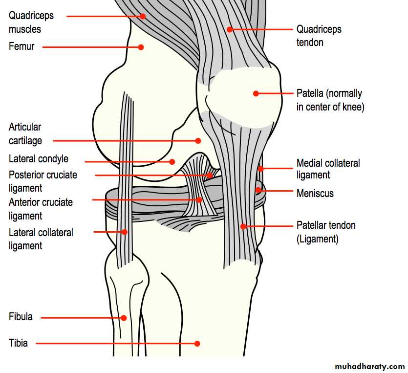
The patellar ligament connects the patella to the tuberosity of the tibia.
It is also occasionally called the patellar tendon because there is no definite separation between the quadriceps tendon (which surrounds the patella) and the area connecting the patella to the tibia. This very strong ligament gives the patella its mechanical leverage and also functions as a cap for the condyles of the femur.
Jabir ibn Hayyan
MEDICAL UNIVERSITY
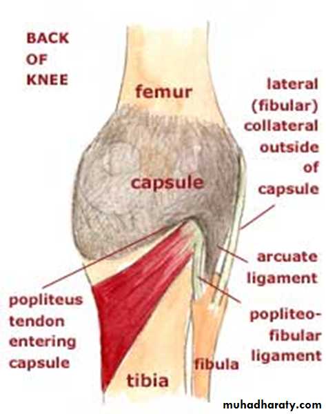
The oblique popliteal ligament is an expansion from the tendon of semimembranosus that blends with the capsule at the back of the joint and ascends laterally to the intercondylar fossa and lateral femoral condyle. The popliteal artery lies on it, and genicular vessels and nerves penetrate it.
Jabir ibn Hayyan
MEDICAL UNIVERSITY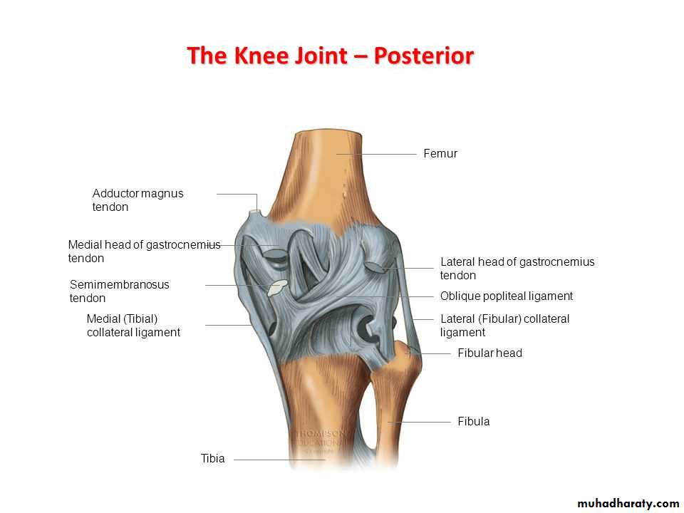
The arcuate popliteal ligament is a Y-shaped thickening of posterior capsular fibres. The stem of the Y is attached to the head of the fibula. The medial limb arches over the tendon of popliteus to the posterior edge of the tibial intercondylar area. Some popliteus muscle fibres are attached to it. The lateral limb ascends to the lateral femoral condyle with the popliteus tendon.
Jabir ibn Hayyan
MEDICAL UNIVERSITY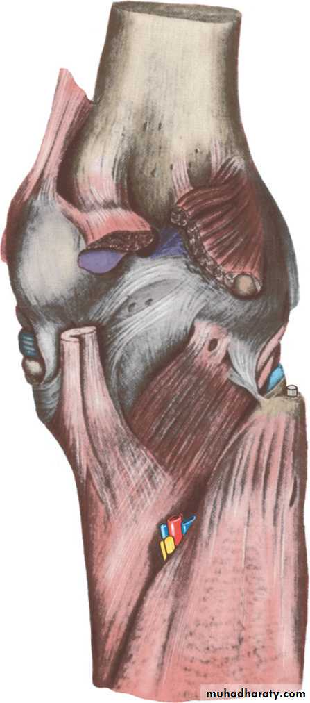
Intracapsular ligaments
The cruciate ligaments are a pair of very strong ligaments connecting tibia to femur. They lie within the capsule of the knee joint, but not within the synovial membrane. It is as though they had been herniated into the synovial membrane from behind, so that they are covered by synovial membrane on their front and sides but not posteriorly.The anterior cruciate ligament is attached to the anterior part of the tibial plateau between the attachments of the anterior horns of the medial and lateral menisci. The ligament ascends posterolaterally, twisting on itself, and is attached to the posteromedial aspect of the lateral femoral condyle.
Jabir ibn Hayyan
MEDICAL UNIVERSITY
The posterior cruciate ligament is stronger, shorter and broader, and less oblique. It is attached to a smooth impression on the posterior part of the tibial intercondylar area which extends to the uppermost part of the posterior surface of the tibia. The ligament ascends anteromedially and is attached to the anterolateral aspect of the medial femoral condyle. The cruciate ligaments cross each other like the limbs of the letter X, the anterior ligament lying mainly anterolateral to the posterior ligament.
Jabir ibn Hayyan
MEDICAL UNIVERSITY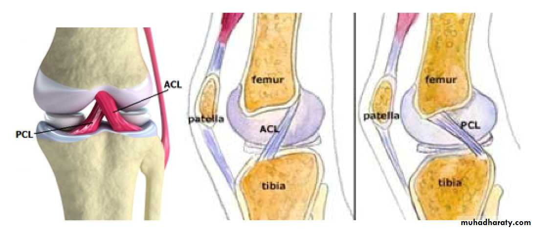
menisci
The menisci—formerly called semilunar cartilages— are crescentic discs of fibrocartilage comprising mainly collagenous fibrous tissue that lie on and are attached to the tibial plateau. They are triangular in cross-section, being thicker at their convex periphery. Their distal surfaces are flat, while their proximal surfaces are concave and articulate with the convex femoral condyles. The menisci are mainly avascular but their peripheral zone is vascularized by capillaries from the capsule.
The medial meniscus is almost a semicircle and is broader posteriorly. Its anterior horn is attached to the intercondylar area in front of the anterior cruciate ligament, while the posterior horn is similarly attached in front of the posterior cruciate ligament. As mentioned above, the medial meniscus is firmly attached to the capsule and the tibial collateral ligament.
Jabir ibn Hayyan
MEDICAL UNIVERSITY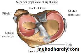
The lateral meniscus is about four-fifths of a circle and is of uniform width. Its anterior horn is attached in front of the intercondylar eminence of the tibia, behind the anterior cruciate ligament with which it partly blends. The posterior horn is attached behind the intercondylar eminence, in front of the posterior horn of the medial meniscus. From the posterior convexity of the lateral meniscus fibrous bands pass upwards and medially to the medial femoral condyle, in front of and behind the posterior cruciate ligament . These are the anterior and posterior meniscofemoral ligaments (of Humphry and Wrisberg). More medially some fibres of the popliteus muscle are attached to the posterior convexity of the lateral meniscus. The transverse ligament is a variable band that connects the anterior convexity of the lateral meniscus to the anterior horn of the medial meniscus.
Jabir ibn Hayyan
MEDICAL UNIVERSITYSynovial membrane
The synovial membrane is the most extensive in the body but the amount of synovial fluid in a normal joint is only 0.5 ml—a mere capillary film. It is attached to the articular margins of the femur, tibia and patella and lines the deep aspect of the capsule, but it is separated from the capsule by the popliteus tendon and the cruciate ligaments, which are thereby excluded from the synovial cavity. Anteriorly the membrane is separated from the patellar ligament by the infrapatellar fat pad, which herniates the membrane into the joint cavity as a pair of folds, the alar folds; these converge in the midline into a single fold, the infrapatellar fold, which extends posteriorly and is attached to the femoral intercondylar fossa .Jabir ibn Hayyan
MEDICAL UNIVERSITY
The synovial membrane does not cover the surfaces of the menisci. It is continuous with the lining of the suprapatellar bursa. Posteriorly a small pouch of membrane projects distally between the tendon of popliteus and the upper ends of the tibia and fibula; it may communicate with the superior tibiofibular joint. The synovial cavity of the knee joint often communicates with the bursa deep to the medial head of gastrocnemius, and sometimes communicates with the bursa deep to the lateral head of gastrocnemius.
Jabir ibn Hayyan
MEDICAL UNIVERSITY
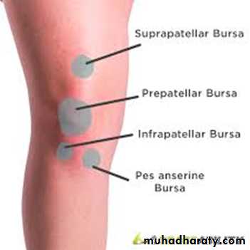
Bursae
Bursae related to the knee jointnumerous synovial sac , act as cushions between tendon and bone, ligament and bone, or skin and bone.
There are a dozen bursae related to the knee joint: four on the anterior aspect; two on either side; and four at the back.
Jabir ibn Hayyan
MEDICAL UNIVERSITY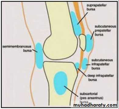
Bursae
Anterior bursae are present in front of knee1. Suprapatellar bursa beneath the quadriceps.
2. Prepatellar bursa
lies in the subcutaneous tissue between skin & the front of the lower half of the patella & the upper part of ligamentum patellae.
3. Superficial infrapatellar bursa
lies in the subcutaneous tissue between the skin and the
front of the lower part of the ligamentum patellae.
4. Deep infrapatellar bursa lies between the ligamentum patellae and the tibia
Lateral bursa
1. Between LCL & biceps femoris tendon
2. Between the LCL & the capsule , where it overlies popliteus tendon
Medial bursa
1. between MCL & the tendons of sartorius, gracilis & semitendinosus
2. Between the MCL & the tibia & joint capsule
Posterior bursae
1-Popliteal bursa : Under popliteus tendon
2-Semimembranosus bursa
3-Between joint capsule & medial head of gastrocnemius
4-Between joint capsule & lateral head of gastrocnemius
Jabir ibn Hayyan
MEDICAL UNIVERSITY
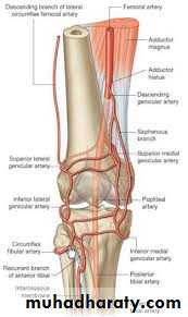
blood supply
The knee joint receives blood supply from extensive genicular anastomosis mainly from popliteal, anterior and posterior tibial artery. Popliteal artery in its course through the
fossa, it gives off medial and lateralsuperior genicular arteries to form
collateral circulation around the knee joint
Also medial and lateral inferior genicular
arteries encircle the leg and formanastomosis around the knee joint.
1. Superior medial & lateral genicular arteries
2. Inferior medial & lateral genicular arteries
3. Descending genicular artery from ( femoral artery )
4. Recurrent branch of anterior tibial artery
Jabir ibn Hayyan
MEDICAL UNIVERSITY
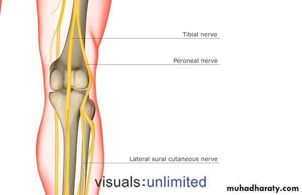
Nerve supply
The joint is supplied from the femoral nerve through its branches to the three vasti, from the sciatic nerve by the genicular branches of its tibial and common peroneal components, and from the obturator nerve by the twig from its posterior division, which accompanies the femoral artery through the gap in the adductor magnus into the popliteal fossa. During arthroscopy it must be remembered that, although local anaesthesia affects the overlying skin, the cruciate ligaments remain sensitive (tibial nerve). The horns of the menisci are innervated but their central parts are devoid of sensory fibres.Jabir ibn Hayyan
MEDICAL UNIVERSITY

movements
The principle movements of the knee joint are flexion & extension1. Flexion
Produced by biceps femorus , semimembranosus & semitendinosus Assisted by Gracillus , Sartorius & popliteus
2. Extension
Produced by quadriceps femoris & gluteus maximums
3. Medial rotation
Produced by Sartorius , Gracillus & Semitendinosus
4. Lateral rotation
Produced by biceps femorus
Jabir ibn Hayyan
MEDICAL UNIVERSITY
.
stability
Factors responsible for knee joint stability are :1. Muscle tone : especially in quadriceps
2. Cruciate ligaments :
stabilize femur on tibia
preventing anteroposterior movement
3. Collateral ligaments : assist in medial and lateral stability
4. Iliotibial tract : stabilizes knee joint during extension5. Oblique posterior ligament : prevent hyperextension
Jabir ibn Hayyan
MEDICAL UNIVERSITY
.
knee relations
Anteriorly The prepatellar bursa
Posteriorly
Popliteal vessels tibial & common peroneal nerves
lymph nodes the semimembranosus
semitendinosus
biceps femoris
the heads of gastrocnemius
plantaris
Jabir ibn Hayyan
MEDICAL UNIVERSITY
. Medially
Sartorius Gracilis
Semitendinosus
Laterally
Biceps femoris common peroneal nerve
clinical notes
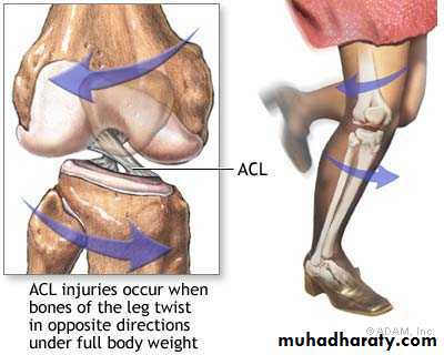
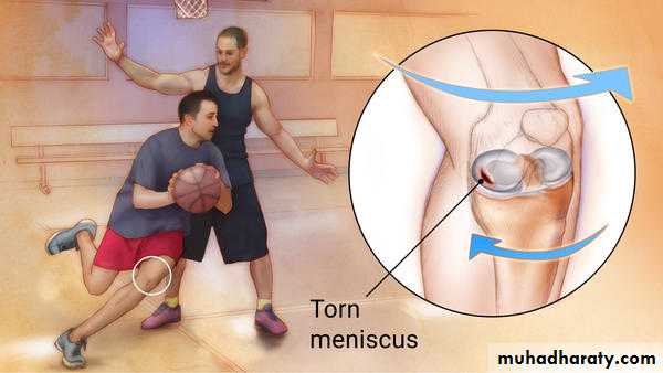
The menisci are liable to injury resulting from twisting strains applied to the flexed weight-bearing knee. The medial meniscus is more prone to damage, probably because it is firmly attached to the capsule and the collateral ligament. Likewise the flat medial ligament, which is attached to the posteromedial capsule, is more likely to be torn than the cord-like lateral ligament, which is unattached to the capsule. A combined rotation and impact injury to the flexed knee, as in sports injuries, may cause rupture of the anterior cruciate ligament. The stronger posterior cruciate ligament is less likely to be damaged, but it may be torn when the tibia is violently thrust backwards in relation to the femur.Jabir ibn Hayyan
MEDICAL UNIVERSITY
.
Jabir ibn Hayyan
MEDICAL UNIVERSITY
THANK YOU
.Jabir ibn Hayyan
MEDICAL UNIVERSITY
.