
Tracheal Tubes 2017
Description:
Tracheal tubes are widely used in anaesthesia to provide gas
transfer between a breathing system and a patient’s trachea. This
module will help you to understand the design and use of standard
tracheal tubes. You will also be introduced to variants of the
tracheal tube which have been modified for use in different clinical
situations.

Session Introduction
Learning Objectives:
• Recognize the purpose of tracheal tubes
• Recognize and describe the design features of
tracheal tubes
• Identify the basic features of specialized
versions of tracheal tubes
• Identify and recognize the basic features of
rigid stylets and bougies
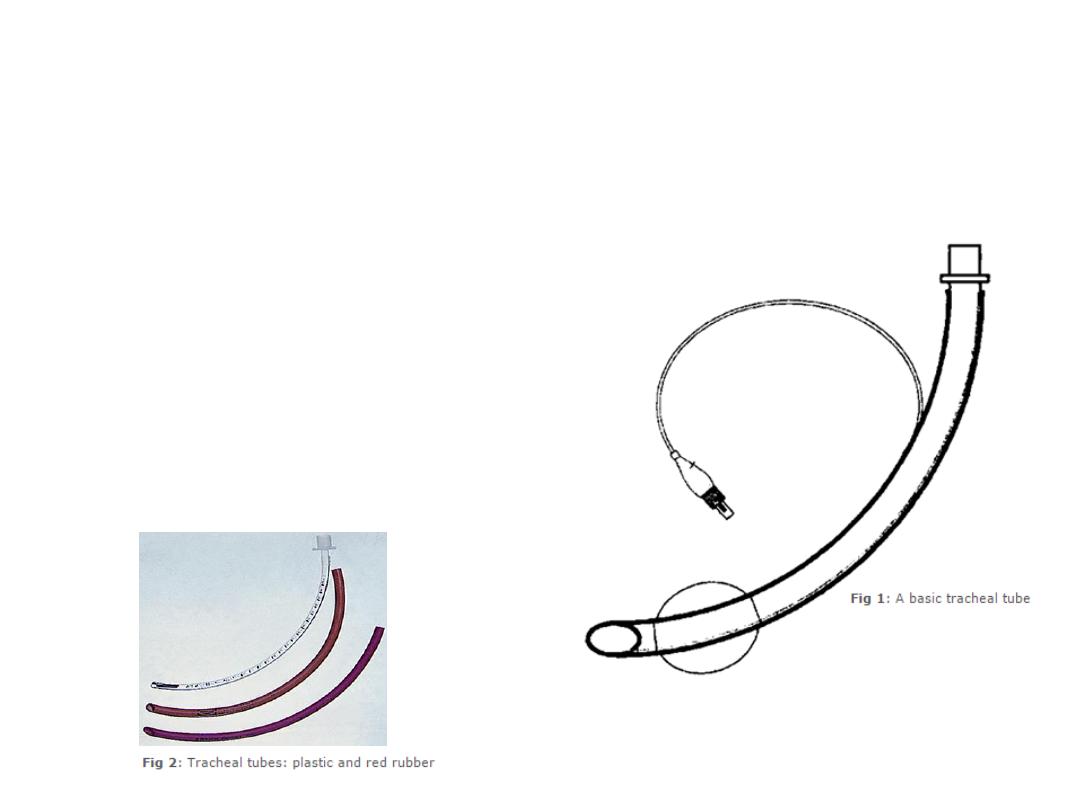
Introduction to Tracheal Tubes
A tracheal tube provides a passage for gases to
flow between a patient’s lungs and an
anaesthesia breathing system. It is a commonly
used device that is critical for patient safety.
Therefore, it is important that you understand its
design and proper use.
Tracheal tubes come in various sizes and
shapes, but the basic concepts underlying them is
the same for all.
The function of tracheal tubes will be described,
followed by a discussion of the many features
that can be attributed to them. Finally, we will
introduce specially designed tracheal tubes that
may be used in specific clinical situations.
This session will end with some self assessment
questions, to test your knowledge of tracheal
tubes and their applications.
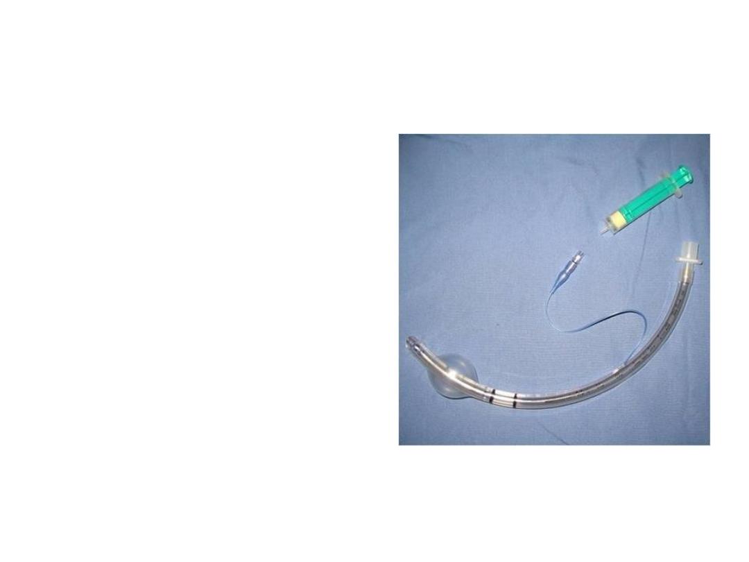
A tracheal tube
What are the Functions of Tracheal
Tubes?
Tracheal tubes provide a route
for gases to flow between a
patient’s trachea and the
anaesthetic breathing system.
In addition, when tracheal
tubes are cuffed (which will be
explained later in the session)
they allow positive pressure
ventilation to be used more
effectively. The cuff of a tracheal
tube also protects the lung from
contamination from gastric
contents and nasopharyngeal
matter such as blood.
Tracheal tube
Basic Design
The basics of tracheal tubes
A tracheal tube has a diameter and a length
and is made out of special materials such as
polyvinyl chloride (PVC) or red rubber. It also
usually has a curvature and markings. It may
have a cuff at the tracheal end to provide a
seal between the tube and the tracheal wall.
If a cuff is present, to inflate and deflate it,
there will be a thin tube attached to a small
one-way valve and balloon.
Basic Design
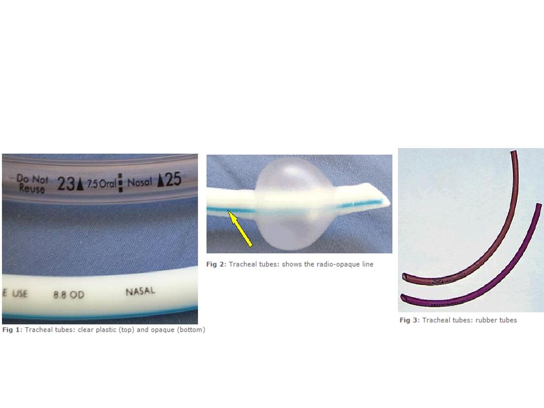
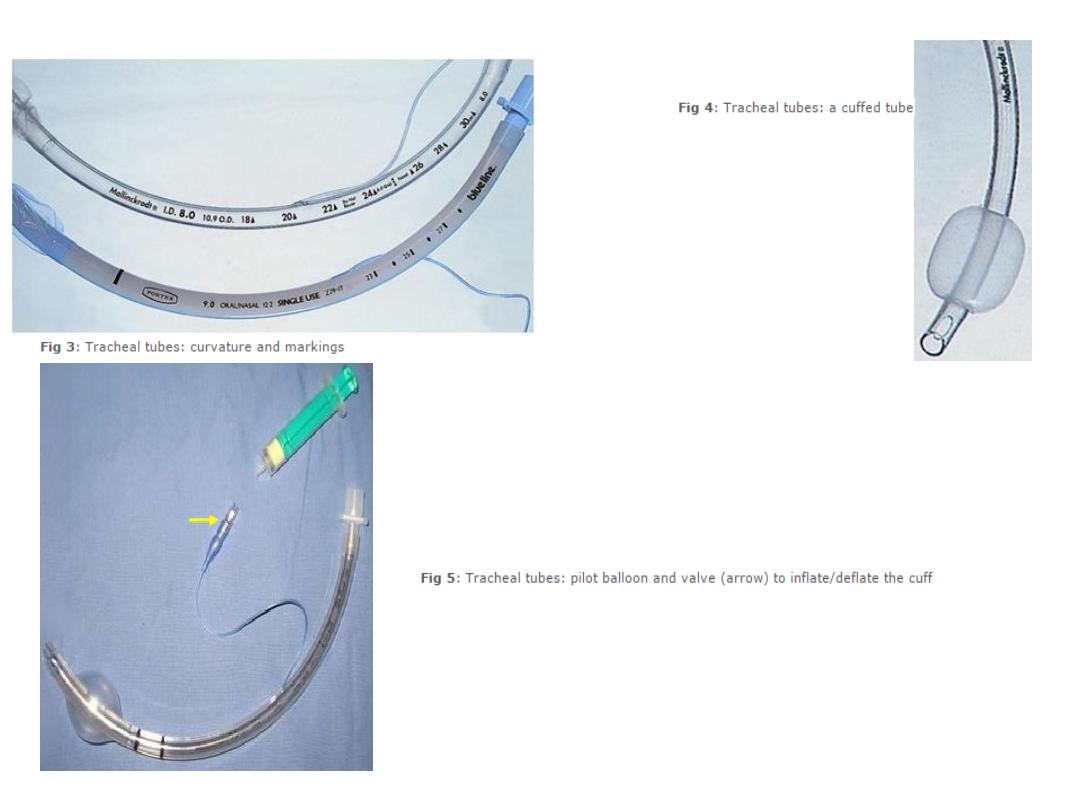
Construction Material
Most tracheal tubes encountered will be made out of polyvinyl chloride (PVC). These are
disposable.
The plastic PVC tracheal tubes may be visually clear or opaque as shown in the image (Fig 1).
Plastic is not radio-opaque (i.e. it is not visible on x-ray) and therefore plastic tracheal tubes
have a line of radio-opaque material that makes them more visible on a chest x-ray (Fig 2).
Tracheal tubes may also be made out of rubber. These may be reused after cleaning and
autoclaving (Fig 3).
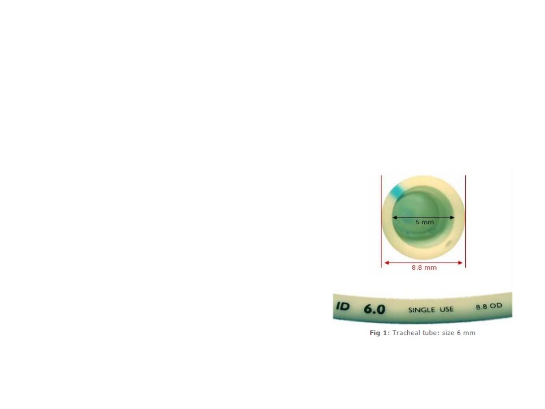
size
Tracheal tubes have an inner diameter and an
outer diameter.
The ‘size’ of a tracheal tube refers to its internal
diameter. Therefore if you ask for a size 6 tracheal
tube, you are asking for one with an internal
diameter of 6 mm.
Narrower tubes increase the resistance to gas
flow. A size 4 mm tracheal tube has 16 times
more resistance to gas flow than a size 8 mm
tube. This can be especially relevant in the
spontaneously breathing patient who will have to
work harder to overcome the increased
resistance. Therefore, the largest suitable internal
diameter tracheal tube should be used.
An average sized male will usually require a size
8.5–9 mm internal diameter tracheal tube and an
average sized female will require a size 7.5–8 mm
internal diameter tube. Paediatric tracheal tubes
are available in much smaller internal diameters
based on the age and weight of the child.
Fig 1 shows a size 6 mm internal diameter
tracheal tube. In this particular tube, the internal
diameter is labelled as ‘ID 6.0’ and similarly, the
outside diameter is labelled as ‘8.8 OD’.
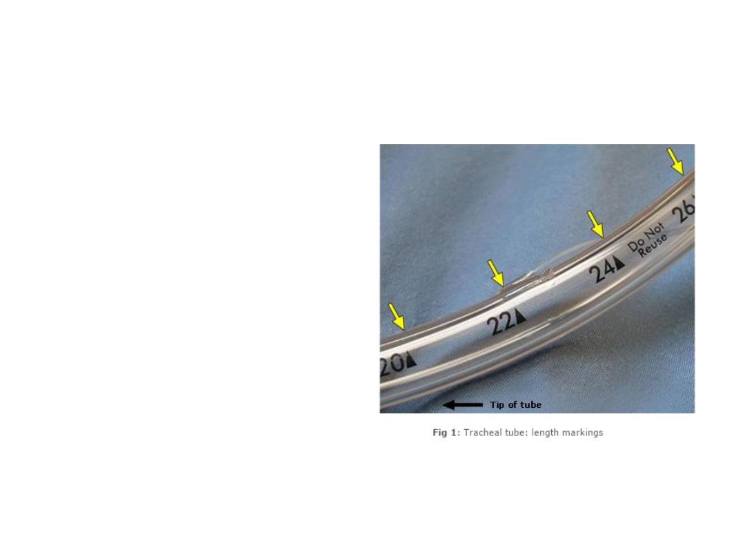
The length of a tracheal tube is
measured from the end that goes into
the trachea and is marked in cm on the
outside of the tube as shown (Fig 1).
After intubation, you should note the
length marking of the tracheal tube with
reference to a landmark such as incisor
teeth or lips. This will help you to
monitor the position of the tracheal tube
and detect if the tube has inadvertently
got pushed too far (into a bronchus) or
pulled outwards (resting on vocal cords).
A tracheal tube that is too long for the
patient maybe more prone to kinking
and becoming obstructed. It can be cut
to a more appropriate length if
necessary.
Length
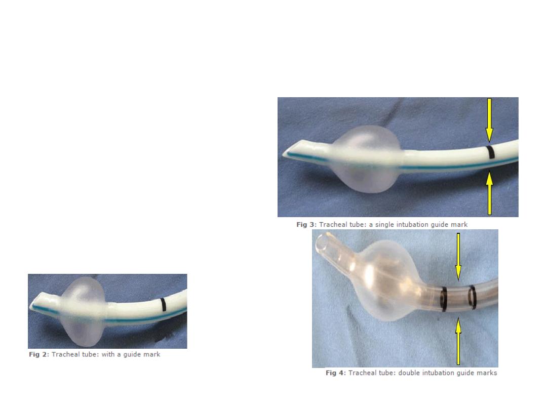
Length
To help the accurate placement of the
tracheal tube tip within the trachea, some
tracheal tubes have black intubation depth
markings located proximal to the cuff (Fig 2).
The vocal cords should be at the black mark in
tracheal tubes with one mark (Fig 3). Or
should be between marks if there are two
such marks (Fig 4). However, these are only
rough estimates and correct tube position
depth should always be confirmed by
auscultation.
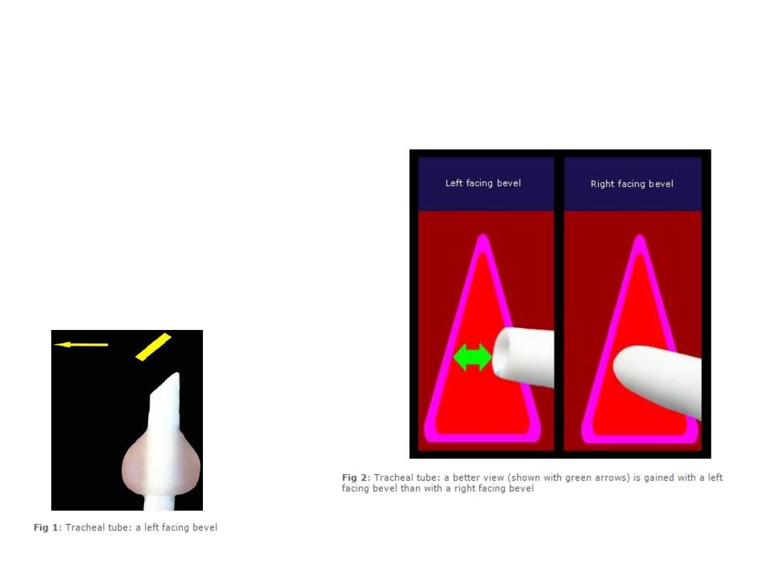
Bevel
To make it easier to pass the tracheal tube
through the vocal cords and to improve the
view of the vocal cords during intubation,
tracheal tubes have a 'slant' called a bevel
(Fig 1).
As the tracheal tube is advanced near the
cords, the left-facing bevel gives a better
view (Fig 2).
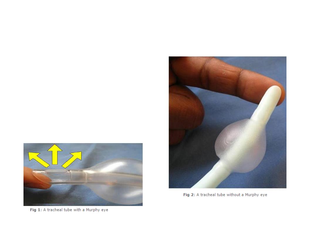
Murphy Eye
Some tracheal tubes have an additional hole
at the tip called a Murphy eye. If the main
opening of the tracheal tube gets blocked by,
for example abutting against the tracheal wall
(represented in Fig 1 by the finger) gas flow
can still occur via the Murphy eye.
Without the Murphy eye the tracheal tube
would have been completely obstructed (Fig
2).
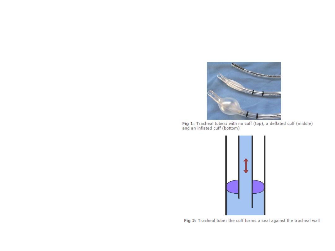
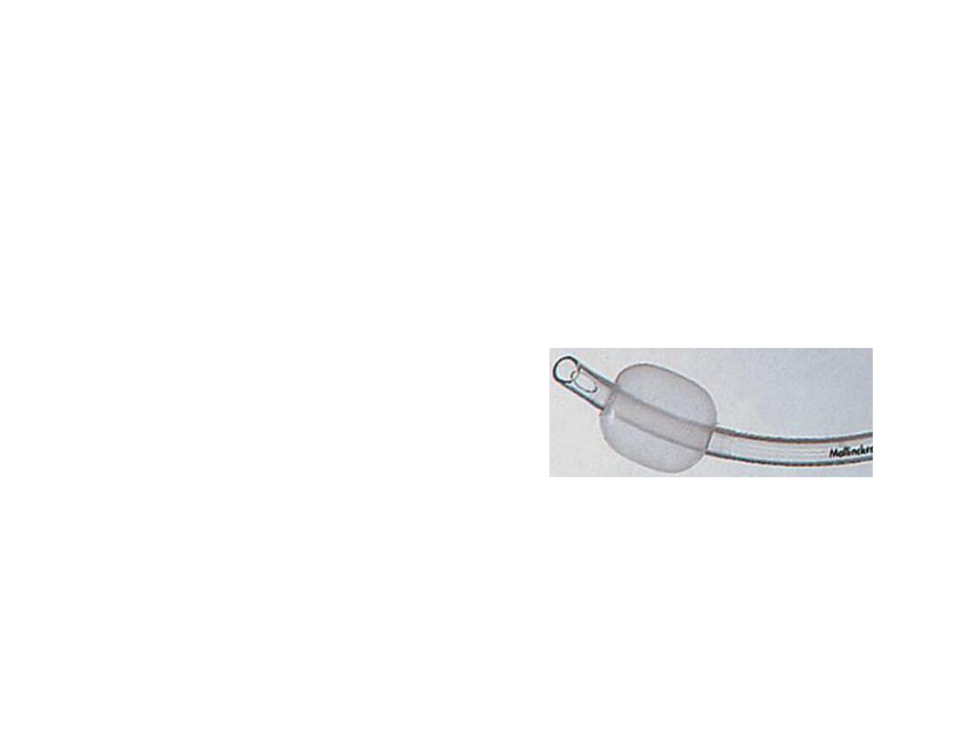
Cuff
A cuff is an inflatable region at the patient
end of a tracheal tube. Tracheal tubes may or
may not have a cuff. The tracheal tube (top)
does not have a cuff. The tracheal tube
(middle) has a cuff that is deflated, and the
tracheal tube (bottom) has an inflated cuff (Fig
1).
The inflated portion forms a seal against the
tracheal wall (Fig 2). This seal prevents gases
from leaking past the cuff and allows positive
pressure ventilation. The seal also prevents
matter such as regurgitated gastric contents
going into the trachea.
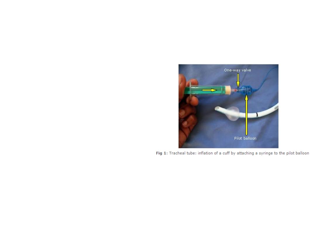
Cuff
After intubation, the cuff is inflated with air.
This is done by attaching a syringe to the pilot
balloon (Fig 1). The pilot balloon is connected to
the cuff by a thin tube. As the syringe supplies
pressurized air, the pilot balloon and cuff inflate.
Cuff inflation has to be done gently to prevent
over-inflation and exerting too high a pressure on
the tracheal mucosa. Continuous listening for an
air leak should be used and the inflation should
stop when the leak stops.
Once the cuff is inflated the syringe is removed.
Air does not leak out as there is a one-way valve
at the pilot balloon. By feeling the pilot balloon,
the amount of pressure in the cuff can be
estimated. If the cuff is leaking, e.g. due to
damage by the surgeon during a thyroidectomy,
the pilot balloon will collapse. Cuff pressure can
also be checked using a specially designed
pressure gauge.

Cuff
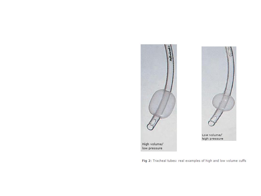
Volume and pressure
Cuffs can either have a high volume
with low pressure or have a low volume
with high pressure (Fig 1).
High volume/low-pressure cuffs
Because of their large volume, these
cuffs have a larger surface area in contact
with the trachea. This means that they
apply a lower pressure against the
tracheal wall and have a lower incidence
of tracheal wall ischemia and necrosis.
The seal is not as good as the seal in
high-pressure cuffs because of the lower
pressures and because the large cuff may
develop wrinkles that allow material
such as regurgitated gastric contents to
pass by the cuff (Fig 2).
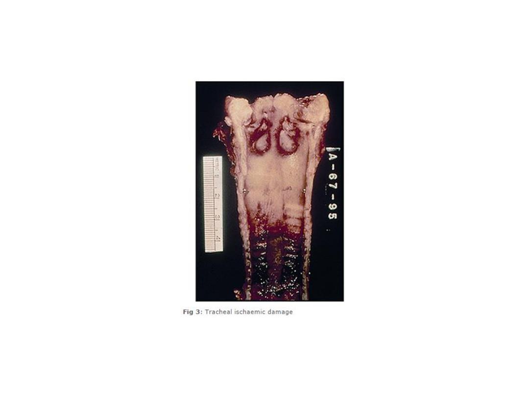
Cuff
Volume and pressure
Low volume/high-pressure cuffs
These cuffs have a lower
volume and the surface area in
contact with the trachea is small.
This results in a high-pressure
seal that is more effective than
the one created by high
volume/low-pressure cuffs.
However, this high pressure is
more likely to cause tracheal
ischaemia and necrosis if used for
a prolonged period of time (Fig
3).
Cuff
Volume and pressure
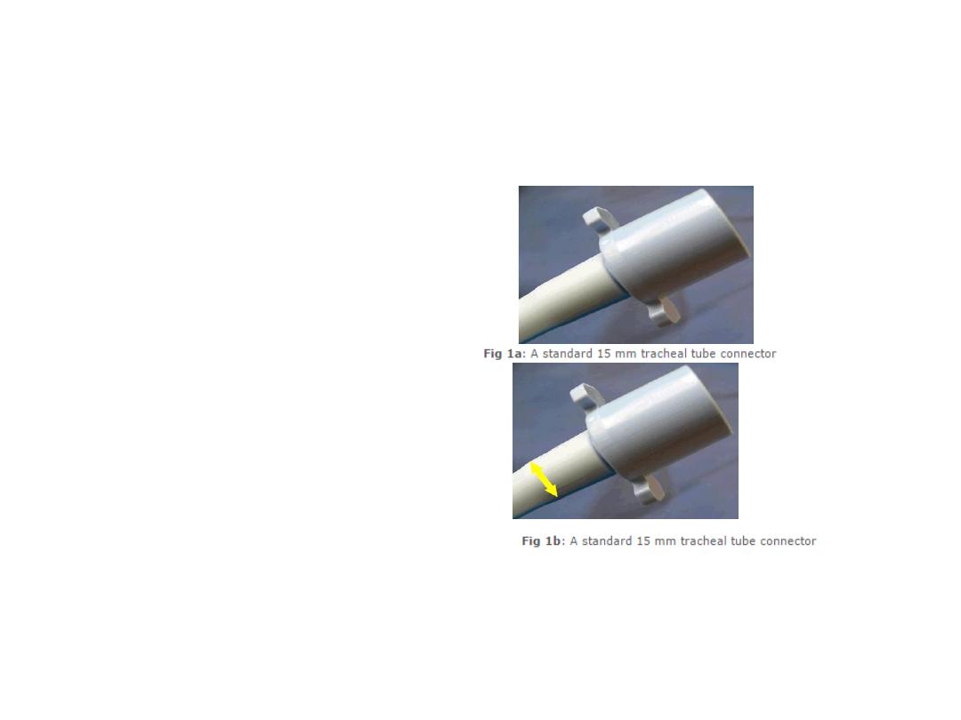
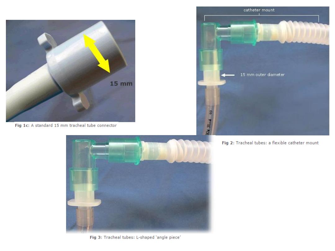
Tracheal Tube Connectors
Tracheal tube connectors connect the
tracheal tube to the breathing system
(Fig 1a). One end of the connector
connects to the tracheal tube and this
end has a diameter that depends on the
size of the tube; see small arrows (Fig
1b). The other end connects to the
breathing system and has a 15 mm outer
diameter (British Standard) (Fig 1c).
Often tracheal tubes are not directly
connected to breathing systems. Instead,
to provide a more flexible connection,
tracheal tubes are often connected to a
flexible ‘catheter mount’ (Fig 2). The
catheter mount is connected to the
tracheal tube via an L-shaped connector
called an 'angle piece' (Fig 3).
Tracheal Tube Connectors
Specialized Versions of Tracheal Tubes
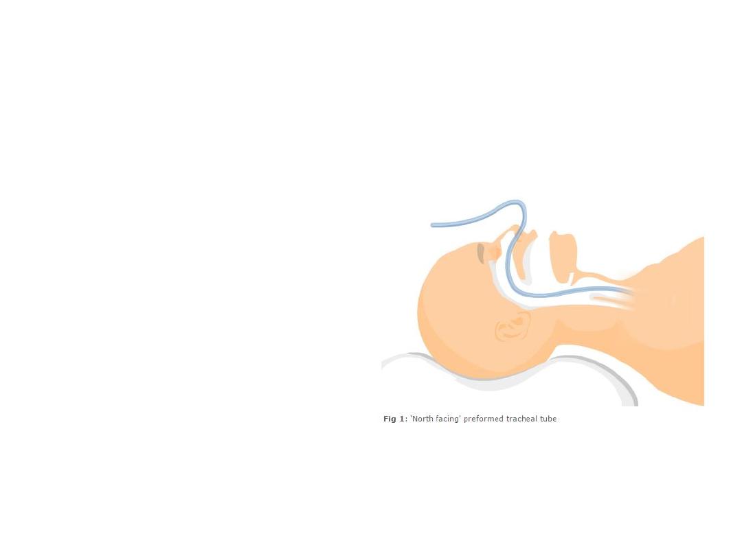
Preformed tracheal tubes
are moulded into special
shapes that permit good
surgical access into the oro-
nasal area. A north-facing
tracheal tube emerges from
the patient and faces
towards the patient’s
forehead (Fig 1). Such
preformed tracheal tubes
provide very good access to
the mouth for dental work.
Specialized Versions of Tracheal Tubes
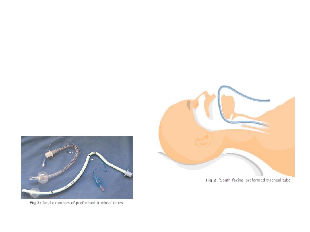
Similarly, south-facing preformed
tracheal tubes provide good access for the
ear, nose and throat (ENT) surgeon needing
to work in the nasal passages (Fig 2). For
some real examples of preformed tracheal
tubes, see Fig 3. The tube marked ‘South’
shows a RAE tube, named after its inventors
Ring, Adair and Elwyn.
Specialized Versions of Tracheal Tubes
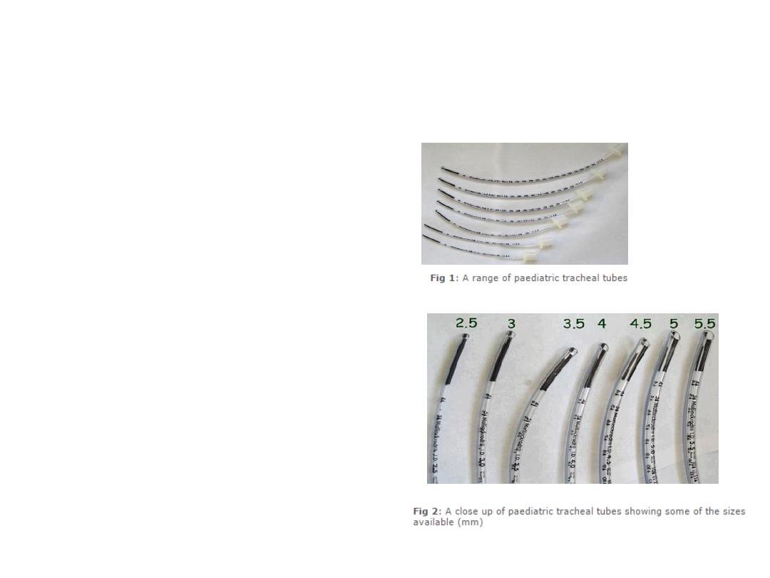
Tracheal tubes for paediatric
patients are smaller than those
meant for adults. Because the
paediatric trachea is very
susceptible to damage by
pressure, many paediatric
tracheal tubes are uncuffed.
However, cuffed versions
similar to adult tracheal tubes
do exist and when used, must
be inflated with care.
A wide range of sizes are
available (Fig 1) and (Fig 2).
Specialized Versions of Tracheal Tubes
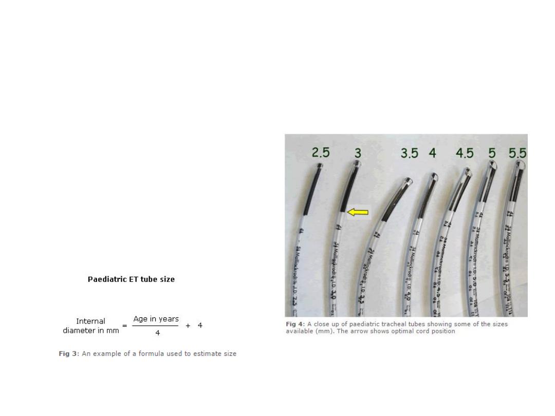
Paediatric tubes
Tables and formulae are available to guide size
selection (Fig 3). After intubation, depending
on whether the fit is too tight or too loose, a
different sized tracheal tube may have to be
used. For this reason, a wide range of
paediatric tracheal tube sizes should be readily
available. The tracheal tubes may have a mark
to guide optimum depth of placement. In
these tubes, the vocal cords should be at the
junction of the black area and the rest of the
tube (Fig 4)
Specialized Versions of Tracheal Tubes
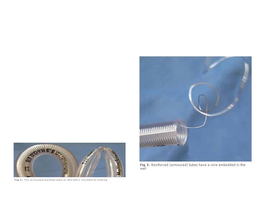
Armoured tracheal tubes
Reinforced or armoured tracheal tubes are
specially designed to resist kinking (Fig 1).
They achieve this property by having a spiral
of wire embedded into the wall of the tracheal
tube to give it strength and flexibility at the
same time (Fig 2). These tracheal tubes are
particularly useful for head and neck surgery
where the tracheal tube may be sharply bent
or compressed by the surgeons. Armoured
tracheal tubes can be easily bent away from
the area of surgery and thus improve surgical
access.
Basic Features
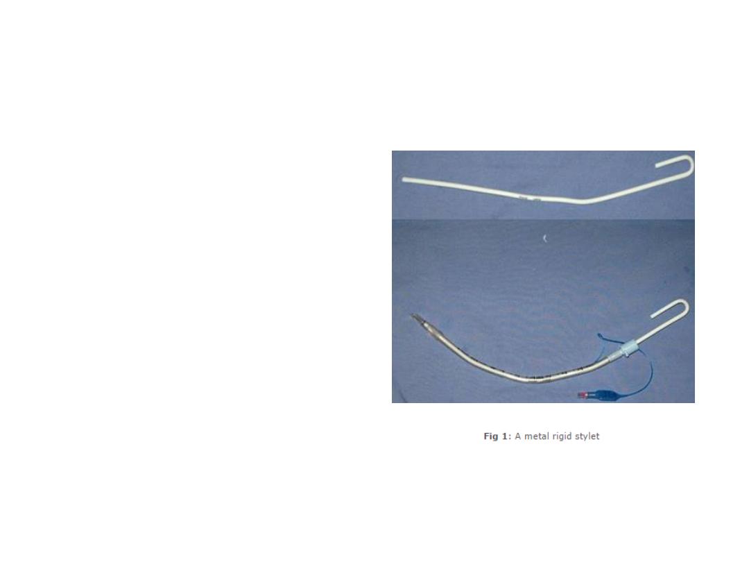
Rigid stylet
Sometimes it is useful to stiffen a
tracheal tube by inserting a metal rigid
stylet into the lumen of the tube prior to
intubation. The rigid metal stylet enables
the tracheal tube to be bent in a
direction more suited to the patient.
They are also often used with armoured
tracheal tubes, which because they are
naturally 'floppy', often need to be made
rigid using a stylet. Once intubation is
achieved, the rigid stylet is removed,
taking care not to dislodge the tracheal
tube in the process. Unlike a bougie
(discussed later), a rigid stylet must
never project beyond the tip of an
tracheal tube. A protruding rigid stylet
can cause serious damage to airway
structures.
Basic Features
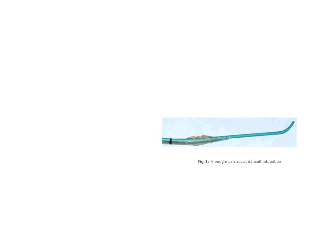
Bougie
A bougie is a relatively flexible
stylet that can assist difficult
intubation. It has a curved tip that
can help intubate an anterior
trachea. If only the epiglottis is
seen, it can even be passed
‘blindly’ into the trachea.
Once the bougie is in the
trachea, the tracheal tube is
‘railroaded’ over it. The bougie is
held in place, while the tracheal
tube is pushed into the trachea.
Once in the trachea, the tracheal
tube is held in place while the
bougie is pulled out.

Session Key Points
• Tracheal tubes provide a path for gas flow,
protect the airway, and allow positive pressure
ventilation for the patient
• The 'size' usually refers to the internal diameter
• The cuff may be high volume/low pressure or low
volume/high pressure
• Paediatric tracheal tubes are smaller and their
size must be chosen carefully
• Specialized versions exist such as preformed,
armoured, double lumen, and laser resistant

Session Summary
Learning Objectives:
•
Recognize the purpose of tracheal tubes
•
Recognize and describe the design features of tracheal tubes
•
Identify the basic features of specialized versions of tracheal tubes
•
Identify and recognize the basic features of rigid stylets and bougies
When you next work in theatre, have a look at the tracheal tubes on the airway
trolley. Ask your operating department practitioner to show you the specialized
tracheal tubes