Surgery of the Digestive System
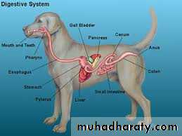
Digestive system
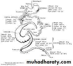
Dog first developed directly from Wolf about 15.000 year agoDog is carnivores
Dog have hinged jaws and large teeth meant to ingest large chunks of meat, it has shortest digestive tract of all mammals, digestion take about 8-9 hr.Stomach play major role in digestion, so it need high level of HCl
Small intestine extract nutrient from food
Large intestine ferment and element unassimilated food and water
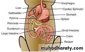
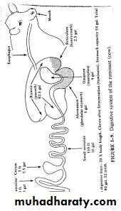
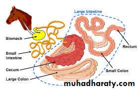
Equine Digestive System
Relatively small stomach efficient in the digestion of grain
Large cecum and colon are adapted for utilizing of fiber and roughaes
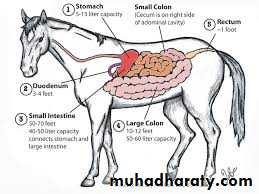
Muscular hollow tube that extends from mouth to anusA little over 100 feet in length
Varies in diameter from 1 inch in the small intestine to 8 inch in large intestine
Loops back on it itself many times
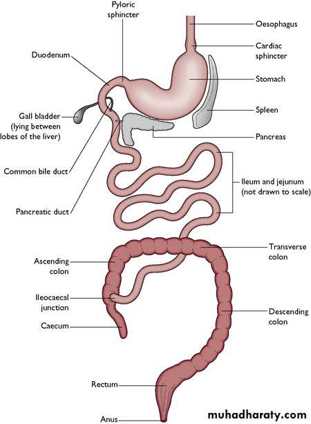
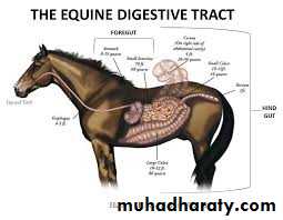
THE ANATOMY OF THE HORSE’S DIGESTIVE SYSTEM
The process of digestion in the horseThe digestive system is responsible for the intake (selection, intake and grinding), and digestion (processing and absorption), of food. The process is complete when the nutrients have been absorbed and the waste is eliminated.
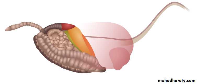
The horse’s digestive system starts at the mouth and ends at the anus. It is one continuous length and is extremely long so it must loop and coil to be able to fit inside the small amount of space left over by the large lungs and diaphragm.
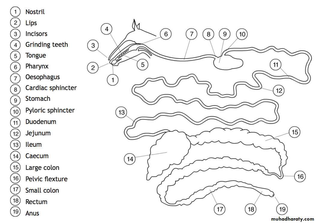
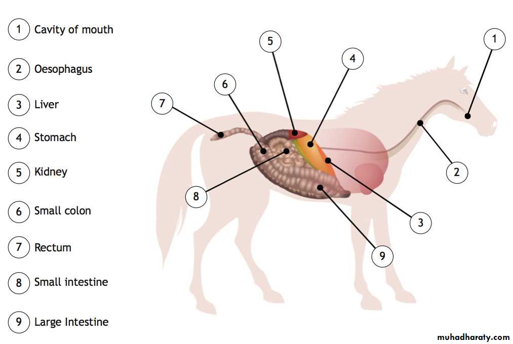
Nostril --The horse has an acute sense of smell and will be selective when choosing food
Lips --The digestive process starts at the lips. He used his lips and the whiskers around the muzzle to select and gather up his foodIncisors -- the incisor teeth grasp and bite off the selected portion
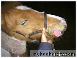
Tongue--The tongue has taste buds which provide the horse with essential information about the food. The tongue and cheek muscles manipulate the food to the back teeth. The tongue rolls the bolus to the back of the mouth
Grinding teeth--to the grinding teeth (premolars and molars) where the food is ground up and mixed with saliva. This chewing action is very important as it is this action that stimulates the production of saliva
Pharynx – passes through into correct tube see next slide
Oesophagus -- This tube is about 1.5 metres in length. It is a passageway from the mouth to the stomach and no digestion occurs hereCardiac sphincter-- is a strong band of muscle called a sphincter muscle. This acts as a one way valve into the stomach
Stomach -- The stomach is relatively small, about the size of a rugby ball
Pyloric sphincter—the exit from the stomach to the small intestine
Small intestine -- The small intestine can be divided into 3 areas. The duodenum about 1 meter long, the jejunum about 20-24 metres long and the ileum about 1 – 1.5 meters long , in all 20m – 27 metres long and has a capacity of 55-70 litres
Caecum--The caecum is a large vat which has a blind end
Large colon-- about 3 – 4 meters long and has a capacity of 90-110 litres. It has to fold into 4 to fit into the remaining space in the horse’s abdomen
Small colon-- The small colon is roughly 4 meters in length and has a capacity of 16 litres
Rectum--The rectum is a short tube used as a storage area that holds waste
Anus--The anus is another sphincter muscle that empties the rectum when there is a build-up of waste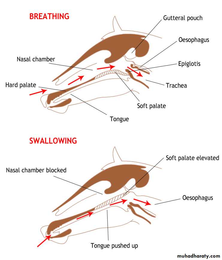
The Pharynx
The pharynx is the muscular passage between the mouth and the oesophagus.In this area air and food pass down different tubes to the lungs and stomach.
The trachea takes air to the lungs and the oesophagus takes the bolus of food to the stomach.
These tubes lie next to each other and there are 2 safety mechanisms to ensure food does not enter the lungs.
The soft palate and the epiglottis provide a moveable passageway for the food and air to move down the correct tube. When breathing the soft palate is depressed forming a constant air way and the epiglottis is open.
When eating and swallowing food the soft palate becomes raised and opens the passage to the oesophagus. The epiglottis folds back to cover the opening to the trachea, preventing food from entering the trachea which could prove fatal.
The second mechanism is the cough reflex, which can propel any stray food violently from the trachea.
The Stomach
Once the food has passed through the pharynx it enters the oesophagus. This tube is about 1.5 metres in length. It is a passageway from the mouth to the stomach and no digestion occurs here.At the end of the oesophagus there is a strong band of muscle called a sphincter muscle. This acts as a one way valve into the stomach. The horse cannot vomit and this sphincter muscle prevents food or gases escaping back into the oesophagus.
The entry to the stomach is via the cardiac sphincter. The stomach is a j shaped sac. It is here that chemical digestion of the food begins.
The stomach is relatively small, about the size of a rugby ball and can expand to hold roughly 9-18 litres. The horse is a trickle feeder and the stomach is constantly being topped up. Over feeding the horse can result in food being forced into the small intestine before it has been broken down properly by the gastric juices, with a risk of colic or other digestive upset. As the horse eats little and often the stomach is never empty.
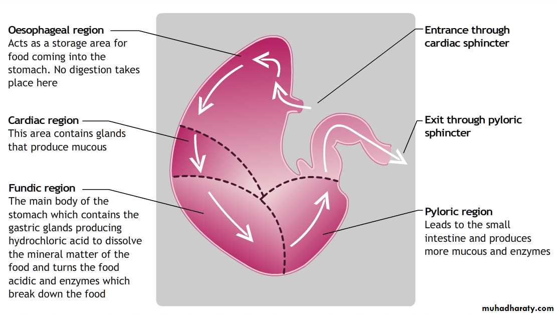
The stomach has four distinct regions:
The StomachThe fact the stomach is never empty is important, as hydrochloric acid is constantly produced, (It is not triggered by food). If the stomach were empty the acid would attack the lining of the stomach causing ulcers.
Food can stay in the stomach for between 30 minutes and 3 hours depending on the type of food. The majority of feed spends about 45 minutes to 1 hour in the stomach. It leaves the stomach as a soupy liquid called chyme which contains all of the ingested food and acidic gastric juices.
The chyme passes out of the stomach via another sphincter muscle called the pyloric sphincter into the small intestine. The chime is passed along the small intestine by wave like muscular contractions called peristalsis.
The Small Intestine
The small intestine can be divided into 3 areas. The duodenum about 1 meter long, the jejunum about 20-24 metres long and the ileum about 1 – 1.5 meters long , in all 20m – 27 metres long and has a capacity of 55-70 litres.The duodenum forms an s shaped curve from the stomach which contains ducts from the pancreas and the liver. These are situated close to the stomach exit and produce pancreatic fluid and bile. The chyme is turned alkaline by these fluids.
The jejunum and the ileum lie to the left of the horse’s abdomen and are looped and coiled to fit the space. The small intestine can move quite freely in this space as they are only attached at the stomach and the caecum.
The small intestine is where the non fibrous food stuff is broken down and absorbed. (The concentrate portion of the diet.) The inside lining of the small intestine has finger like protuberances which increase the surface area. Peristalsis forces the chyme to the walls so that breakdown and absorption are more effective. There are glands which are responsible for producing digestive enzymes.
The Hind Gut
The horse’s natural diet of grass contains large amounts of complex insoluble carbohydrates or fibre and this is not broken down until the chyme reaches the hind gut.This consists of the caecum, the large colon and the small colon, the rectum and anus
The caecum is a large vat which has a blind end. The entrance to the caecum lies near the horses right hip and runs forward and down 0.6-0.9 metres to finish midway along the horses belly at the base of the abdomen. It contains vast numbers of bacteria which can break down the fibre by a fermentation process. Energy is released from this fermentation process in the form of volatile fatty acids.The resulting waste is passed into the large colon. This is about 3 – 4 meters long and has a capacity of 90-110 litres. It has to fold into 4 to fit into the remaining space in the horse’s abdomen. At the pelvic flexture the large colon narrows and turns back on itself and this can cause problems as it is susceptible to blockage at this point. There is further absorption of nutrients and water in the large colon. The food takes 36 to 48 hours to travel the length of the large colon.
The small colon is roughly 4 meters in length and has a capacity of 16 litres.
By the time the food reaches here all of the digestion and absorption of the nutrients has been carried out and the job of the small intestine is to absorb water. The waste is non-digestible food matter and as it passes along the small colon it is formed into loose balls.The rectum is a short tube used as a storage area that holds waste until it is ready to be passed out via the anus.
The anus is another sphincter muscle that empties the rectum when there is a build-up of waste.
Affections of lips
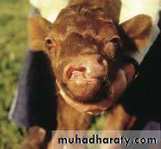
1- Hare lip: - this is a cleft in the upper lip which often runs into the nostrils. It may be uni-or bilateral and is often associated with cleft palate and split jaw. It is most common in calves and occurs due to genetic origin.Treatment
Under G.A. the edges of the cleft are excised so that the mucous membrane and the skin are separated. Then the M.M. edges are sutured together, using silk or nylon, while the skin edges then united by simple interrupted sutures using the same suture material.
2- Trauma:-
occurs due to car accidents, protruding objects or attacks by dogs. Because of the excellent blood supply healing is usually rapid. In severe laceration or loss of tissue plastic surgery is needed to preserve the normal function of lips and checks.
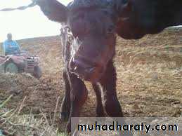
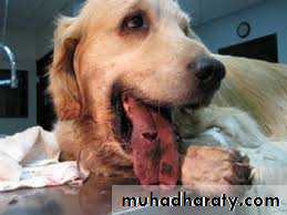
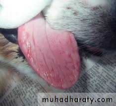
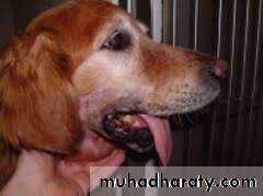
Affections of the Tongue
Trauma:- trauma of the tongue occurs in all species of animals, and all the affected animals will show the following sings.Inability to protrude the tongue for feeding.
Salivation.
Halitosis.
Treatment
Cleaning of the area after insertion of mouth gag and application of local antiseptics and antibiotics.
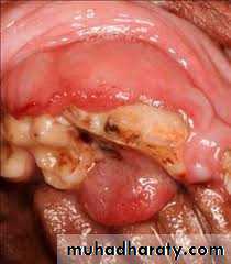
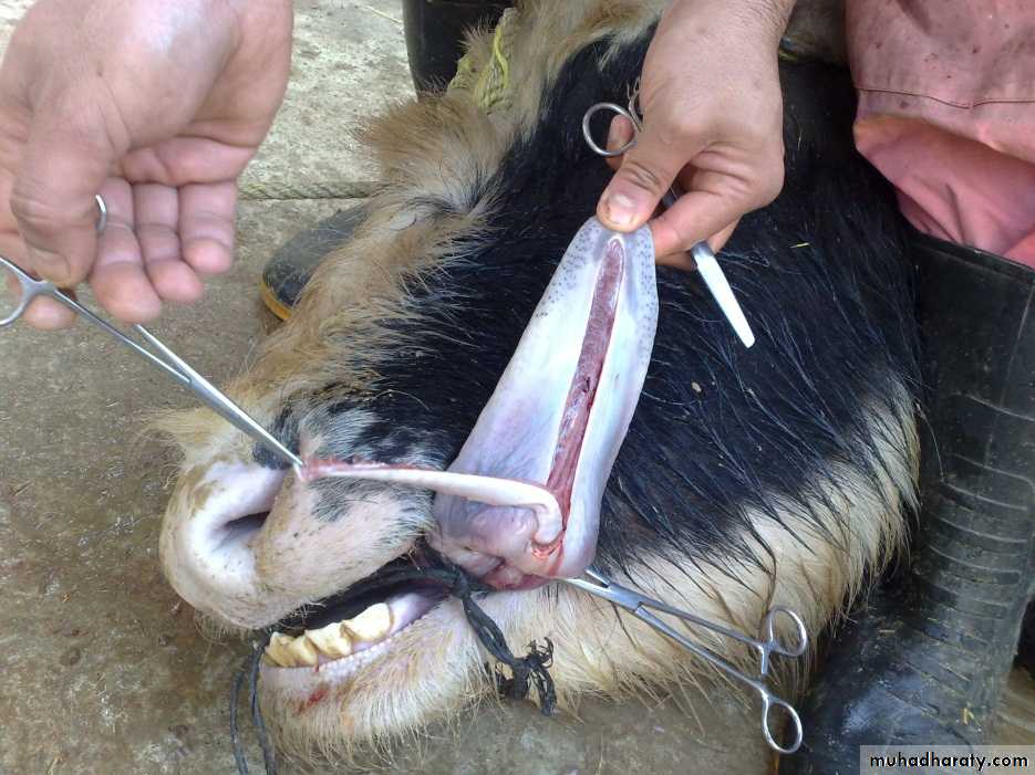
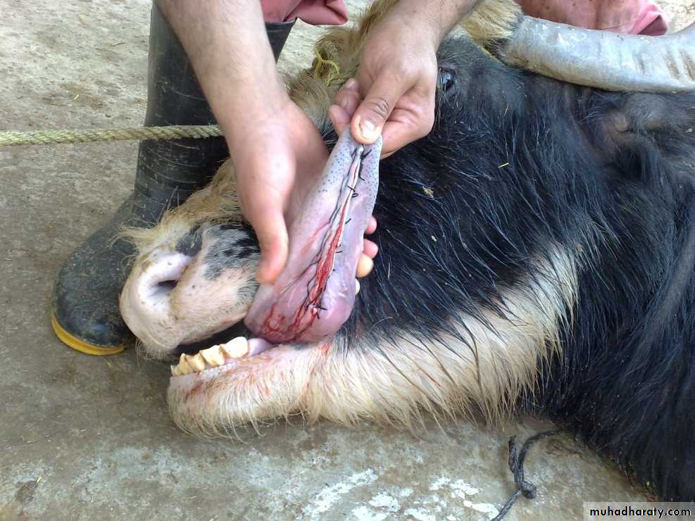
Lacerations:- can occur after street accident or by sharp tooth fragment, if laceration are deep, suture under G.A. and a tension suture (vertical mattress) and non absorbable suture material are used. In case of severe laceration partial glossectomy was done. Amputation in cattle must be avoided because of the loss of prehensile function of the tongue.
Surgical procedure of partial glossectomy
1-The animal is anesthetized and placed in lateral recumbency.2- A tourniquet (rolled gauze) is applied proximal to the intended transaction site.
3- The tongue is transected dorsal and ventral to the lacerated site( the incision will take V shape).
4- The dorsal and ventral aspects are sutured together with an interrupted horizontal mattress pattern with no. I or2 absorbable sutures.
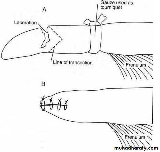
Transect dorsal and ventral to the lacerated site.
Dorsal and ventral aspects are sutured together.Snake bite:-
The tongue can be bitten by a snake as it protruded to bring grass or hay into the mouth.Clinical sings
gangrene and septicemia.
Swelling of the inter mandibular region.
Swelling of the tongue and discolored and protruded stiffly from the mouth.
Treatment
immediate local I.M. injection of antivenin and antibiotics (penicillin and streptomycin) is essential.Giving water and feed.
Incise the tongue to drain away exudates.
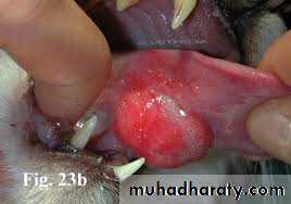
Paralysis of the tongue (glossoplagia):
Paralysis of the hypoglssal nerve and the tongue shown one side of the mouth. In bilateral paralysis the whole tongue is flaccid and hangs out of the mouth.Causes
infection or neoplasm along the course of the nerve.
Lead poisoning and botulism.
Treatment
tonics.
Water and nutrients must be administered by stomach tube.
Neoplasia:-
tumor of the tongue are very uncommon, but the one usually seen is the squamous cell carcinoma. The treatment is excision of the tumor or amputation if it is possible.
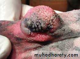
Self suckling
If nose ring with burr is not successful for preventing self suckling a partial glossectomy is used. The technique alter the tongue contour to prevent from forming a U shaped tongue for suckling.Surgical procedure
The operation is performed with sedation and local infiltration of Lidocaine or under general anesthesia.Two techniques could be performed:
Ventral glossectomy: an elliptical incision is made that is approximately 5cm at its widest part and start rostral to the frenulum attachment on the tongue and extends rostrally 2.5cm caudal to the tip of the tongue. The edges closed with horizontal mattress suture pattern.
Lateral glossectomy: removes half of the tip(5cm) of the tongue.
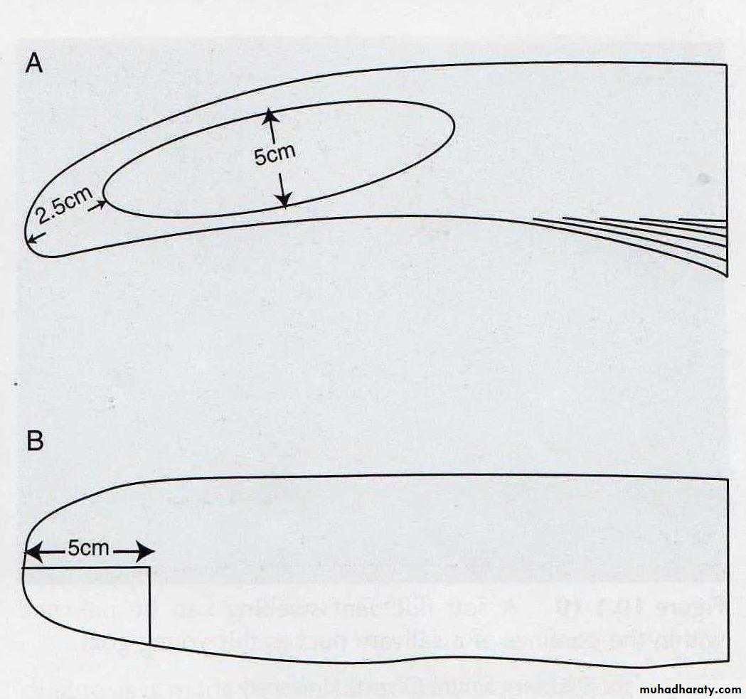
-Lateral glossectomy
-Ventral glossectomy.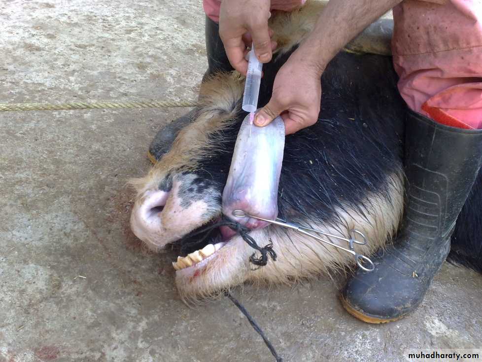
Affections of the salivary glands:
Salivary glands affection divided into 2types: congenital and aquired.Congenital abnormalities of the salivary glands are associated with agenesis or atresia of the parotid ducts, resulting in a fluid- filled swelling proximal to the obstruction site.
Aquired diseases are usually secondary to lacerations or other trauma that ruptures the salivary glands or ducts.
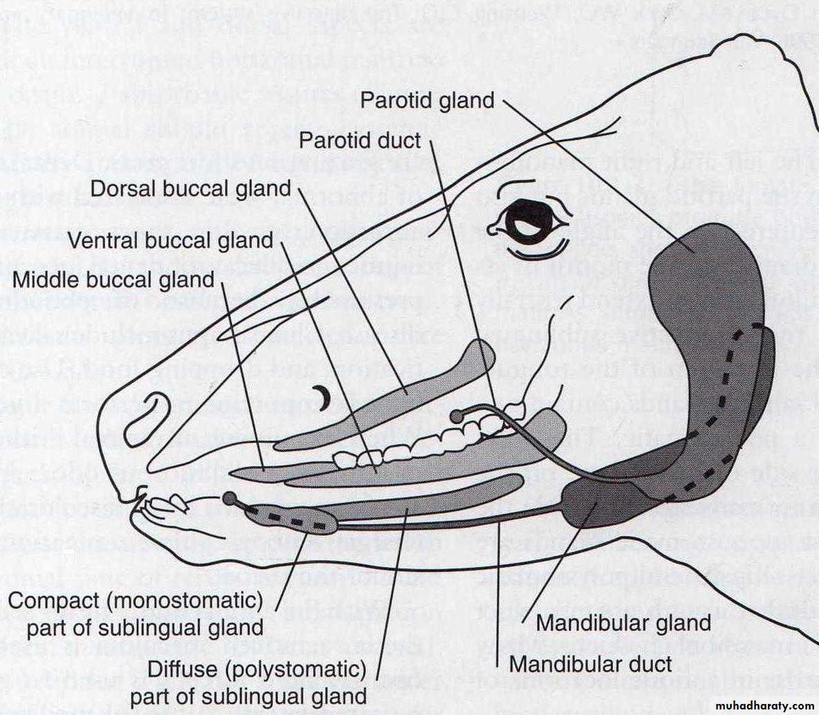
In horses and ruminants there are 3 main pairs of salivary glands: parotid, mandibular, and sublingual glands.
In dogs there are 4 main pairs of salivary glands:
the parotid, mandibular, sublingual, and the zygomatic glands, which are the dorsal buccal glands in other animals.The parotid gland is the largest of these glands and occupies the space between the vertical ramus of the mandible and the wing of the atlas. The parotid duct (Stenson's duct) is formed by three or four radicles and leaves the gland ventrally about 2.5cm above the external maxillary vein.
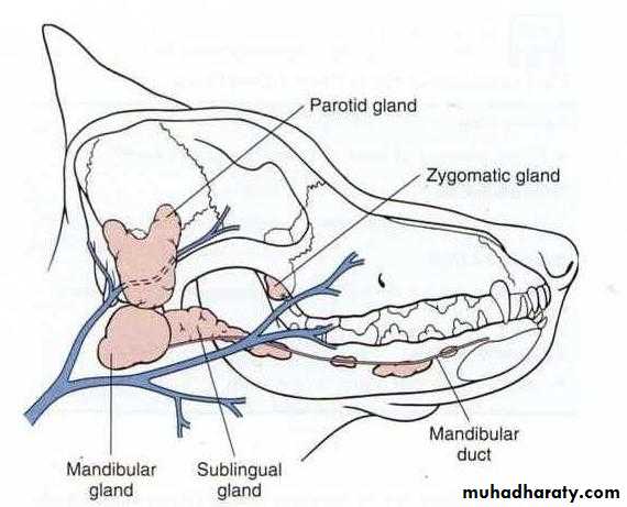
Salivary glands in the dog
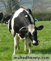
Salivary glands in the cow
Trauma:-Fresh wound of the gland must be sutured after thorough cleaning and debridement, the leaking wound has a great tendency toward spontaneous closure.
Close the wound where the fistula has been excised in several layer with cat gut, the skin wound is closed with interrupted silk suture. The tube facilitates normal drainage of saliva.