Lec . 2 Periodontic Dr.Hussein AL Dabbagh
Microscopic anatomy of gingiva:
The gingiva consists of fiberous connective tissue known as lamina properia covered by stratified squamous epithelium.
The gingival epithelium covering the gingiva may be differentiated as follows:
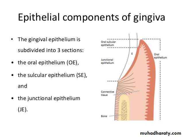
The oral epithelium faces the oral cavity, sulcular epithelium faces the tooth without touching it, junctional epithelium which provides the contact between the gingiva and the tooth surface.
Oral epithelium:
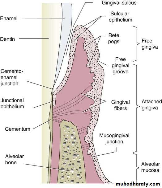
It covers the crest and the outer surface of the marginal and attached gingiva, it is either keratinized (no nuclei) or parakeratinized (retained nuclei), the boundary between oral epithelium and underlying connective tissue has a wavy course.
The projection of the epithelial cells into connective tissue are known as rete pegs , the connective tissue portions which project into the epithelium are called connective tissue papillae.
This alternating patterns of depression and proturberances of connective tissue papillae and epithelial rete pegs is thought to give the attached gingival the “stippled appearance’’.
The characteristic feature of the O.E and O.S.E is the presence of rete pegs while these structures are lacking in J.E.
The oral epithelium consists of 4 layers of cells:
Stratum basale.Stratum spinosum.
Stratum granulosum.
Stratum corneum.
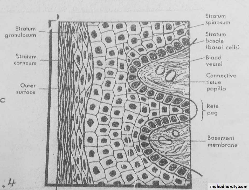
In the outer cell layers the nuclie are lacking such epithelium called orthokeratinized , but often the cells of st.corneum contain remnants of nuclie and in such cases the epithelium is denoted parakeratinized.
Other cells of OE:
Melanocytes : pigment producing cells.
Langerhans cells : play a role in defeanse mechanism.
Merkel cells : located in deeper layers , harbor nerve endings, they have been identified as tactile receptors.
Keratinocytes cells : keratin producing cells which comprise about 90% of total cell population.
Under normal conditions there is complete equilibrium between cell renewal and desquamation (cell turn over), it takes approximately 3-4 weeks for keratinocytes to migrate from basal layer until reach the outer epithelial surface.
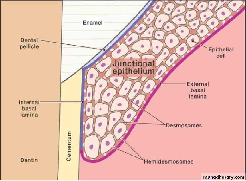
The basal cells are found immediately adjacent to the connective tissue and seperated from this tissue by a basment membrane (basal lamina).
The epitheial cells are attached to each other by a structure called desmosomes which is composed of two hemidesmosome seperated from each other by granulated material.
Hemidesmosomes composed from following structures:
The outer leaflets (OL): of the cell membrane of two adjoining cells.
The thicker inner leaflets (IL): of the cell membrabe.
The attachment plaque (AP): which represent granular and fibrillar material in cytoplasm.
Sulcular epithelium:
It lines the gingival sulcus, it is thin non-keratinized stratified sequamous epithelium, extend from coronal limit of the junctional epithelium to the crest of gingival margin.The sulcular epithelium is important because it is thin and may act as a semipermaible membrane through which tissue fluid from the gingiva seeps into sulcus and makes easier for bacterial products of dental plaque to penterate into connective tissue and stimulate inflammation and tissue destruction, this is why sulcular epithelium considered as a poor barrier againts bacterial infection.
Junctional epithelium :
The epithelium that attach the gingiva to the tooth surface, it is consists of stratified sequamous non-keratinized epithelium.
Iy is quite permeable and thus serve as a pathway for diffusion of products of plaque bacteria to the connective tissue , there is also diffusion in the opposite direction moving towards the sulcus of host defense substance.
Diffrence between J.E and O.E & S.E:
The size of the cells in the J.E is larger than O.E.
The intercellular space in the J.E is wider than in the O.E.
The number of desmosomes is fewer than O.E, this may explain the susceptaibilty to tear during probingand it is greater permeability to migrate cells fluid.
The junctional and sulcular E. Are not thick as O.E because they are not keratinized and in health have no rete pegs.
Cells of J.E turnover rate is very high (4-6 days) comapared to O.E that has longest turnover rate (6-12 or up to 40 days).
J.E forms the attachment of the gingiva to the tooth surface while O.E and S.E have no attachment to the tooth surface.
Connective tissue (CT) :
The connective tissue of the gingiva is known as lamina propria and consists of 2 layers:The papillary layer : it consists of papillary rojection between the eithelial rete pegs.
The reticular layer : it is contiguous with the periosteum of the alveolar bone.

The major components of the CT are :
Collagen fibers (60%) 2- fibroblast (5%) 3- vessels , nerves, and matrix (35%).Cellular elements of the gingival CT :
Fibroblast : the most predominant cells of the CT (65%), they synthesize collagen and elastic fibers as well as the CT matrix and regulate collagen degradation.
Mast cells : it is responsible for the production of certain components of the matrix , they also produces vasoactive substance which may control the flow of blood through the tissue.
Macrophages : have phagocytic function and are involved in the defense of the tissue againts irritating substances.
Inflammatory cells : include various types such as polymorphonuclear leukocytes, lymphocytes , plasma cells, these cells have different immunological functions.
Fibers of CT :
Collagen fibers : the most predominant type of fibers in CT.
Reticular fibers.
Oxytalon fibers.
Elastic fibers.
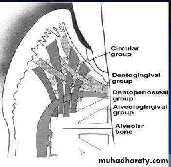
Classification of gingival fibers :
According to their insertion and course in the tissue , the gingival fibers can be divided into the following groups :
Circular fibers (CF) : are fibers encircling the tooth, and run through the CT of the marginal and interdental gingiva.
Dentogingival fibers (DGF) : the are embeded in the cementum of the supra-alveolar portion and project in a fan-like configuration out into free gingival tissue of the facial , lingual, and interproximal surfaces.
Dentoperiosteal fibers (DPF) : they extend from the cementum in apical direction to the periosteum of the alveolar bone and terminate in the attached gingiva.
Transseptal fibers (TF) : located interproximally , these are horizontal bundles that extend between the cementum of the adjacent teeth.
Matrix of the CT :
Matrix is roduced by fibroblast , but some constituents are produced by mast cells and blood, the matrix is the medium in which the CT cells are embedded and the transportation of water , electrolytes, nutrients, etc, to and from the individual CT cells occurs within matrix.Thank you