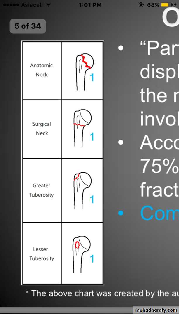
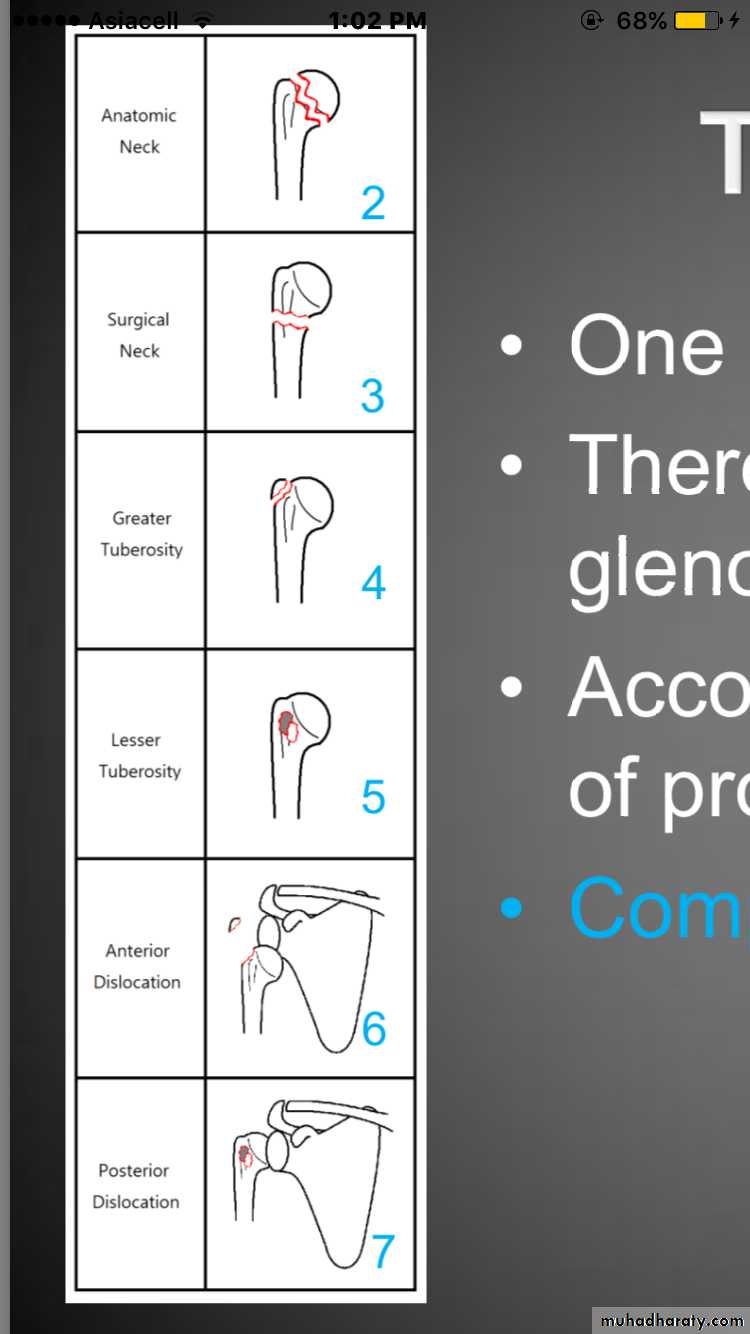
Neer classification
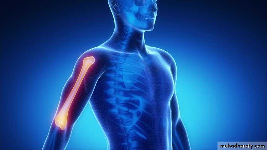
Fracture of proximal humerus1
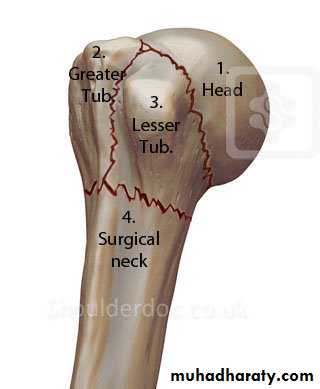
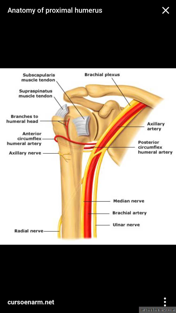
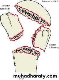
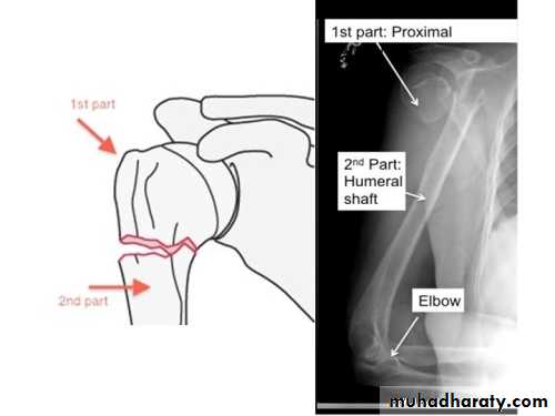
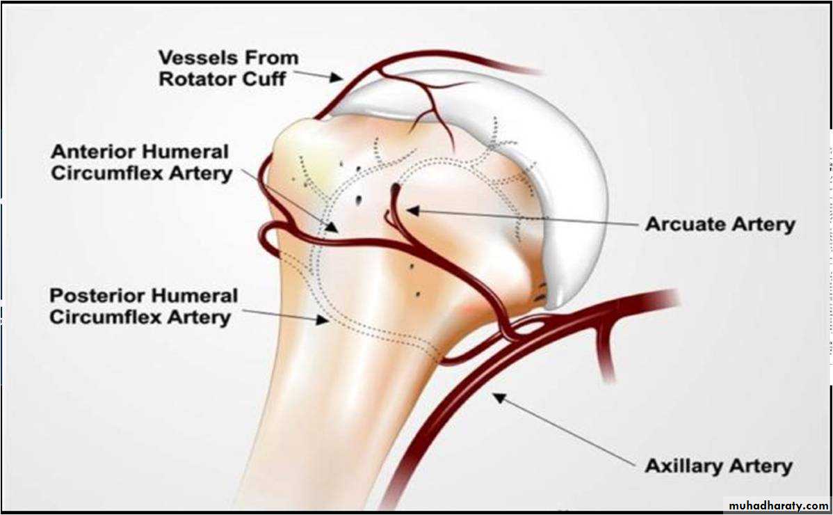
Anatomy of proximal humerus
Learn the epidemiology of proximal humeral fractureMechanism of injury and clinical feature
Understands the fracture patterns with majour emphasis on neer classification
Learn various treatment options
Majour complication
2
objectives
Anatomy of the humerus
3
5% of all fracture
Higher incidence in elderlyFemale 2:1 greater incidence than male
85% are minimally or non displaced & generally treated non operative
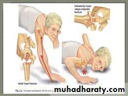
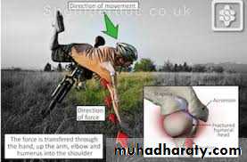
MECHANISM OF INJURY
Low energy (most common)
Fall from a standing height
Outstretched arm
• Elderly osteoprotic
• High energy (young )
4
epidemiology
5
mechanism
6
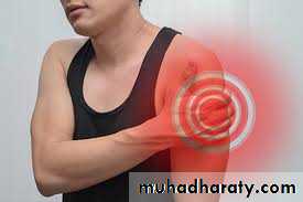
Clinical feature
Two main components
Number of fracture partdisplacement
7
Main components off classification
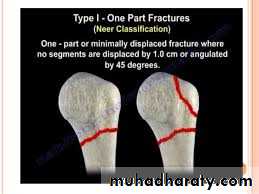
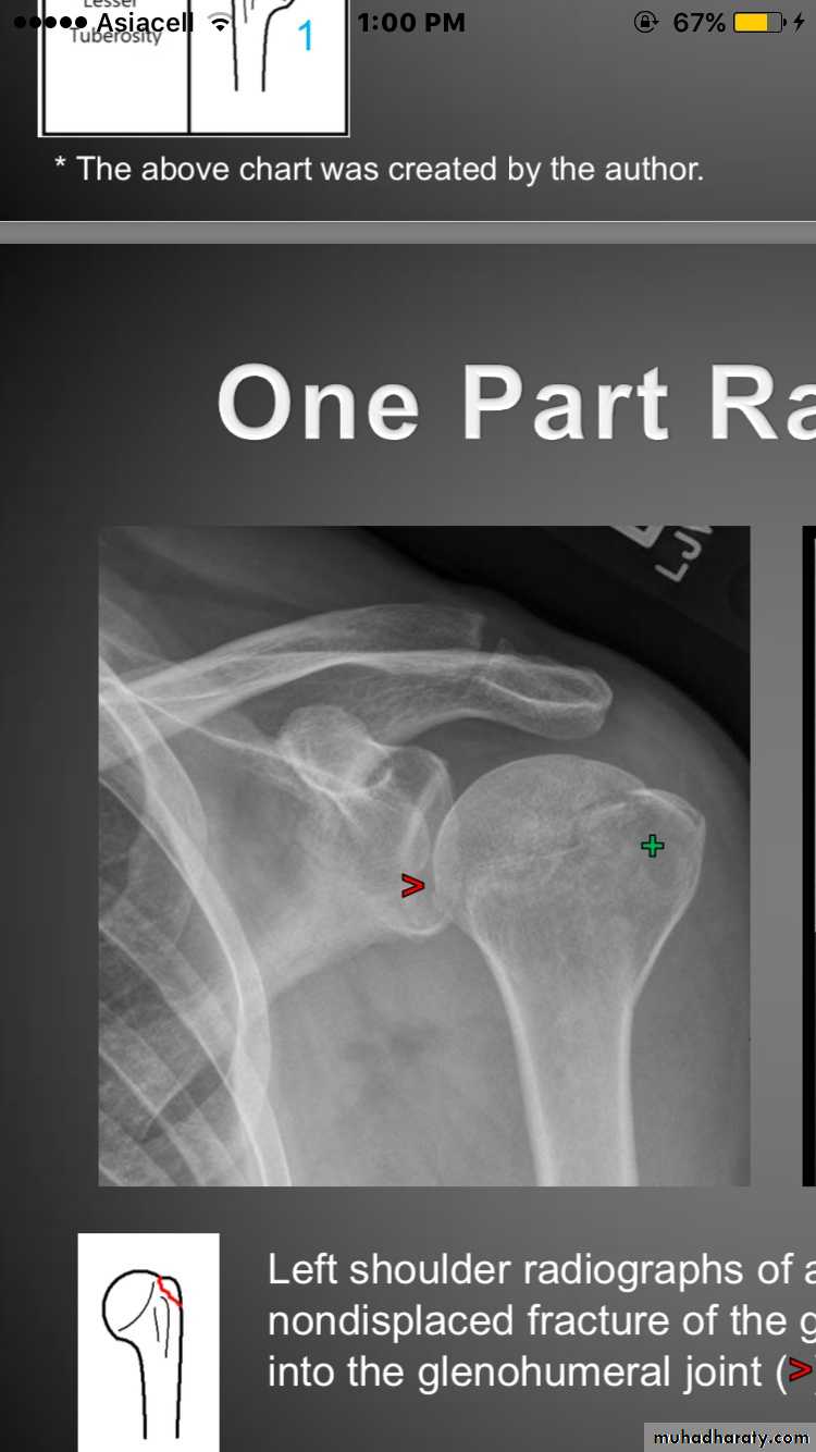
One _part fracture
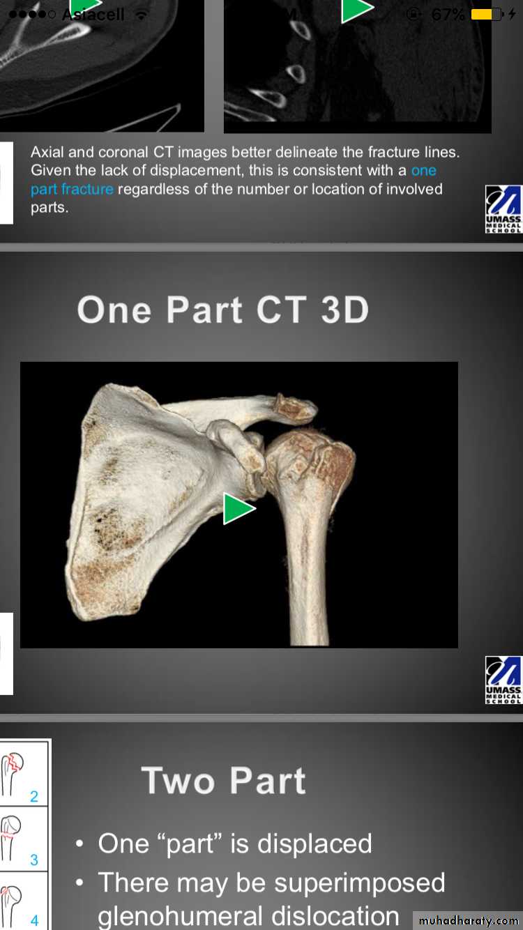
Fracture lines involve 1-4partsNon of the parts are displaced (<1cm&<45degree)
70_80% of all proximal humeral fracture
8
Classification
9
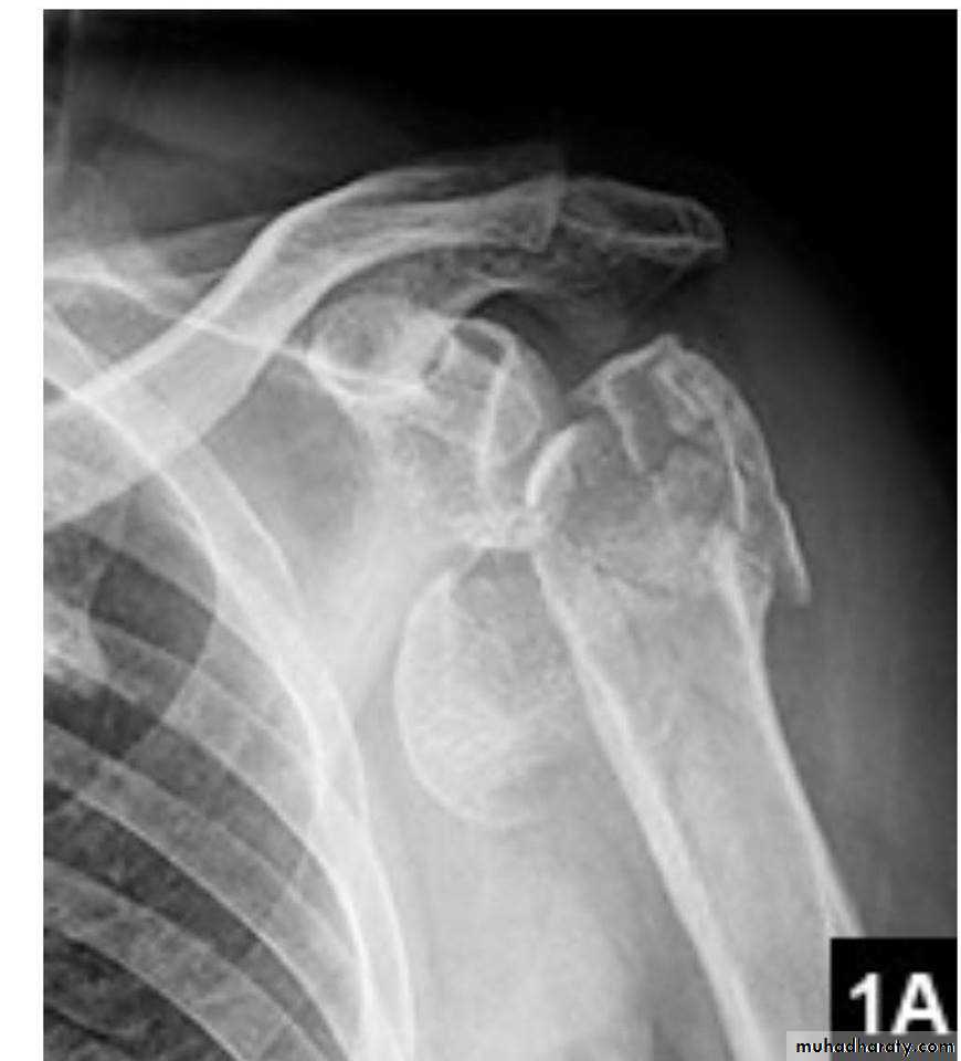
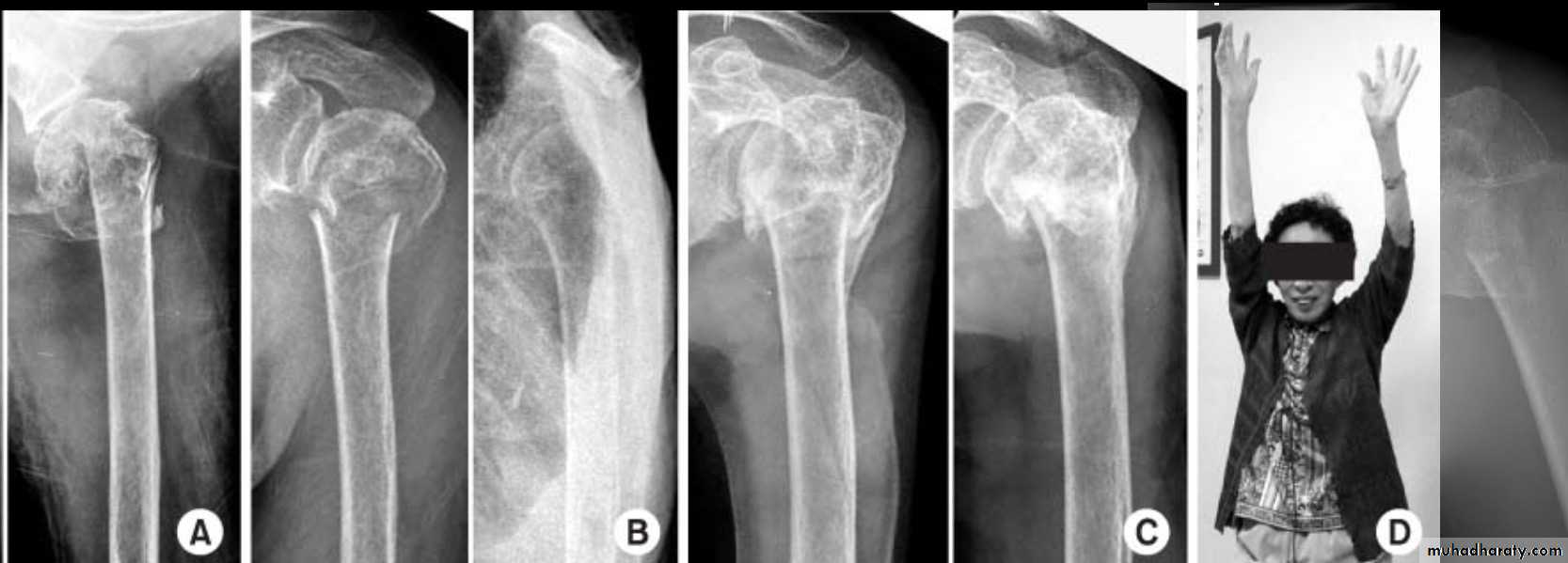
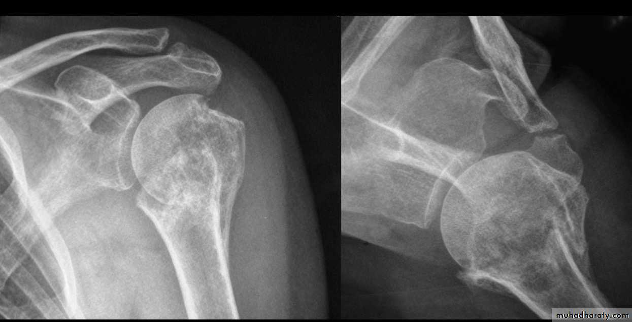
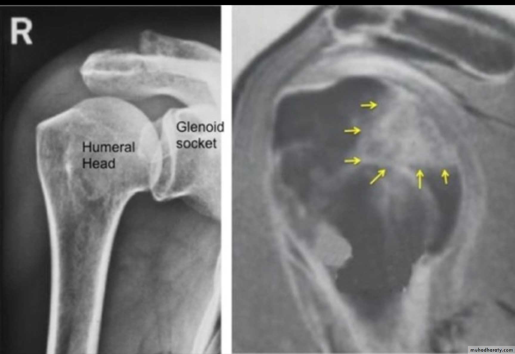
X RAY
10
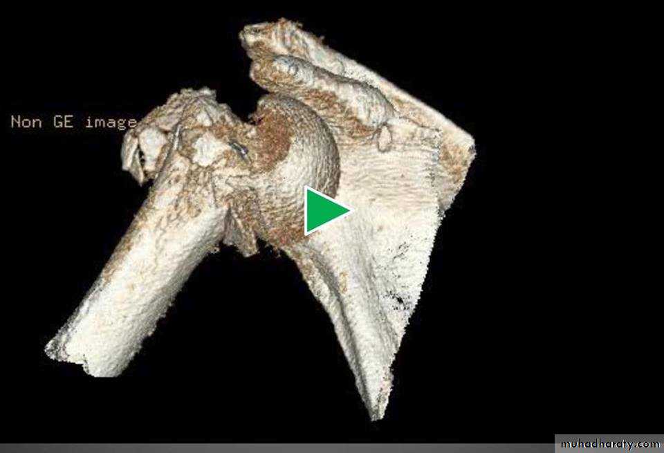
3D CT
11
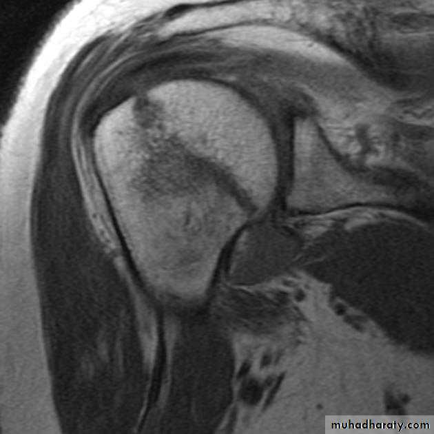
MRI
Conservative
12Treatment type one

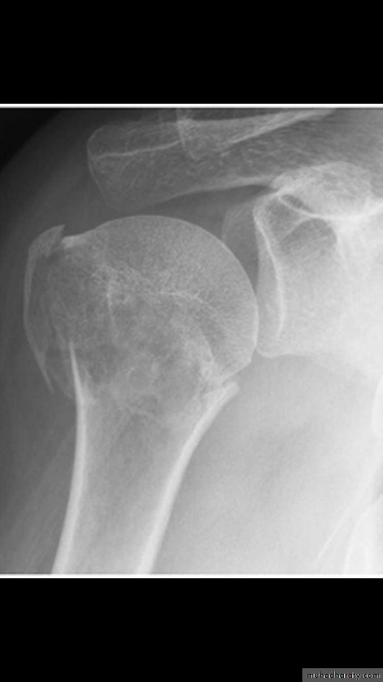
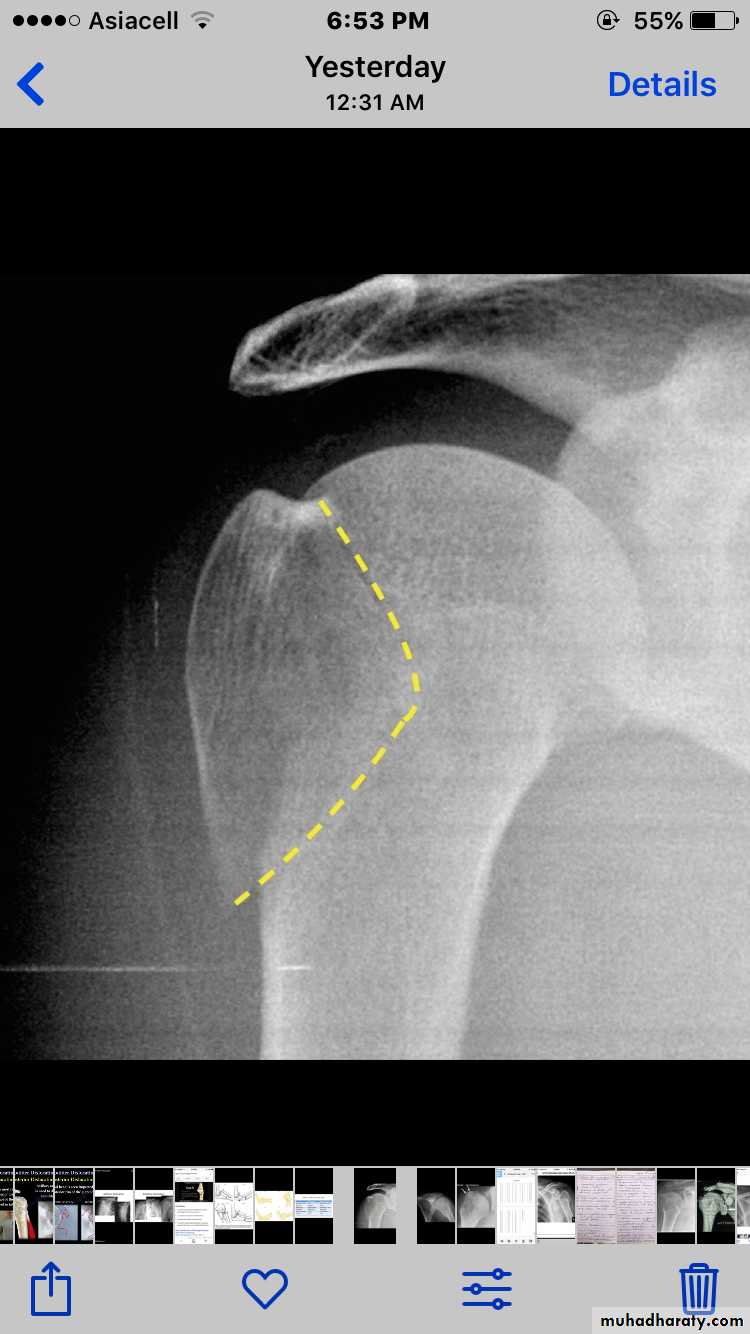
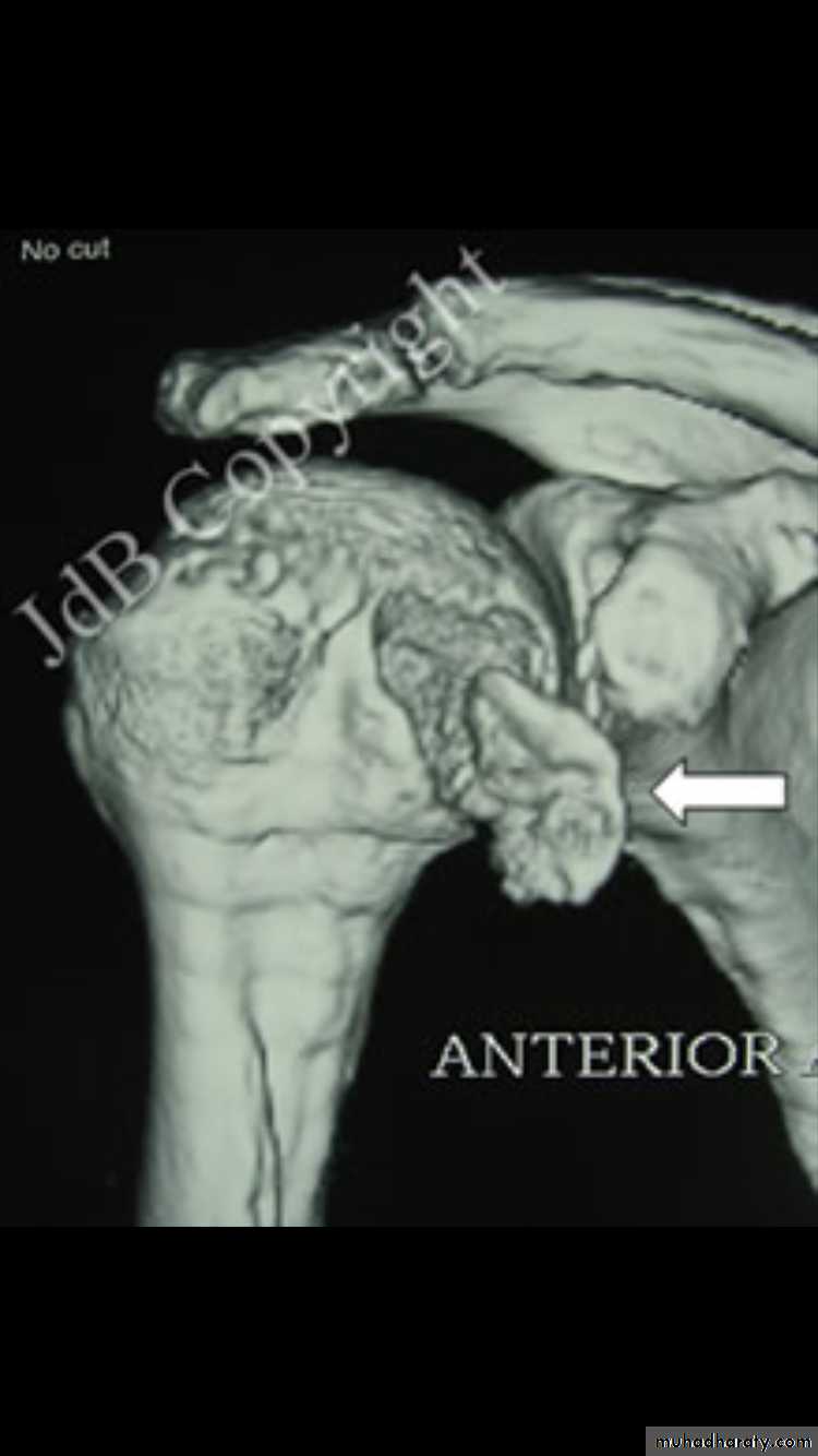
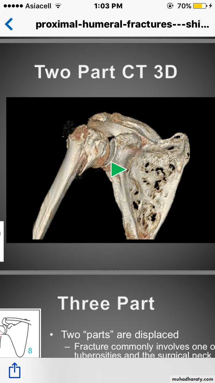
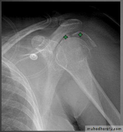
Two part fracture
20% of proximal humeral fractureFracture lines involve 2-4 parts
One part is displaced(>1cm&>45degree)
13
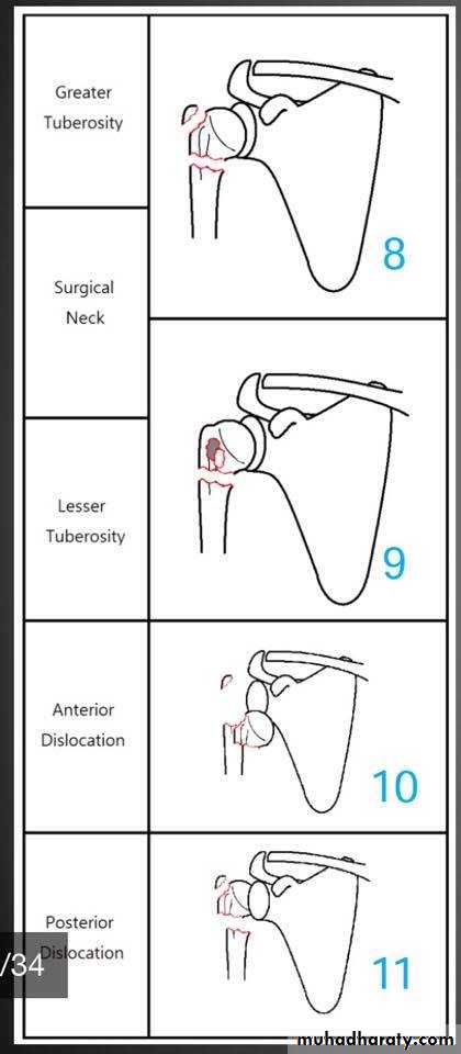
Types of two part fracture
Anatomical neckSurgical neck (most common)
Greater tuberosity
Lesser tuberosity
14
15
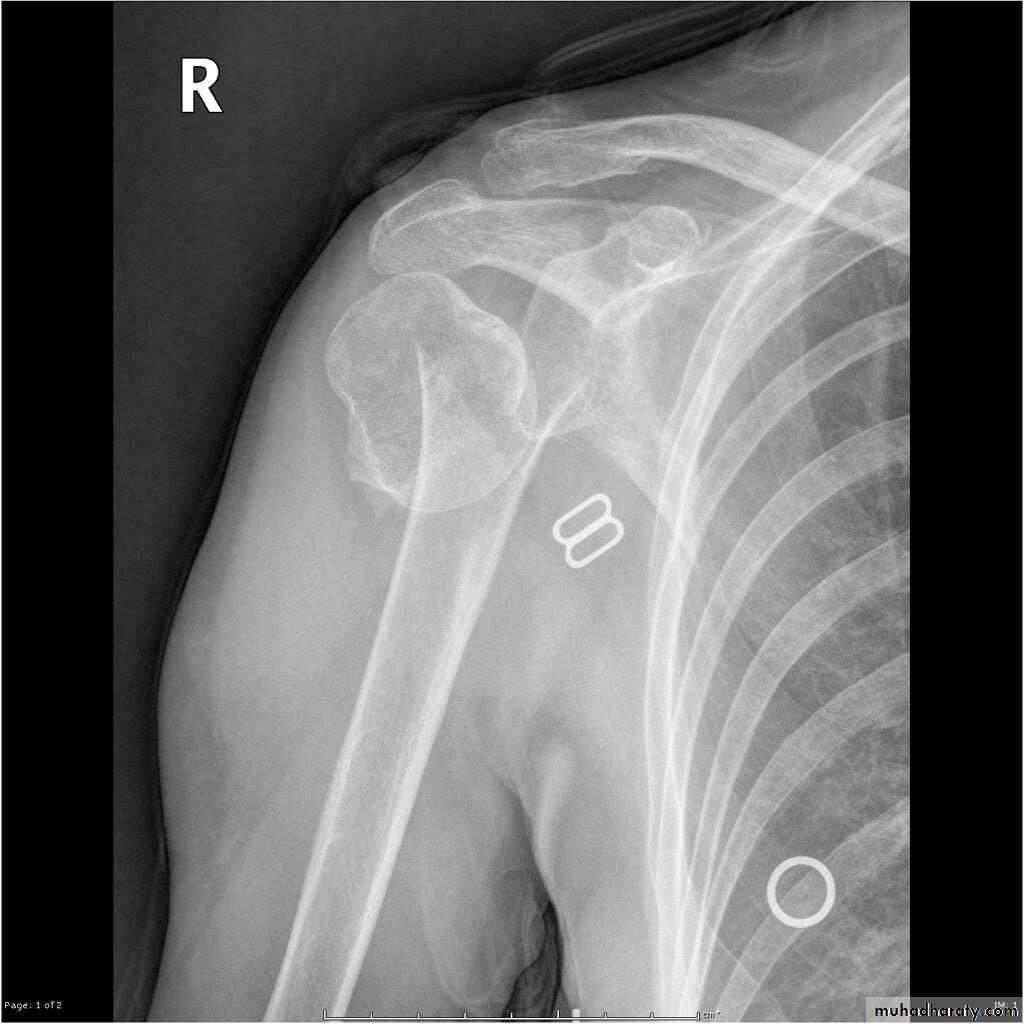
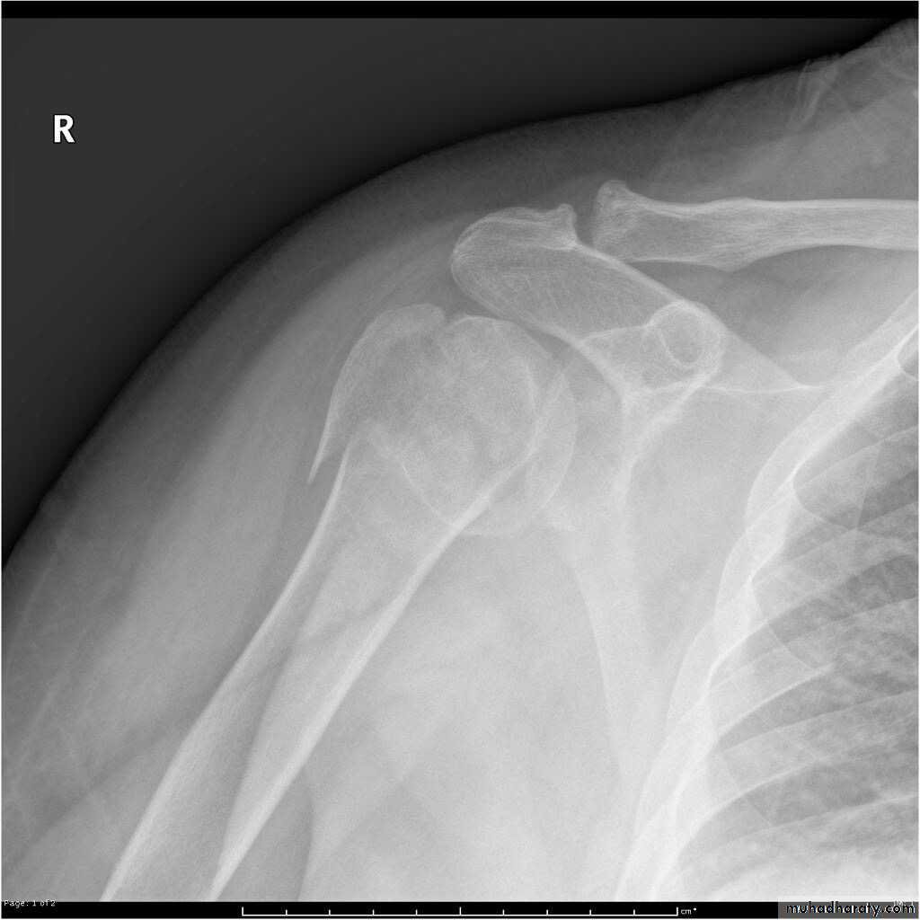
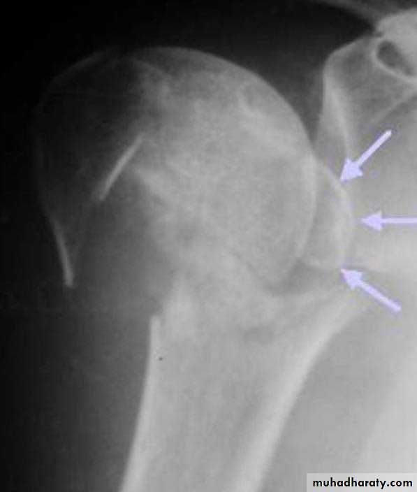
X RAY
16
X RAY
17
X RAY
18
X RAY
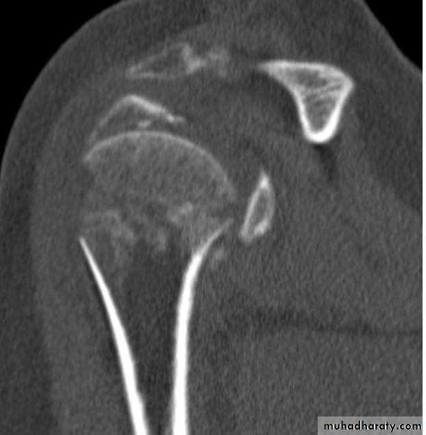
19
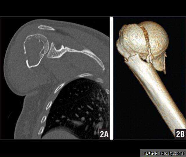
CT
20
3D CT
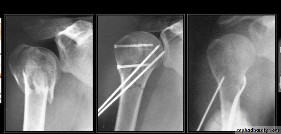
• Close reduction (MUA&sling ) 6weeks and active exercise later if failure
• Open reduction & internal fixation by percutaneous pinning plate and screws or intramedullary nailing21
TREATMENT
22
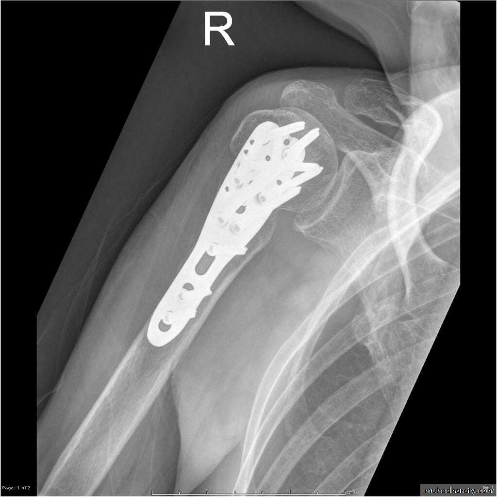
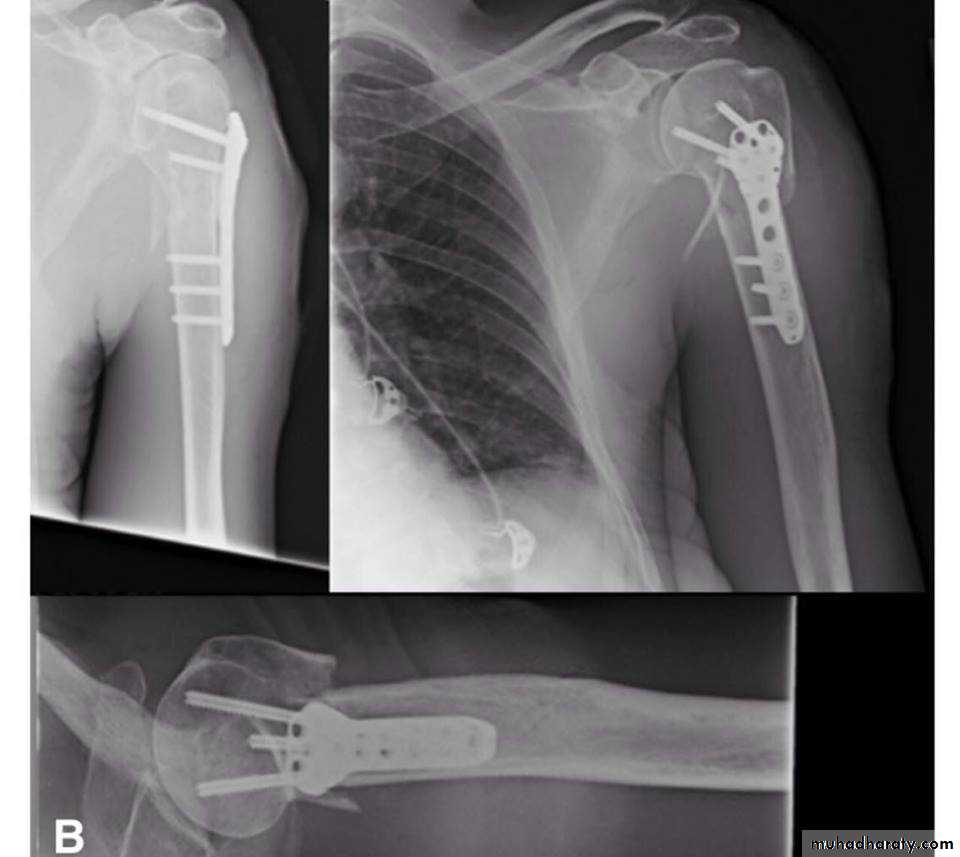
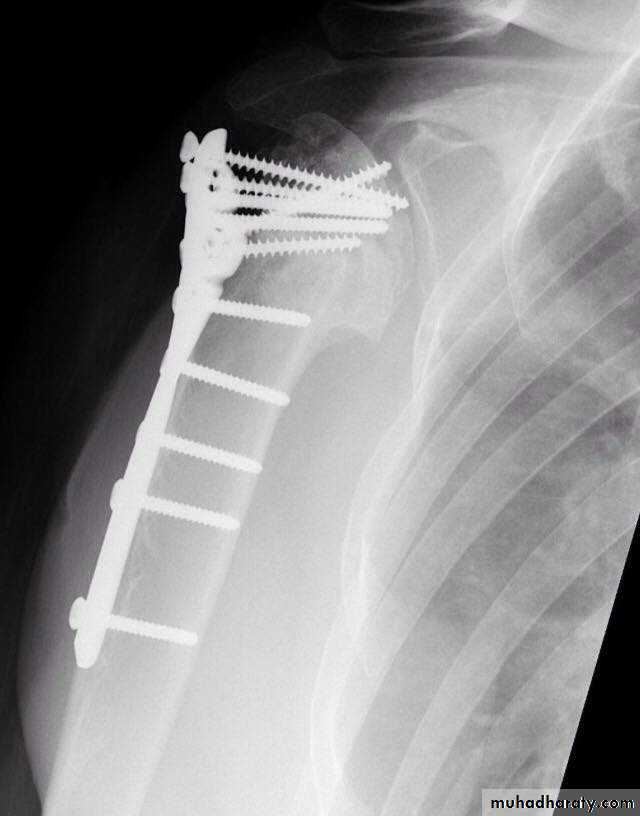
Plate and screws
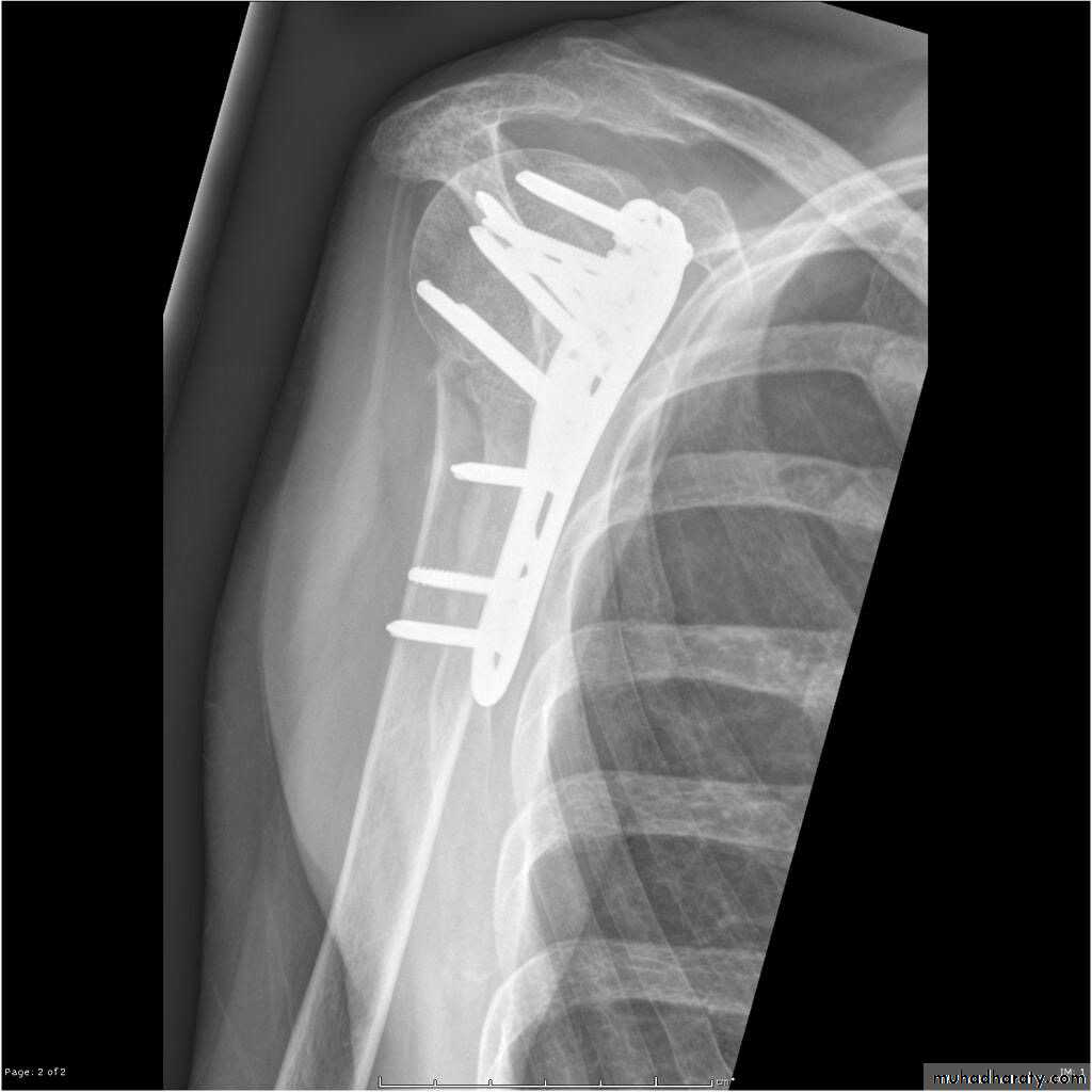
23
Plate AND SCREWS
24
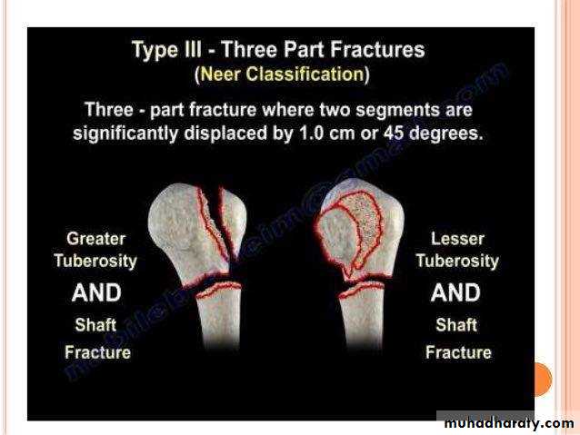
Three part fracture
Its account for 5%of proximal
Humeral fracture
Fracture line involve 3_4 part
Two part are displaced more than
1cm
Or more than 45 degrees
25
26
X RAY
27
X RAY
28
CT
29
TREATMENT
30
ORIF
UNCOOMMON LESS THAN 1%
Three parts are displacedMore than 1cm
Or more than 45degrees
31
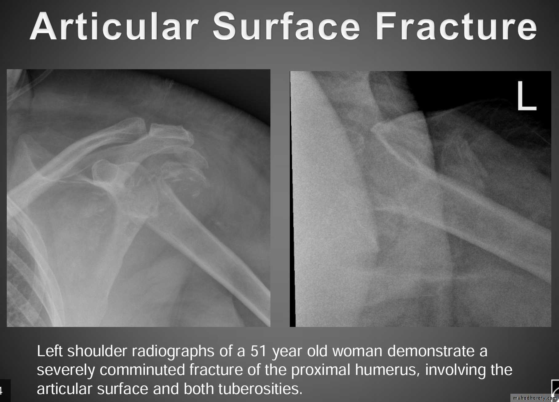
FOUR PART FRACTURE
32
X RAY
33
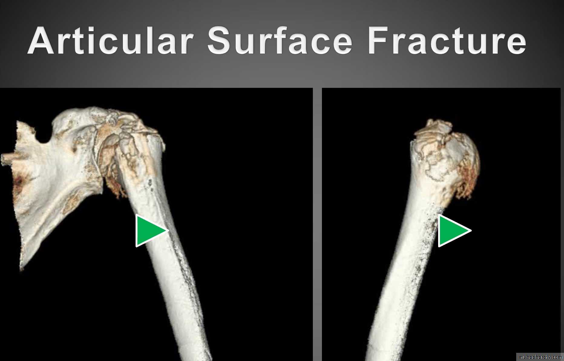
CT
34
TRATMENT IN YOUNG
35
ARTHROPLASTY
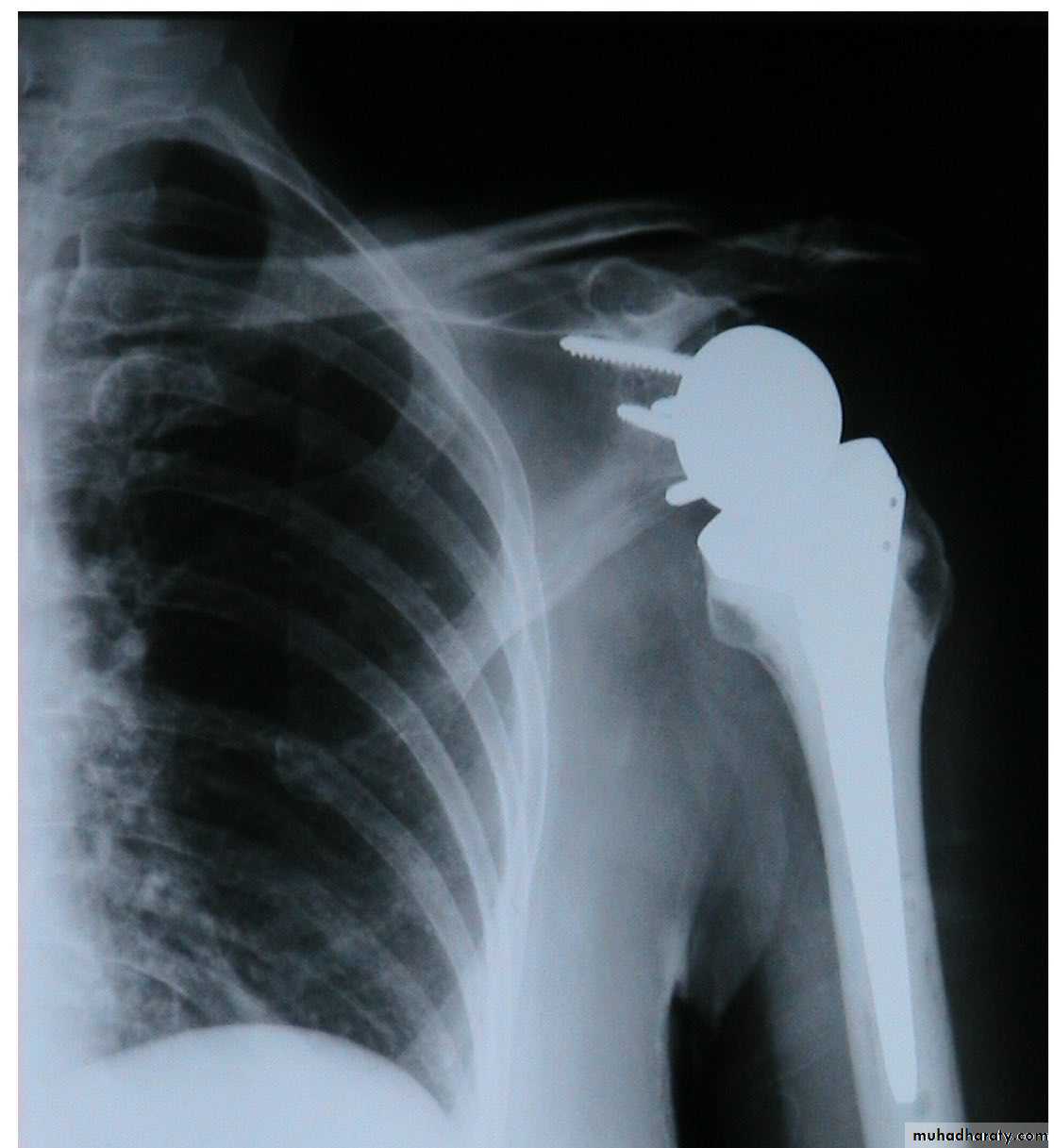
36
37
1-VASCULAR INJURY
38COMPLICATION
39
2-STIFFNESS OF SHOULDER

40
3-MALUNION
41
42
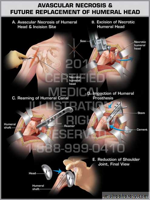
4_AVASCULAR NECROSIS

43
44

45