
Alveolar ridge atrophy
Clinical factors affecting bone resorption:1.Anatomic factors
As the size and shape of the ridge, the ridge relations, and the thickness and character of mucosal covering
2.Metabolic factors
As age, Sex, nutrition, hormonal balance, osteoporosis.
3.Functional factors
Frequency, direction, and amount of force applied to bone.
4.Prosthetic factors
The type of denture base, the form and type of teeth, the interocclusal distance and etc.
Etiology of RR
• Type of bone: cancellous bone is more prone to resorption than is cortical bone.• Size and shape of the ridge: Thin narrow ridges will resorb more than well-formed broad ridges, as the force received per unit.
• Ridge Relationships: Long faces have more alveolar bony ridges which will decrease rate of atrophy.
• amount and quality of the mucoperiostum, muscle contour, depth of the vestibule.
I. Anatomic factors
• 1. Age: RR generally increases with age.
• 2. Sex: RRR occurs more in females. This usually occurs during menopause, as a result of hormonal disturbances.
• 3. Nutritional deficiency: Calcium deficiency, decrease in vitamin C and/or protein utilizanation and/or dysfunction of carbohydrate metabolism.
• 4. Loss of natural teeth due to periodontal diseases.
• 5. systemic diseases: as uncontrolled diabetes.
• 6. Radiation therapy.
II. Biologic / Metabolic factors
• 1. Long-term wearing of dentures without serviceability.
• 2. Improperly constructed dentures with improper vertical dimension of occlusion, centric relation, non-balanced occlusion and incomplete coverage of basal seat area.• 3. Continuous wearing of the dentures without rest to the underlying tissues.
• 4. Porcelain teeth and/or anatomic teeth with high cusp angles transmit more force to the underlying ridge.
• 5. Force duration and frequency.
• 6. Bruxism.
• 7. Overdiplaced supporting tissues.
• 8. Unfitting denture.
III. Prosthodontic factors:
Gross mandibular atrophy:
Multifactorial biomechanical disease resulting from a combination of anatomic, metabolic, and mechanical determinant varying with time from patient to patient in an infinite number of combinationsCauses of mandibular atrophy and alveolar bone loss:
1.Disuse atrophy.
2.Localized excessive pressure during incising and unilateral function under a denture .
3.Periodontal bone loss before extraction of the teeth.
4.Hyperparathyroidism.
5.Deficiencies and tissue resistance to stress.
Class I
This classification level describes the stage of edentulism that is the most to be successfully treated by conventional prosthodontic techniques with complete denture prosthesis.Residual bone height of 21 mm or greater measured at the least vertical height of the mandible
Class II
This classification level distinguishes itself with the noted continuation of the physical degradation of the denture supporting structures and in addition is characterized with the early onset of systemic disease interactions, localized soft tissue factors and patient management/lifestyle considerations.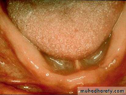
Class III
This classification level is characterized by the need for surgical revision of denture supporting structures to allow for adequate prosthodontic function.
Additional factors now play a significant role in treatment outcomes.
Class IV
This classification level depicts the most debilitated edentulous conditionSurgical reconstruction is almost always indicated but can not always be accomplished due to the patient’s health, desires, past dental history and financial considerations
When surgical revision is not selected, prosthodontic techniques of a specialized nature must be used in order to achieve an adequate treatment outcome
Problems as a result of extensive changes in the facial and intraoral tissues following the loss of permanent dentition:
• 1.Morphological changes:
• caused by either reduction in facial tissue support due to resorption and remodeling of the alveolar tissues.
• 2.Neuromuscular changes:
• Resulting in indefinite occlusal positions
Facial morphological changes:
Changes in facial contourFacial support
Rest facial height
Changes in facial muscles
Loss of support for the facial musculature
Muscle attachment
Changes in temporomandibular joints.
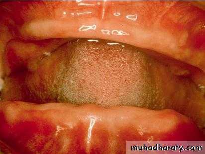
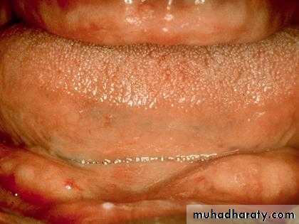
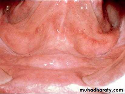
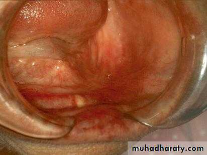
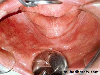
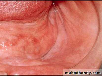
Intraoral morphological changes:
-Apparent loss of sulcus width and depth-Muscle attachment
-Bony prominence
1. Sharp, spiny ridges
2. Uneven alveolar bone
3. Prominent mylohyoid and internal oblique ridge.
4. Sharp mentalis eminence.
5.Enlarged genial tubercle.
The problem of the Mandibular reduced residual ridge:
1.The average maxillary denture bearing area is 23cm², while the average mandibular denture bearing area is only 12cm² and the mandible is susceptible to resorption four times than the maxilla.
2. The surface contour of the resorbed ridge may prejudice denture support and the superfacial aspect of the mylohyoid ridge may also be sharp, irregular, and prominent which makes it unfavorable for support due to painful loading of the covering mobile mucosa. In cases of nerve dehiscence and ridge irregularity the master cast should be relieved before construction of the conventional denture base, where surgery is thought to be inappropriate
3. Lack of retention and stability of the conventional mandibular complete denture is commonly a complaint of patient’s with reduced residual ridges because of the unfavorable flat ridge from which does not provide any resistance to anteroposterior or lateral movements. Chronic mucosal irritation, discomfort, and the inability to properly masticate are usually attendant history findings as well.
4. As a result of the reduction of the residual ridge, the floor of the mouth becomes relatively superficial.
5. Severe mandibular atrophy will result in the genial tubercle and attached muscle becoming sufficiently superficial to interfere with the lingual flange.
6. On the labial surface of the anterior region several muscles show proximity to the crest of the ridge, especially in badly resorbed ridges. These muscles should not be impinged on because their action is nearly at right angles to the flange.
7. The influence of the lip on lower denture stability becomes more critical as resorption of the ridge increases or as the patient becomes older. The lip instead of being everted as in young individual becomes thinner and inclines backward into the mouth.
8. The large intermaxillary space that results from excessive bone loss creates prosthesis problems of esthetics related to loss of facial support, occlusion, and the patient ability to control the prosthesis.
9. These cases with grossly resorbed lower ridges often have the crest of the ridge at the level of the mental foramina, in which the nerves and blood vessels are impinged on easily. This causes paresthesia of the lower lip occurring during mastication.
Treatment :
• Ideal denture supporting ridge:
• 1.Adequate bone support for dentures.
• 2.Bone covered by adequate soft tissue.
• 3.No undercuts or hanging protuberances.
• 4.No sharp ridges.
• 5.Adequate buccal and lingual sulci.
• 6.No scar bands to prevent normal seating of a denture.
• 7.No muscle fibers or frenula to interfere with the periphery of the prosthesis.
• 8.Satisfactory ridge relationships between the maxillae and mandible.
• 9.No soft tissue folds, hypertrophies on the ridge or sulci.
• 10.A ridge free of neoplastic disease.
Management of RRR
Prosthetic Management With Surgical InterventionProsthetic Management Without Surgical Intervention
?????
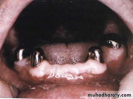
1. Preventive prosthodontics:The greatest way to preserve the mandibular anterior ridge comes from the maintenance of one or more endodontically treated roots and the placement of an overdenture.
The advantages of the overdenture over the conventional denture are:
The advantages of the overdenture over the conventional denture are:
1.The denture bearing mucosa of the residual ridges are spared abuse.
2.Maintenance of the alveolar bone.
3.Sensory feedback.
4.Minimal load thresholds.
5.Tactile sensitivity discrimination.
6.Masticatory performance.
7.Reduction of Psychological trauma.
2.Prosthodontic Treatment:
Many techniques have been developed to deal with the problem of the compromised ridge.1.Principle of mucostatics.
2.Using metal bases for snugness of fit of the mandibular denture.
3.Implanting platinum cobalt magnets to increase mandibular denture stability.
4.The flange technique which provided greater denture-bearing surface for stabilization.
Proper coverage of all available denture-bearing surface is fundamental to good denture construction.
3.Surgical management:
1.Enlagement of denture-bearing areas
a)Vestibuloplasty.
b)Ridge augmentation.
2.Implants
a)Subperiosteal.
b)Transosseous
c)Endosseous.