Strongyloides stercoralis

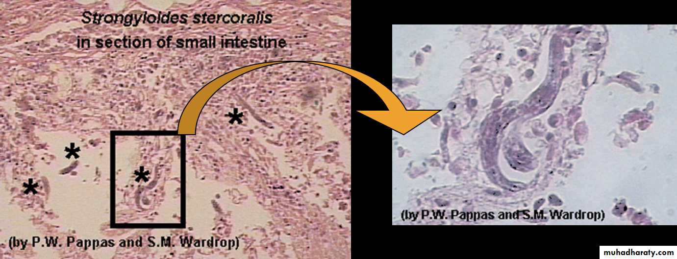
• Habitat: females live in the superficial tissues of the small intestine (duodenum and jejunum)
Definitive host: Human, dogs and cats
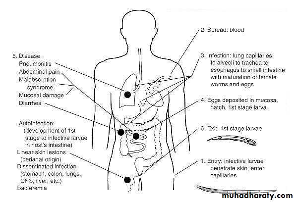
Route of infection: Filariform larvae penetrate the skin of human.
Infective stage: Third stage larvae ( filariform).
Diagnostic stage: First stage larvae(Rhabditiform) in feces.
Geographical distribution: worldwide parasite, mainly in moist and warm areas of low hygiene
• Morphology
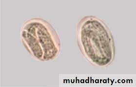
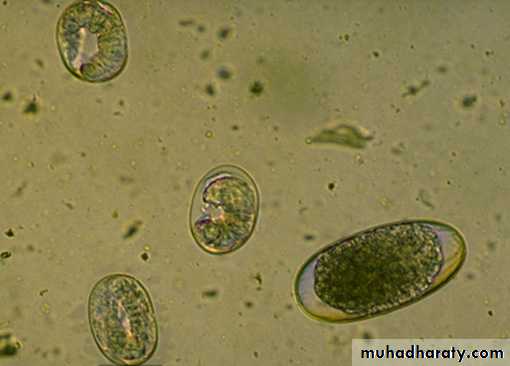
Egg:Size : 55 x 30 um.
Shape: oval . Clear, thin shelled Similar to hookworm but are smaller.
Eggs are laid in the mucosa, hatch into rhabditiform larvae that penetrate the glandular epithelium and pass into the lumen of the intestine and out the feces
(Eggs are seldom seen in stools).
Egg:
Morphology
• Adult:• Male (parasitic or free-living):
• - 0.7 mm in length
• - Rhabditiform oesophagus
• - Posterior end curved ventrally with Spicules
Morphology
• Parasitic female:• - 2.2 mm in length
• - Cylindrical oesophagus (1/3 body length)
• - Posterior end straight
• Free living female:
• - 1 mm in length
• - rhabditiform oesophagus
• - posterior end straight
Morphology of Strongyloides stercoralis
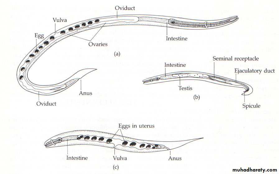
parasitic female
free-living malefree-living female
Since the parasitic females live in the superficial tissues of the small intestine, and can be present in high numbers, they can cause significant pathology.

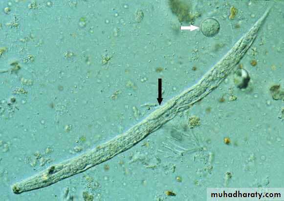
Rhabditiform larvae
220 x 15 um.Short buccal cavity.
Diagnostic stage
appear in stools within 4weeks of infection.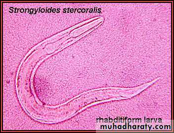
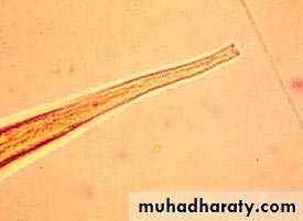
• Filariform larvae posterior part
Filariform larva withnotched tail.
Infective stage:
Size 600 x 20 um.

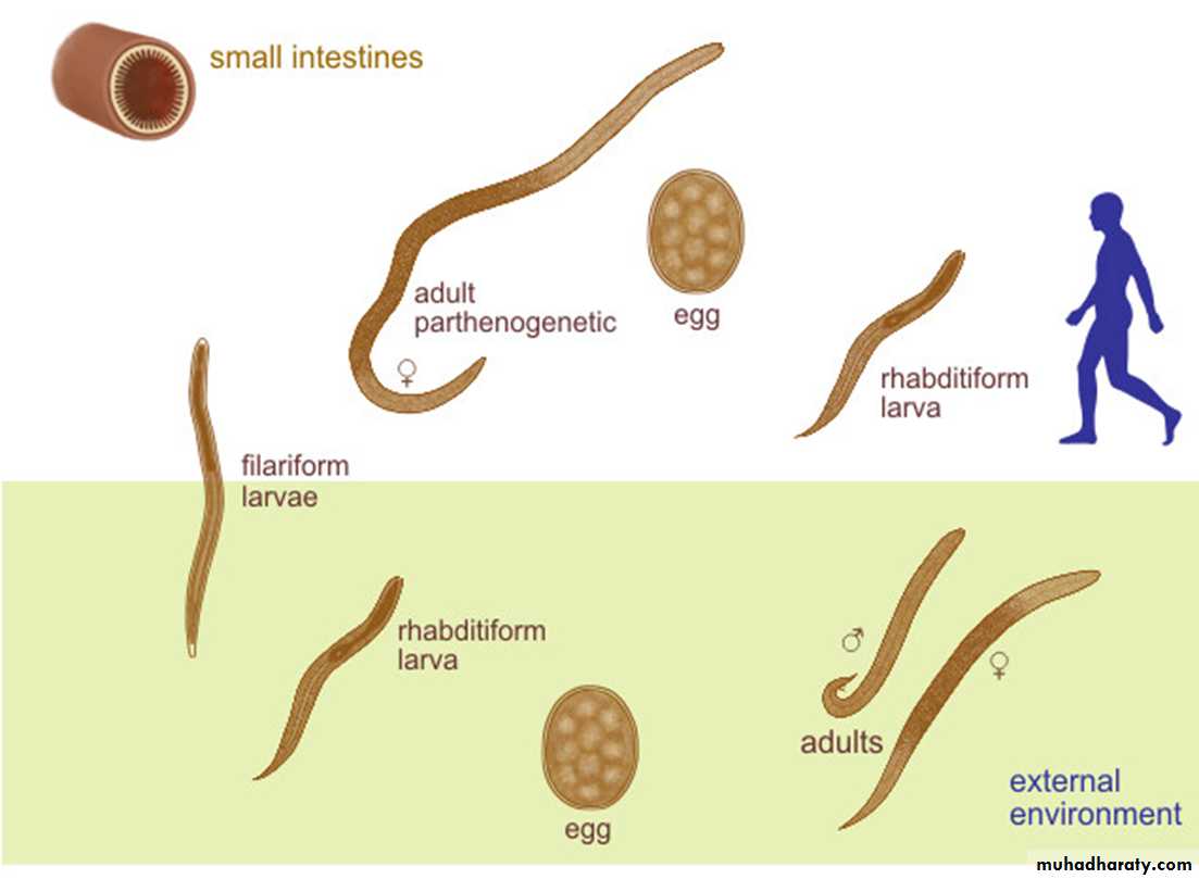
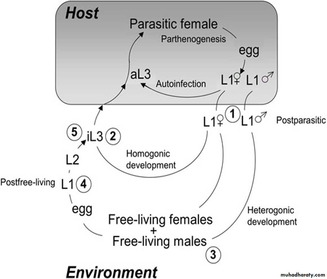
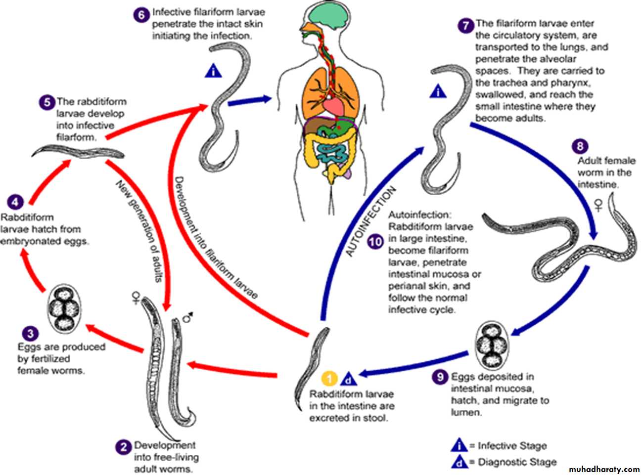
Life Cycle
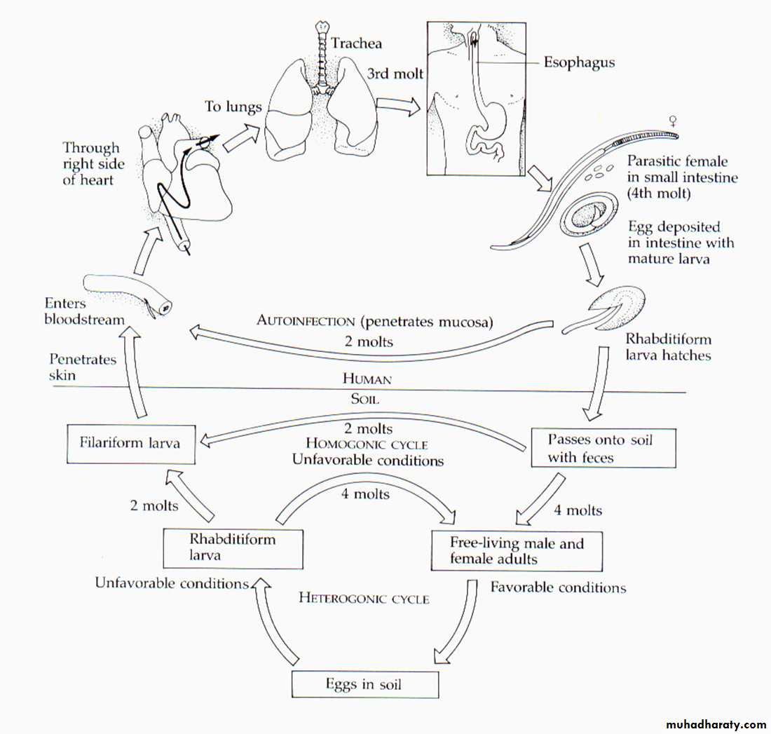
Life Cycle
Laboratory Diagnosis
Direct stool smears (larvae)Cultivation of stool. (Damp charcoal or Harada-Mori mediums).
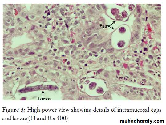
Histological examination of duodenal or jejunal biopsy specimens obtained by endoscopy can demonstrate adult worms embedded in the mucosa.
Eosinophilia, is present in uncomplicated strongyloidiasis, but is lost in hyper infection
For population screening in endemic areas, an ELISA for IgG anfi-Strongyloides antibodies is effective.