Nervous Tissue Histology
Nervous Tissue Composed ofneurons and
supporting cells(Neuroglial Cells)
Peripheral Nervous System (PNS):
Schwann Cells and Satellite CellsSchwann Cells - forms myelin sheath around large nerve fibers in PNS and is also phagocytic (engulfs damaged and dying nerve cells).
Satellite Cells - surround nerve cell body and may aid in controlling chemical environment of neurons.
Central nervous system (CNS):
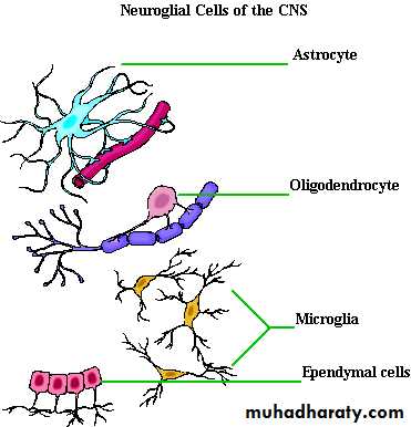
Astrocytes - - half of neural volume; have many projections that cling to neurons and capillaries (therefore connecting neurons to blood/nutrient supply); attached to the outer surface of the capillaries of brain and it acts as Blood-Brain barrier.Microglia - cells with highly branched processes; macrophage that engulfs microbes and dead neural tissue.
Oligodendrocytes - few branches, produce myelin in CNS and wraps extensions around nerve fibers (myelin sheaths).
Ependymal Cells - line the ventricles of the brain and central canal of spinal cord, creating a barrier between CNS cavities and tissues surrounding cavities,their cilia circulates the cerebrospinal fluid
Neurons (nerve cells)Characteristics:
-Conducts messages in form of nerve impulses- Neurons structurally composed of :
cell body and one or more processes
Cell Body
-Most clusters of neuron cell bodies located within CNS and called nuclei.-Few clusters of cell bodies located in PNS and called ganglia
Processes
In CNS :Cellular processes(axon) are called tractsIn PNS : Cellular processes(axon) are called nerves
Dendrites - have large surface area to receive chemical signals as well as conduct electrical signals
Axons - single in each neuron, transmit potential away from cell body to axonal terminal
Myelinated processes form the white matter of nervous tissue unmyelinated processes, nerve cell bodies, dendrites, and axon terminals form the gray matter of nervous tissue.
Classification of Neurons
Functional Classification:According to direction in which nerve impulses travel relative to the CNS
Structural Classification :
Grouped according to the number of processes extending from their cell body
Functional Classification
Sensory (afferent) neurons - transmit impulses from sensory receptors toward CNSMotor (efferent) neurons - carry impulses away from CNS to organs
Interneurons - transmit impulses within CNS (usually sensory to motor); found in CNS only; mostly multipolar
Structural Classification
Anaxonic - axons and dendrites are indistinguishable; found in brain.Multipolar neurons - three or more processes (usually with a single axons); most common type, major neuron in CNS.
Multipolar neurons - cell body located within CNS and neurons form neuromuscular junctions with effector cells
Bipolar neurons - two processes (axon and dendrite) extend from opposite sides of neuron; found in retina and olfactory mucosa to CNS
Unipolar neurons - one process extending from cell body and forms central and peripheral processes.
Unipolar neurons - skin or internal organs to CNS
Central process Peripheral process associated with sensory region (receptor)
SOMATIC SENSES
The somatic senses can be classified either physiologically or according to their locationThree physiologic types:
• The mechanoreceptive somatic senses, which include both tactile and position sensations stimulus by mechanical displacement of some tissue of the body
(2) The thermoreceptive senses, which detect heat and cold
(3) The pain sense, which is activated by any factor that damages the tissues.
Or Classified according to their location
Exteroreceptive sensations are those sensitive to stimulus from the surface of the body.Proprioceptive sensations,are those having to do with the physical state of the body, including position sensations, pressure sensations from the bottom of the feet.
Receptors present in tendon and skeletal muscle
Visceral sensations are those from the viscera of the body(internal organs).
Deep sensations are those that come from deep tissues, such as from fasciae,muscles, and bone. These include mainly “deep” pressure, pain, and vibration.
Sensory Pathways for Transmitting Somatic Signals
The sensory signals are carried through one of two alternative pathways:• The dorsal column–medial lemniscal system.
• (2) The anterolateral system.
These two systems come back together partially at the level of the thalamus.
The dorsal column–medial lemniscal system,
Their fiber come from receptors as first order neuron, enters dorsal root ganglia ,on entering the spinal cord to the dorsal horn they don’t decussate they ascend up word in the dorsal columns to the medulla, where they synapse in the dorsal column nuclei(the cuneate and gracile nuclei). From there, second-order neurons decussate to the opposite side and continue upward through the medial lemniscuses to the thalamus.In this pathway through the brain stem, each medial lemniscus is joined by additional fibers from the sensory nuclei of the trigeminal nerve; these fibers subserve the same sensory functions for the head that the dorsal column fibers subserve for the body.
The medial lemniscal fibers terminatein the thalamic sensory relay area, (ventrobasal complex). From here third-order nerve fibers project, to the postcentral gyrus of the cerebral cortex, which is called somatic sensory area I
These fibers also project to a smaller area in the lateral parietal cortex called somatic sensory area II.
In the dorsal columns of the spinal cord, the fibers from the lower parts of the body lie toward the center of the cord, whereas those that enter the chordate progressively higher segmental levels lie laterally.
Because of the crossing of the medial lemnisci in the medulla, the left side of the body is represented in the right side of the thalamus, and the right side of the body in the left side of the thalamus.
The anterolateral system.
Signals in the anterolateral system, immediately after entering the dorsal horns of the spinal cord, they cross by the anterior commissure of the cord to the opposite anterior and lateral white columns, where they turn upward toward the brain by way of the anterior spinothalamic and lateral spinothalamic tracts.To the brain stem then They terminating in some of the same thalamic nuclei where the dorsal column signals terminate. From here, the signals are transmitted to the somatosensory cortex along with the signals from the dorsal columns
The anterolateral system
-is composed of smaller myelinated fibers-the velocities of transmission are only one third to one half
-has much less spatial orientation
-the gradations of intensities are also far less accurate system.
-transmit pain, temperature, tickle, itch, and sexual sensations, in addition to crude touch and pressure
The dorsal column–medial lemniscal system
-is composed of large, myelinated nerve fibers-has a high degree of spatial orientation of the nerve fibers with respect to their origin.
-transmit rapid and precise sensation (fine touch, vibration, two-point discrimination, and proprioception (position sense
Somatosensory Cortex
The map of the human cerebral cortex is divided into about 50 distinct areas called Brodmann’s areas based on histological structural differences.Somatosensory Areas I and II.
Somatosensory area I lies immediately behind the central fissure, in the postcentralgyrus (Brodmann’s areas 3, 1, and 2).
Somatosensory Association Areas (Somatosensory Areas II).Brodmann’s areas 5 and 7 of the cerebral cortex, located in the parietal cortex behind somatosensory area I play important roles in understanding meanings of the sensory information.It receives signals from (1)somatosensory area I, (2) from the thalamus, (3) the visual cortex, and (4) the auditory cortex.
Each lateral side of the cortex receives sensory information from the opposite side of the body.
Some areas of the body are represented by large areas (the lips the greatest of all, followed by the face and thumb) whereas the trunk and lower part of the body are represented by relatively small areas. This is directly proportional to the number of receptors
Functions of Somatosensory Area I
Widespread bilateral excision of somatosensory area I lead to inability to:1. localize discretely the different sensations in the different parts of the body. However, he or she can localize these sensations crudely, such as to a particular hand, toe major level of the body trunk, or to one of the legs.
2. judge degrees of pressure against the body.
3. judge the weights of objects.
4. judge shapes or forms of objects. This called astereognosis.
5. judge texture of materials
—The Dermatomes(Segmental Fields of Sensation)
A “segmental field” of the skin innervated by spinal nerve called a dermatome.The legs originate embryologically from the lumbar and upper sacral segments (L2 to S3).
One can use a dermatomal map as to determine the level in the spinal cord at which a cord injury has occurred when the peripheral sensations are disturbed by the injury.


