Radiolucent Lesions
Classification according to diagnostic point of view :Lesions at apex of tooth
*Radiolucency at apex of nonvital tooth
Dental granuloma
Radicular cyst
Abscess
*Radiolucency in extraction site
Residual cyst
*Radiolucency at apex of vital tooth
Cementoma(stage 1)
*Radiolucency at apex of endodontically treated tooth
Radicular cyst
Dental granuloma, abscess
*Radiolucency associated with R.C.filling & apical surgery
Apical scar
Lesions in midline of maxilla : *Nasoalveolar cyst(nasolabial cyst)
*Globulomaxillary cyst
*Median alveolar cyst
*Nasopalatine cyst
Lesions in place of missing tooth
*Odontogenic keratocyst
*Ameloblastoma
Lesions around crown of impacted tooth
*Dentigerous cyst
*Odontogenic fibroma and odontogenic myxoma
*Eruption cyst
*Ameloblastoma
*Odontogenic adenomatoid tumor
Soap bubble-like radiolucencies
*Central giant cell granuloma*Aneurysmal bone cyst
*Ameloblastoma
Multiple but separated radiolucent lesions
*Cementoma(stage 1)
*Histiocytosis X
*Hyperparathyroidism
*Multiple myeloma
*Metastatic tumors
Lesions that destroy the cortical plate
*Primary malignant tumors of the jaws
*Metastatic tumors to the jaw
*Osteomylitis
Miscellaneous & rare lesions
*Traumatic cyst
*Gingival cyst
*Lateral periodontal cyst
*Physiologic osteoporosis
*Periodontal disease
*Median mandibular cyst
*Fractures
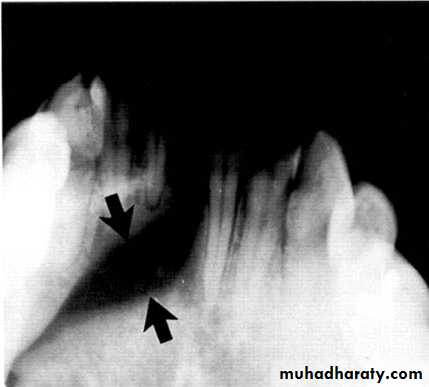
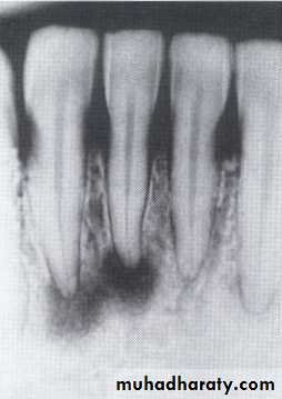
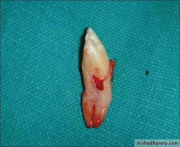
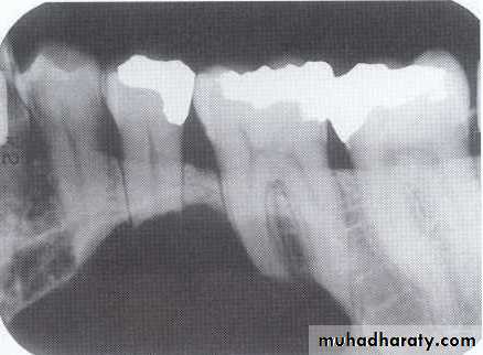
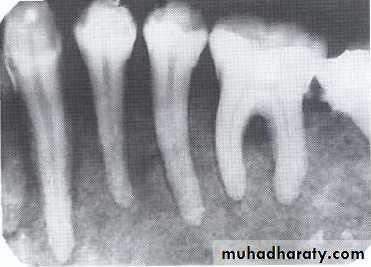
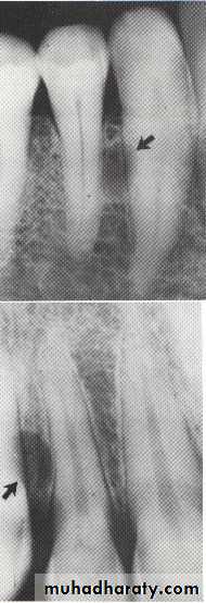
Radiolucency at apex of nonvital tooth
P. A. abscess
P. A. Cyst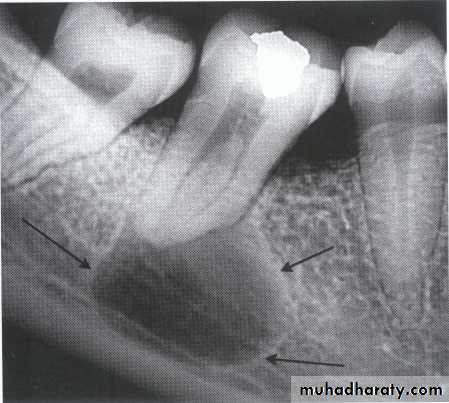
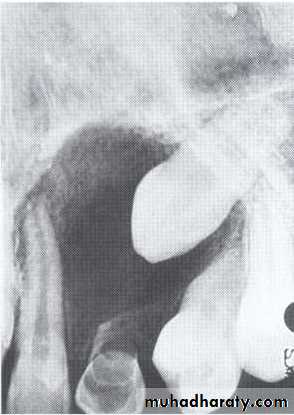
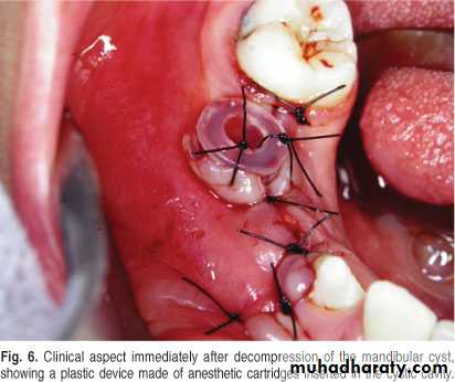
Is a cyst that remains after incomplete removal of the original cyst. The term residual is used most often for a radicular cyst that may be left behind, most commonly after extraction of a tooth.
*Persistent radiolucency in an area where a tooth has previously extracted.
• Residual cyst
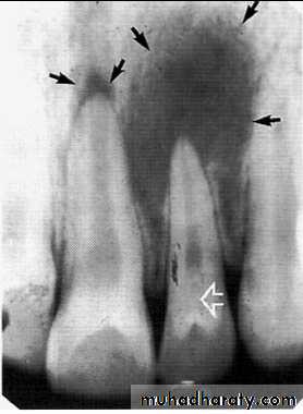
Radiolucency at apex of vital tooth:*Cementoma (stage 1)
Is a localized change in normal bone metabolismin the replacement of the that result
components of normal cancellous bone with
fibrous tissue and Cementum -like material,
(abnormal bone)or a mixture of the two.
The involved tooth is vital and often painful.
The pain seems to vary from patient to patient and can
be relieved by anti-inflammatory drugs.
Treatment. It is self-limiting and rarely recur after enucleation. Simple excision and extraction of the associated tooth are sufficient treatment.
In some cases the tumor may be amputated from the tooth, which is then
treated endodontically.
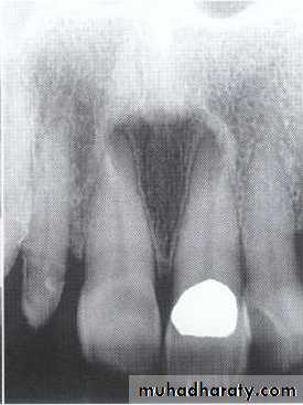
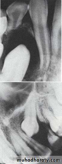
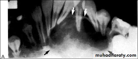
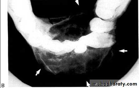
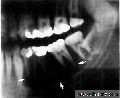
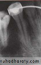
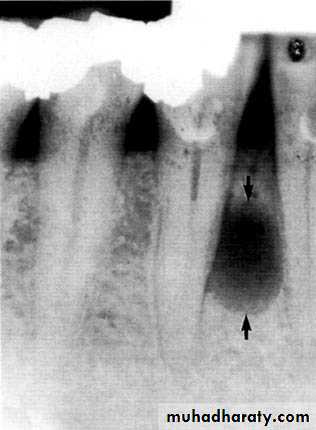
Radiolucency at apex of endodontically treated tooth
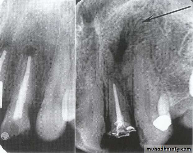
A: Radiolucent apical scar left after successful endodontic treatment..
B:New bone forming from the periphery of theLesion.
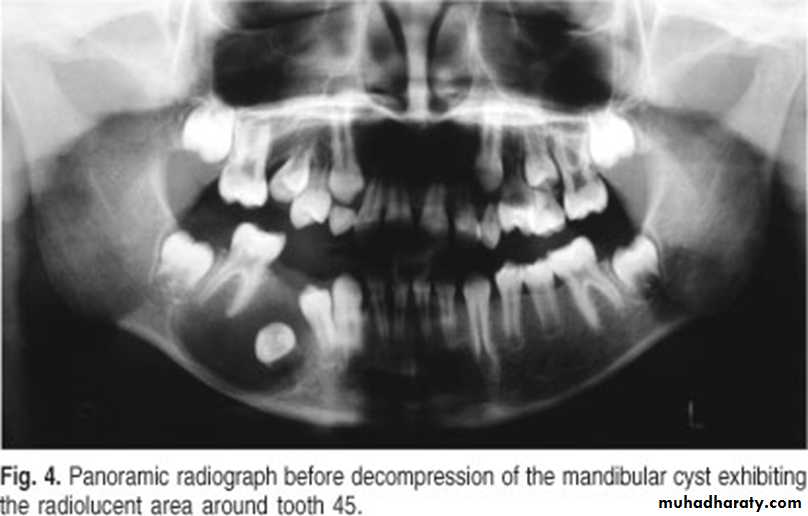
If the R.L.enlarged, pain,sinus,or no regression in the size of lesion after 6months (P. A. Cyst).
A
B
Apical scar
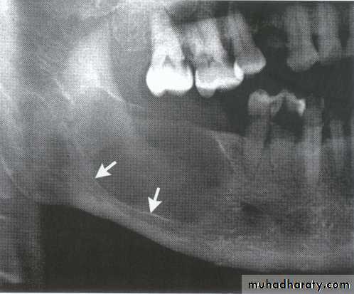
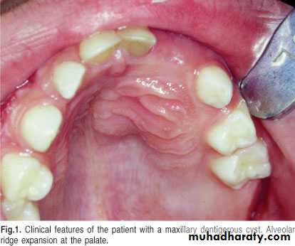
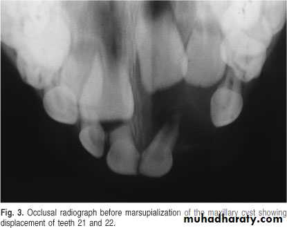
Lesions in midline of maxilla
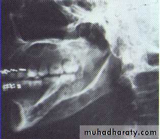
Nasoalveolar cyst (nasolabial cyst)Located in the vestibule of upper lateral & cuspid area , its soft tissue lesion may not produce any radiolucency ,in such case aspiration &/or radiograph with radiopaque contrast media is used to establish the diagnosis. Teeth are vital , clinically involve the lip & the floor of the nose .
The radiograph shows erosion of the alveolar bone (o) and elevation
of the floor of the nasal fossa (arrows).• Globulomaxillary Cyst
• Located between the lateral incisor & cuspid teeth, the teeth are vital . Radiographically;an inverted• pear shape radiolucency
• between the roots of lateral & cuspid teeth, the roots of teeth pushed apart.
• Treatment by excision.
• Median Alveolar Cyst
• It is located between the maxillary central incisors (which are vital). This lesion dose not exist as an entity but it represents an incisive canal cyst.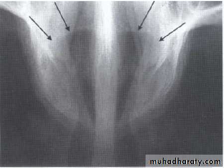
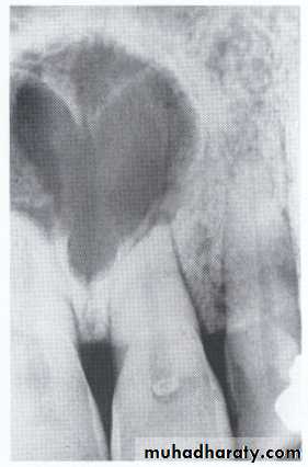
• Nasopalatine Cyst
Also called nasopalatine canal cyst, incisive canal cyst, median palatine cyst , or median anterior maxillary cyst.*It is located in the midline between the upper central incisors. Note the superimposed shadow of nasal
spine causing the cyst to appear heart shaped.
The teeth are vital.
This cyst causes the roots of the central incisors to
diverge, and occasionally root resorption occur.
*Treatment by excision.
Lesions in place of missing tooth:
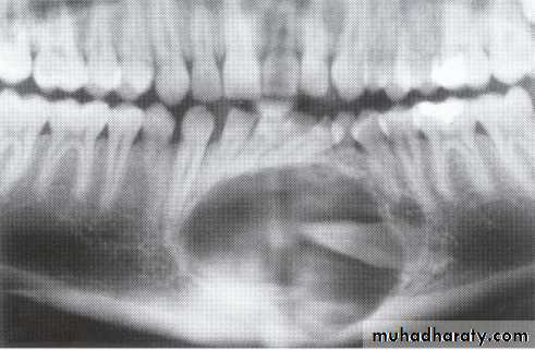
Odontogenic keratocyst:*This cyst develops from the epithelium of the dental lamina instead of the normal tooth which is therefore typically missing from the series.
*The regional teeth are vital.
*Shape: Oval, extending along the body of the mandible with little
mediolateral expansion.
*Minimal displacement of the adjacent teeth , rarely resorbed
Extensive expansion within the cancellous bone.
*Treatment by surgical excision.

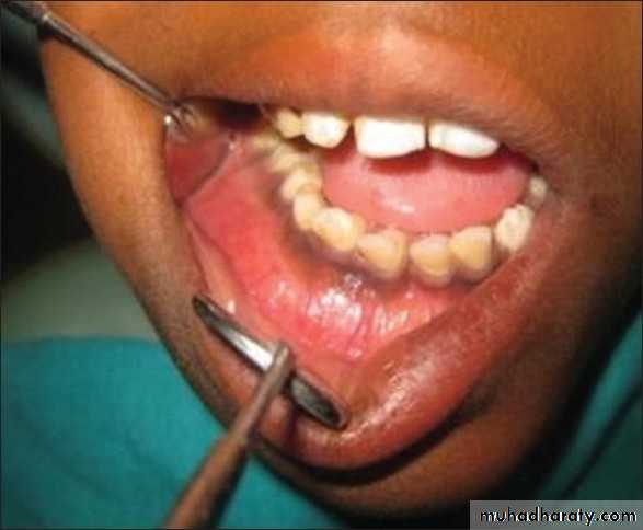
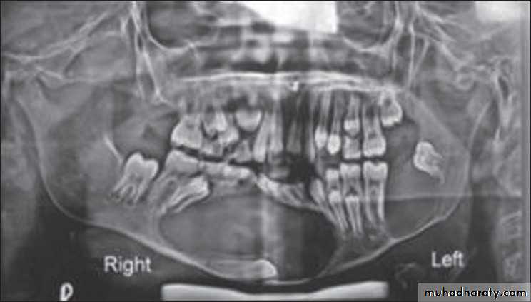
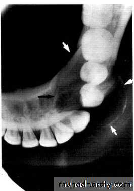
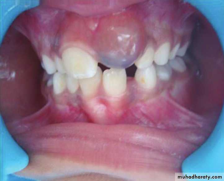
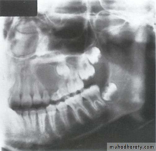
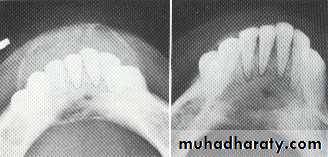
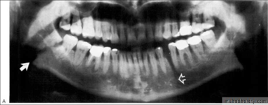
A 11-year-old female with swelling on the right side.
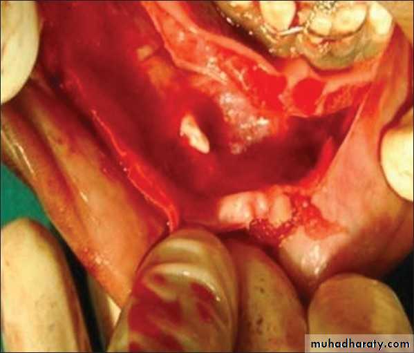
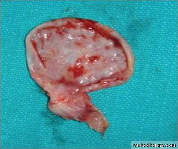
impacted canine
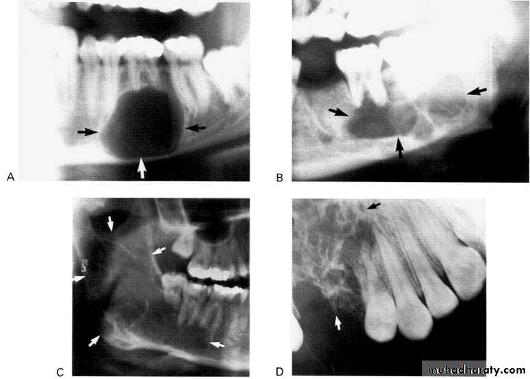
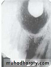
Ameloblasoma*Non-metastasizing tumor originating from remnants of the odontogenic epithelium of the enamel organ or dental lamina. *Associated with the crown of impacted teeth.
*80% located in the bicuspid & molar regions.
*Shape: is either unicystic or soap bubble-like lesion, Expansile, *Tooth migration is common,teeth in the area are vital.
*Adjacent teeth displaced, loosened , often resorbed
Extensive expansion in all dimensions
Maxillary lesions can extend into the paranasal sinuses, orbit or base of the skull.
Male > female.
Age; > 30 years

Lesions around crown of impacted tooth
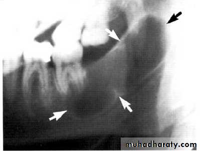
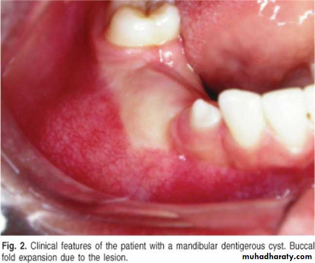
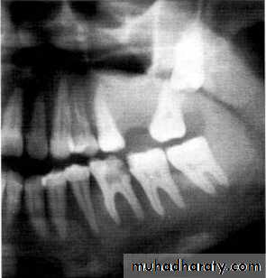
Dentigerous CystIt forms around the crown of an unerupted or supernumerary tooth at CEJ.
Some dentigerous cysts are eccentric, developing from the lateral aspect of the follicle so that they occupy an area beside the crown instead of above the crown.
It displace and resorb adjacent teeth.
It commonly displaces the associated tooth
in an apical direction.
Eruption Cyst
The term eruption cyst is used to describe a dentigerous cyst when it is in the soft tissues overlying the unerupted tooth.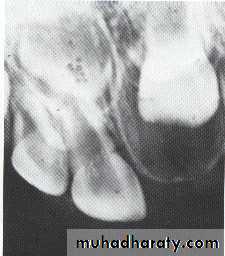
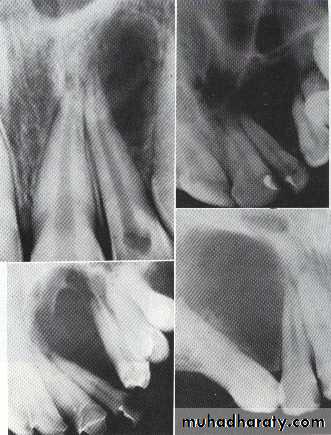
• Odontogenic Fibroma
• Benign tumor appear as a solid mass, around the crown of impacted teeth.• Odontogenic Myxoma
*They are benign, intraosseous neoplasms that arise from the mesenchymal portion of the dental papilla.
*Monolucular or multiloculor radiolucency associated with missing or unerupted tooth.
*Can be distinguished from the Fibroma by histological examination(excessive ground substance myxoid tissue).
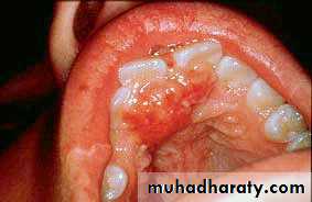
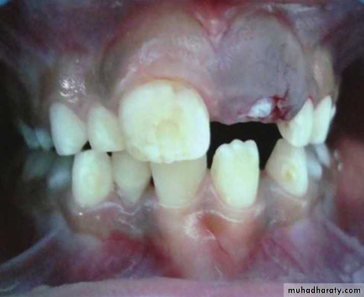
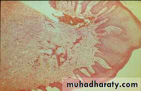
*A 13 year old boy presents with a recurrent friable granular lesion of the anterior maxilla. This is a recurrent lesion after excision.
Differential Diagnosis:
• Pyogenic Granuloma• Peripheral Odontogenic (Ossifying) Fibroma
• Peripheral Giant Cell Granuloma
• Squamous Cell Carcinoma other soft tissue malignancies
Differential Diagnosis:
• Ameloblastoma• Odontogenic Keratocyst
• Odontogenic Myxoma
• Giant Cell Granuloma
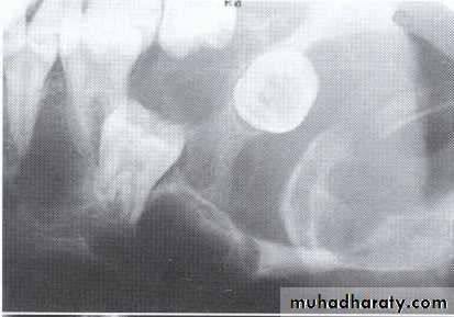
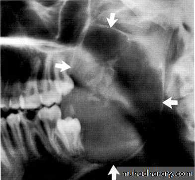
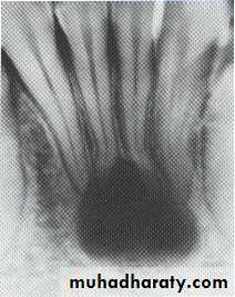
*A 55 year old man with an expansile multilocular radiolucency of the left mandible. The swelling had been noticed by the patient for approximately one year.
A: Odontogenic fibroma
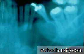
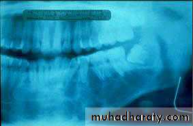
B,C,D:Odontogenic myxoma.Odontogenic Adenomatoid Tumor (OAT)
*Uncommon, nonaggressive tumor of odontogenic epithelium.*Age <20 years.
*Location; cuspid area of the maxilla.
*As the tumor enlarged the adjacent teeth are displaced. Root resorption is rare.
*This lesion also may inhibit eruption of an involved tooth.
*Although some expansion of the jaw may occur, the outer cortex is maintained.
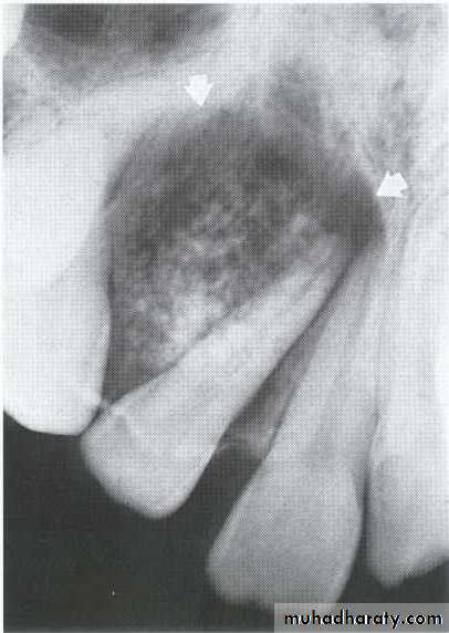
The maxillary left canine and premolar have been displaced superiorly by the tumor.
The canine and lateral incisor have been displaced by the lesion.Soap bubble-like radiolucencies
Central giant cell granuloma*A relatively uncommon, non- neoplastic mass in the jaws (intraosseous) producing an expansile radiolucent soap bubble appearance.
*Age; <20 years. Female > Male.
*Teeth are vital, with some root resorption & migration of teeth.
Multilocular appearance, expansion (arrowed) and considerable displacement of the adjacent teeth.
Buccal and lingual expansion (arrowed) and the undulating cortical border.
• Aneurysmal Bone Cyst
•
Large multilocular aneurysmal bone cyst
in the ramus with marked expansion andthe displacement of/8.
It is non-neoplastic ,exaggerated , localized, proliferative lesion of vascular tissue, containing giant cells.
It’s a variation of the central giant cell granuloma , can be diagnosed only by histological examination
Multiple but separated radiolucent lesions
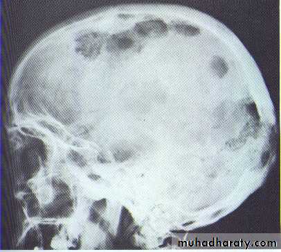
Histiocytosis-X (Langerhans cell disease):They are three manifestations produce tumour-like lesions in bone, caused by proliferation of Langerhans cells and eosinophilic leucocytes.
Radiographically :
• Multiple radiolucent areas in the interdental & interradicular bone.
• teeth seem to hang in air. Exfoliation of teeth & teeth germ are common, ulceration of overlaying mucosa.
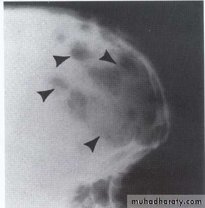
• Multiple radiolucent foci seen in the skull & long bone of the skeleton, lesion in the skull give appearance of geographic map , with skull enlargement .
• Histiocytosis-X
• Eosinophilic Granuloma (2nd-3rd decade of life).• Hand-Schüller-Christian disease (1stdecade of life).
• Letterer-Siwe disease (before 2 years of age).
•
• Hyperparathyroidism
Increased secretion of parathormone, causes generalized skeletal bone resorption leading to osteopenia (generalized decrease in bone density).Single or multiple radiolucency in the maxilla & mandible.
• Loss of normal bone trabeculation & replaced by fine poorly calcified bone spicules that give ground glass appearance.
• Missing of lamina dura.
• D.D.by elevation of serum calicum & alkaline phosphatase levels.
•
Loss of the lamina dura and the granular texture of the bone pattern.
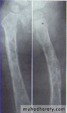
• Multiple Myeloma
*Is a malignant tumor of the bone marrow.Age; > 40 years, male > female.
Radiographically; multiple punched-out in the jaw & skull, the peripheral border is smooth & irregular in comparison with the Histiocytosis-x.
Miscellaneous & rare lesions
Traumatic cyst*Mandible > Maxilla. <20 years of age.
*Asymptomatic, expansile, associated with history of trauma. *Extend between the roots of teeth with scalloped border .
*Aspiration is not productive, empty cavity.
• Gingival Cyst
• * Arises from attached gingiva & with growth cupping the cortical plate.• *The teeth are vital.
•
• Lateral Periodontal Cyst
*It is a developmental cyst and not an inflammatory cyst ,developed from the cell rests of the dental lamina.*Occur at the lateral surface of the roots of vital teeth in the lower canine/premolar region or upper lateral incisor region.
*It is the intrabony counterpart of the gingival cyst in the adult .
•
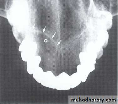
• Median Mandibular Cyst
• *Located in the midline of the mandible. *Teeth are vital. Arise from the fissural epithelia.•
FRACTURES