Cestodes
Echinococcus granulosusMedically important Cestodes (Tapeworms) of human beings
Class Cestoidea• Order Pseudophyllidea
Order CyclophyllideaDiphyllobothrium latum
Echinococcus Spp.Taenia saginata
Taenia solium
Hymenolepis nana
Hymenolepis Diminuta
Diphyllobothrium caninum
Echinococcus Spp.
Genus Echinococcus include three different species:
Echinococcus granulosus
E. multilocularisE. vogeli
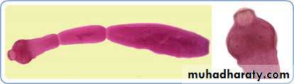
E.granulosusMorphology of adult worm :
2-6 mm long. hermaphrodite
The body consist of:
head (scolex) with 4 suckers and rostellum with two rows of hooks.
three segments (proglottids):Immature, mature and gravid.
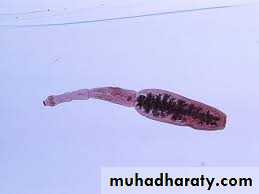

The mature segment
contain fully developed male and female sexual organs .The gravid segment:
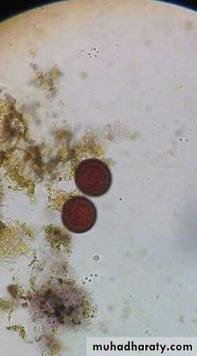
contain only uterus full with eggs.Morphology of the eggs
Spherical, 35-45um in diameter .Hexacanth Embryo centrally located .
Radially staited shell .
Infective stage to human, sheep & cattle .
The same egg of Echinococcus Spp., Taenia saginata & T. solium.
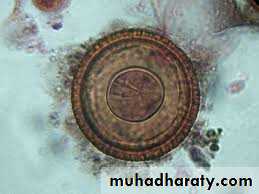
Taenia spp. Eggs
Life cycle
Echinococcus granulosusdefinitive(final) hosts: dogs or other canids( cats, foxes and wolves).
intermediate host : sheep, goat, cattle and man.
infective stage : egg
Diagnostic stage : Hydatid Cyst
Site or Inhabit of the definitive(final) hosts:
upper part of small intestine
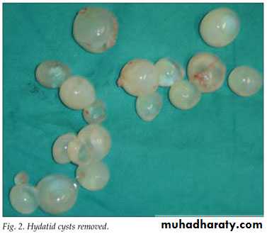
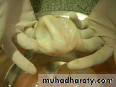
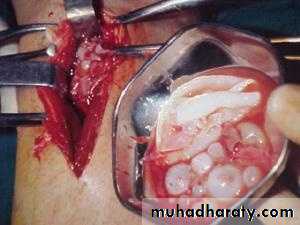
Hydatid disease
Types of hydatid cystsUnilocular
OsseousMultilocular or alveolar
Morphology
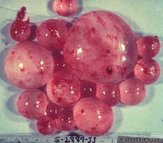
Unilocular hydatid cyst consist of three layers with central fluid contain protoscolices.
The majority of human H. cysts are unilocular type. and single cavity
The three layers are :1- Inner germinal layer
2- non-nucleated laminated layer
3- Outer (fibrous layer)
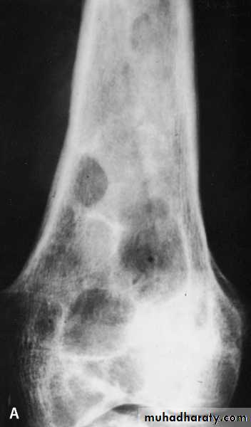
osseous H. cyst
When the embryo of E. granulosus reaches bony tissues it will develop to osseous hydatid cyst.It is occur in the ends of long bones and pelvic arch
sterile never produces brood capsule and protoscolices with little or no fluid and no fibrous capsule.
Multilocular or Alveolar Hydatid Cyst
It is the larval stage of E. multilocularis, it is composed of numerous small spaces or cavities, separated from each other by connective tissue.occasionally it may contain protoscolices.
The germinal and laminated layers are poorly developed, it has no fibrous capsule.
It is occurs usually in the liver and rarely in lung. Because of its fast growth, it is usually fatal.

Diagnosis
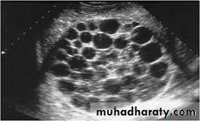
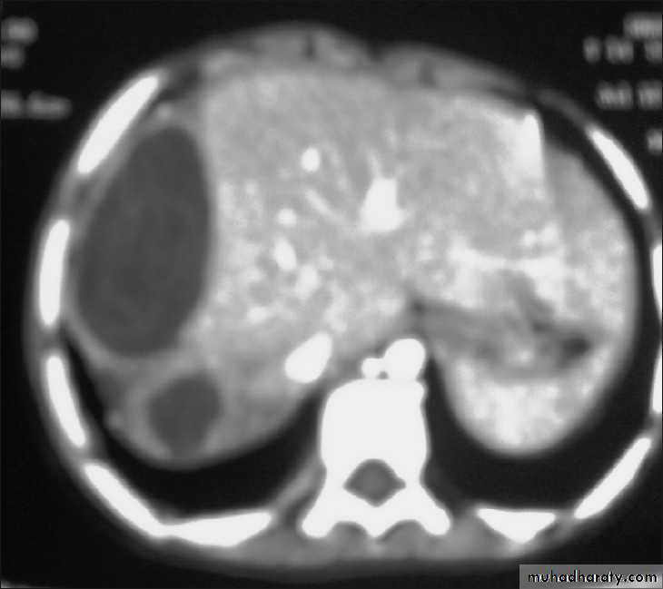
• Clinical manifestations.• 2. Imaging techniques:
• X-ray picture: useful to detect the calcified cyst.
• Ultrasound scan.
• MRI & CT scan.
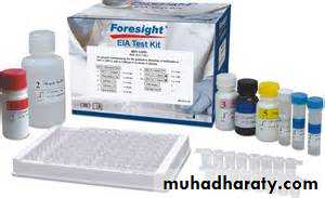
3. Serology tests :
Indirect immunofluescence.Enzyme-linked immunosorbent assay (ELISA).
Polymerase chain reaction (PCR).