Diagnosis of skin diseases
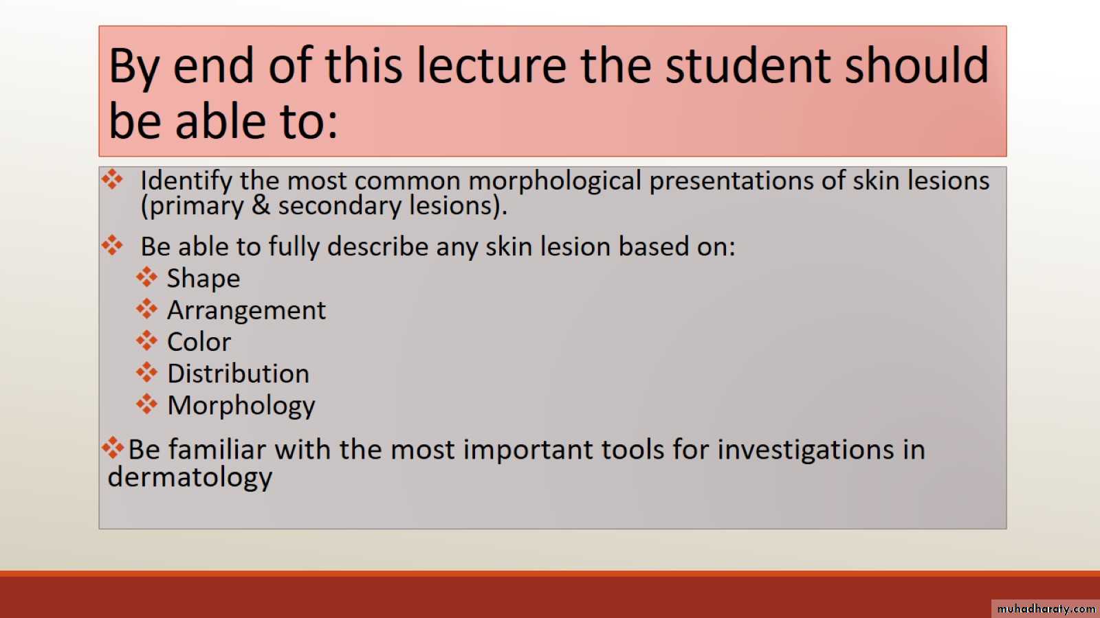
By end of this lecture the student should be able to:
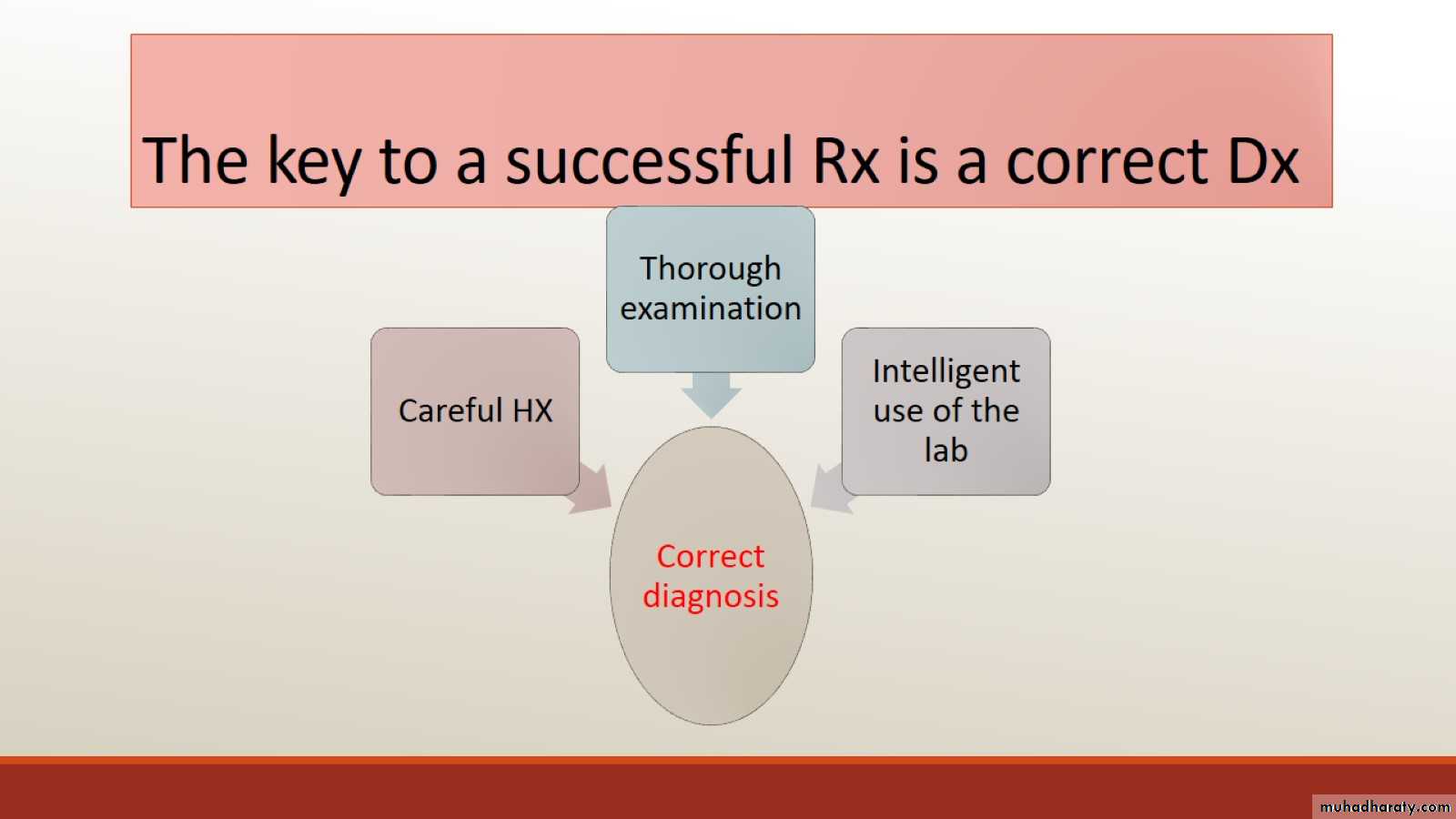
The key to a successful Rx is a correct Dx
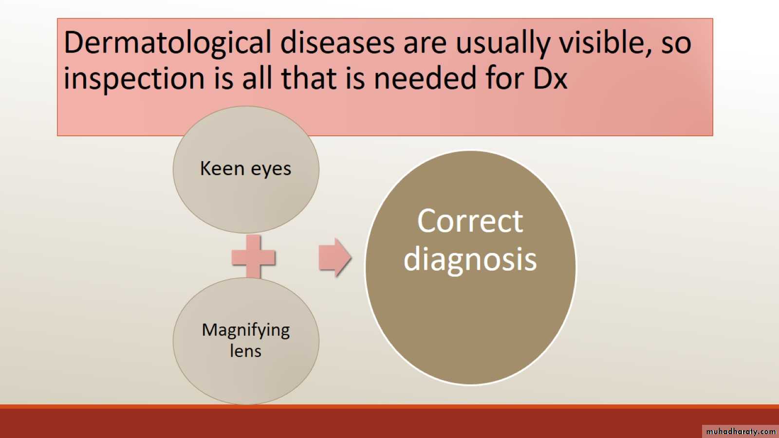
Dermatological diseases are usually visible, so inspection is all that is needed for Dx
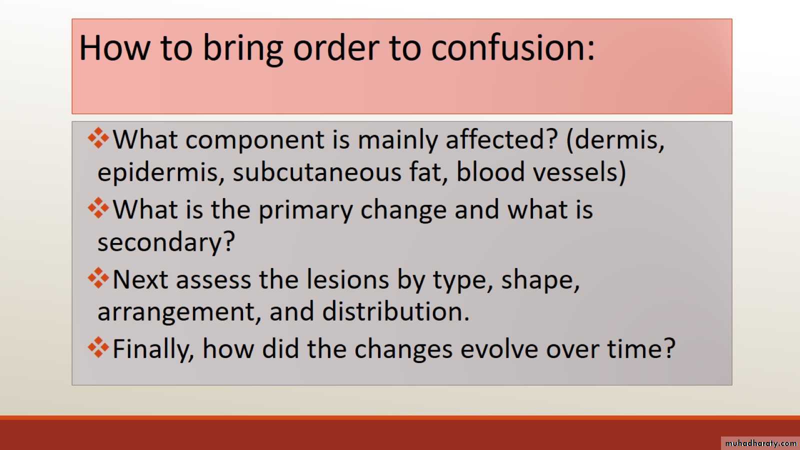
How to bring order to confusion:
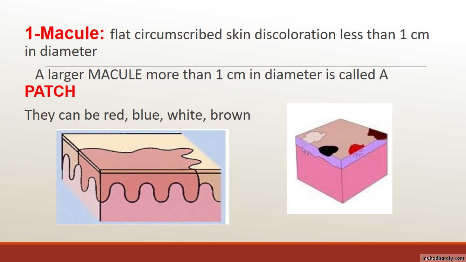
Types of lesions
Primary skin lesions
2---
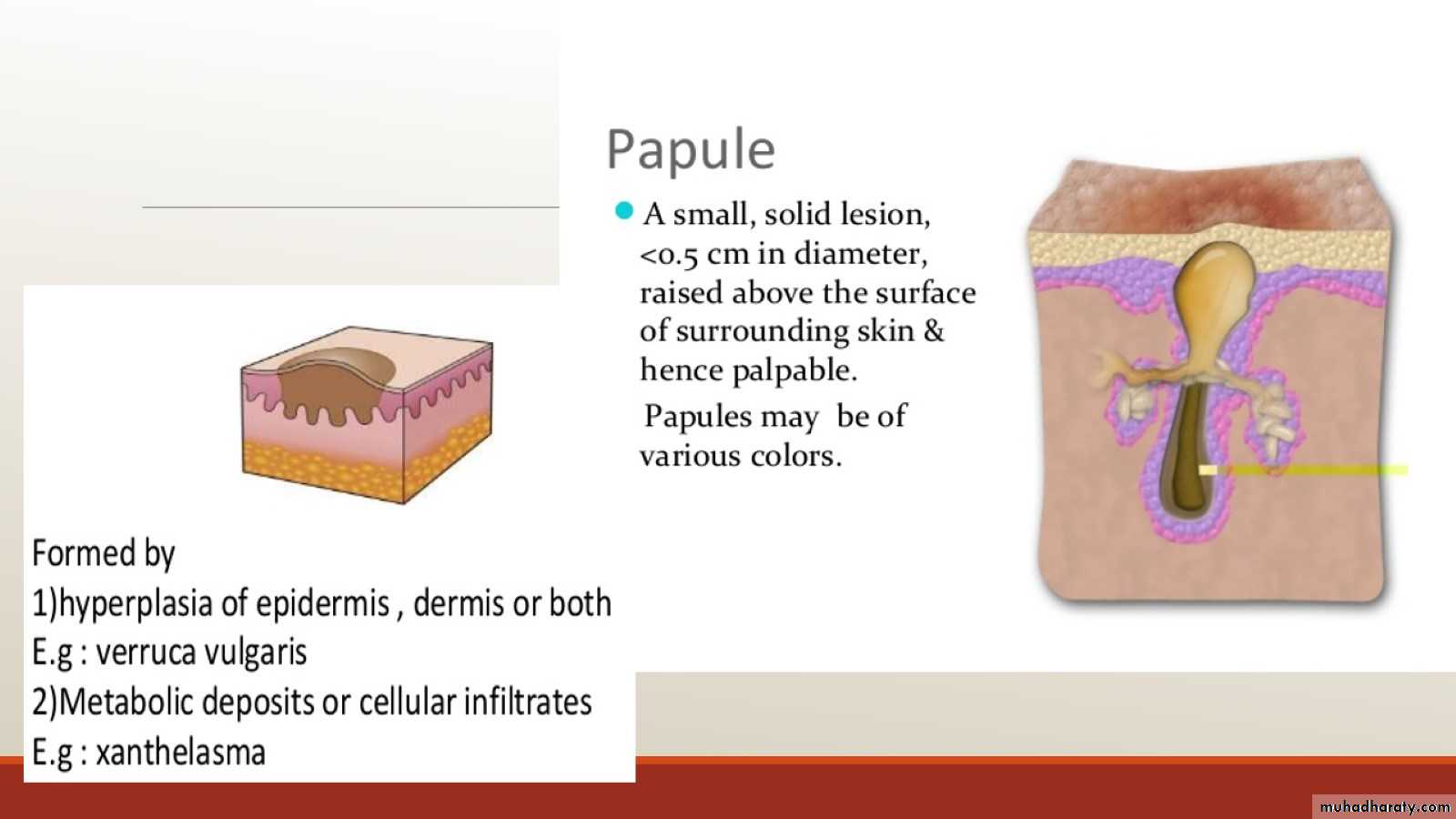
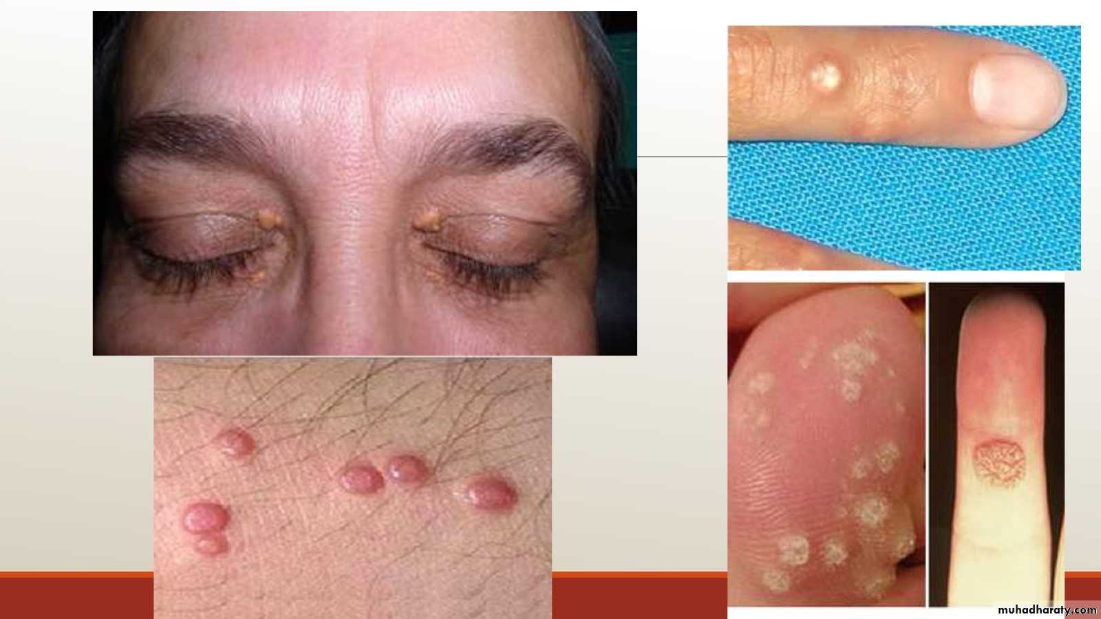
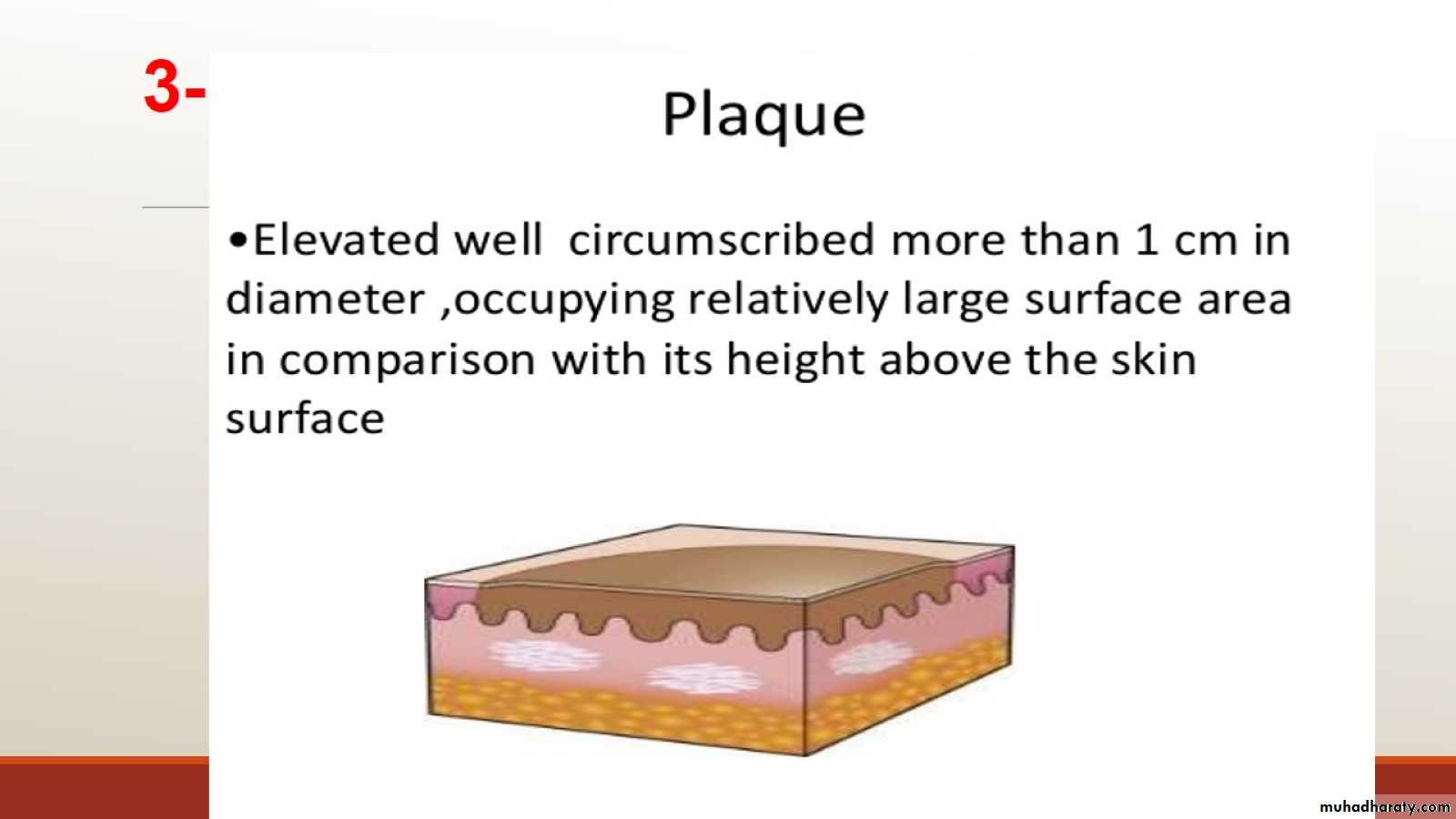
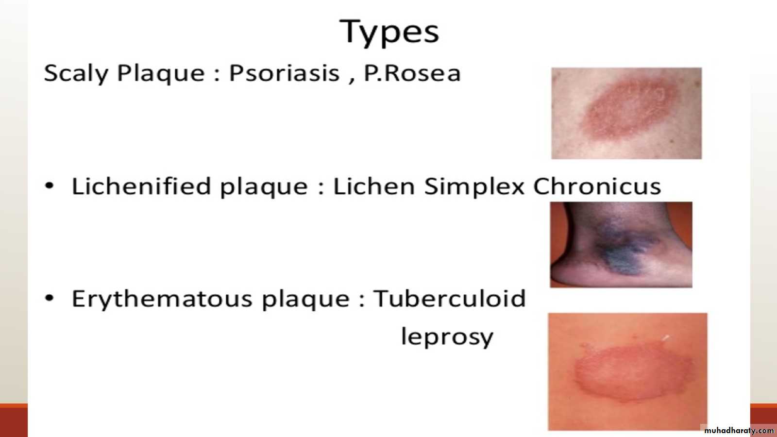
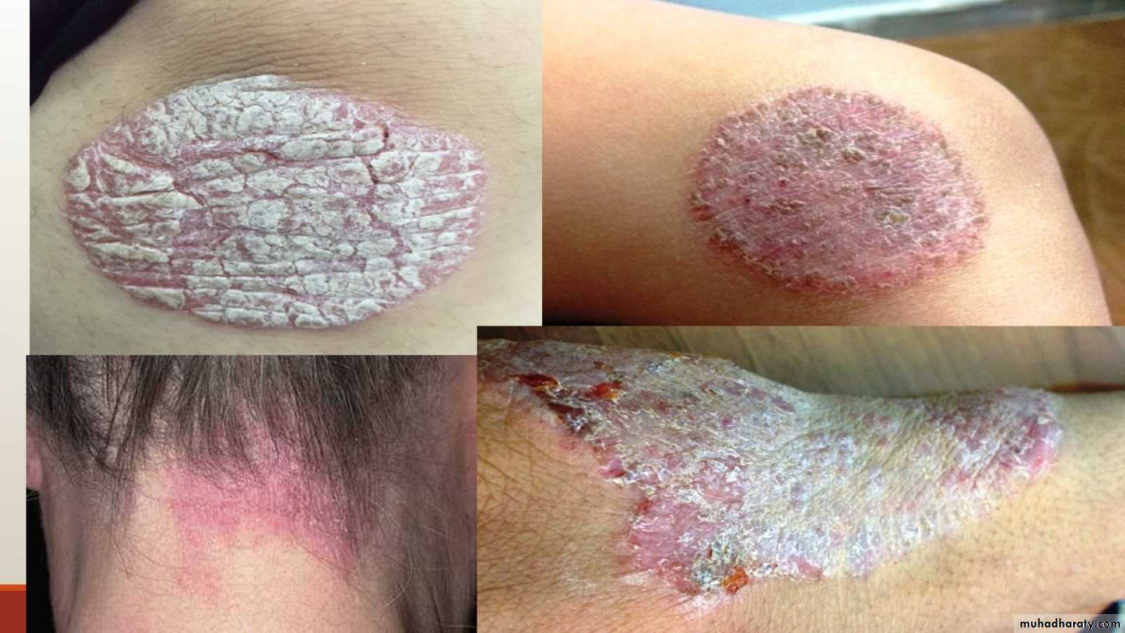
3-
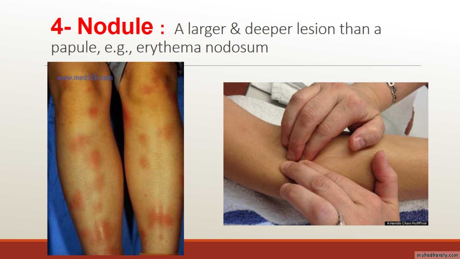
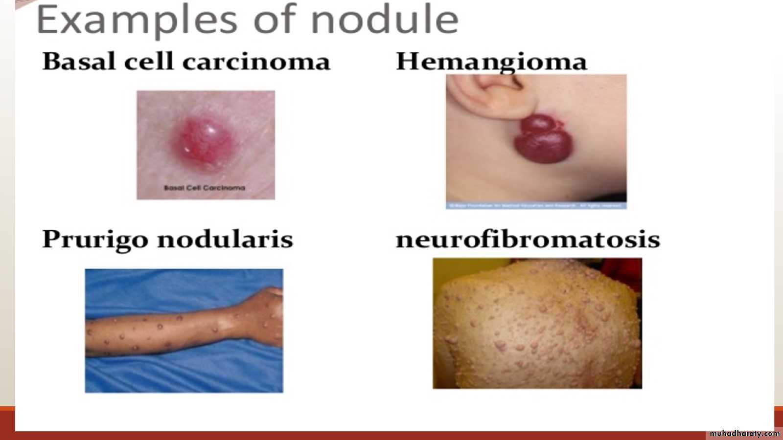
4- Nodule : A larger & deeper lesion than a papule, e.g., erythema nodosum
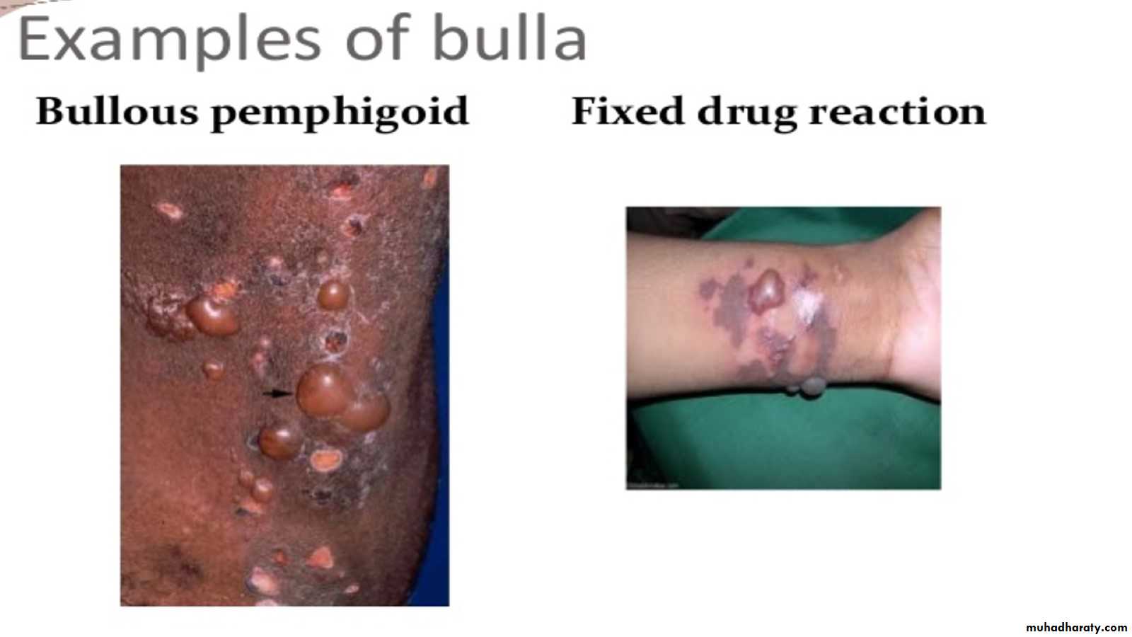
5- Blisters
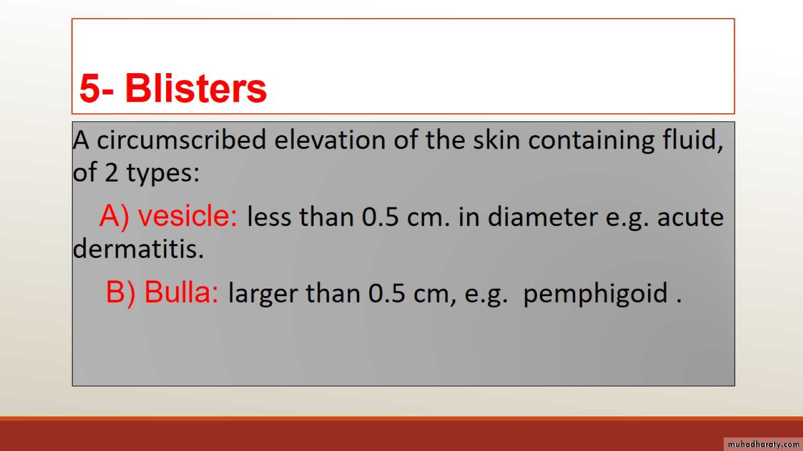
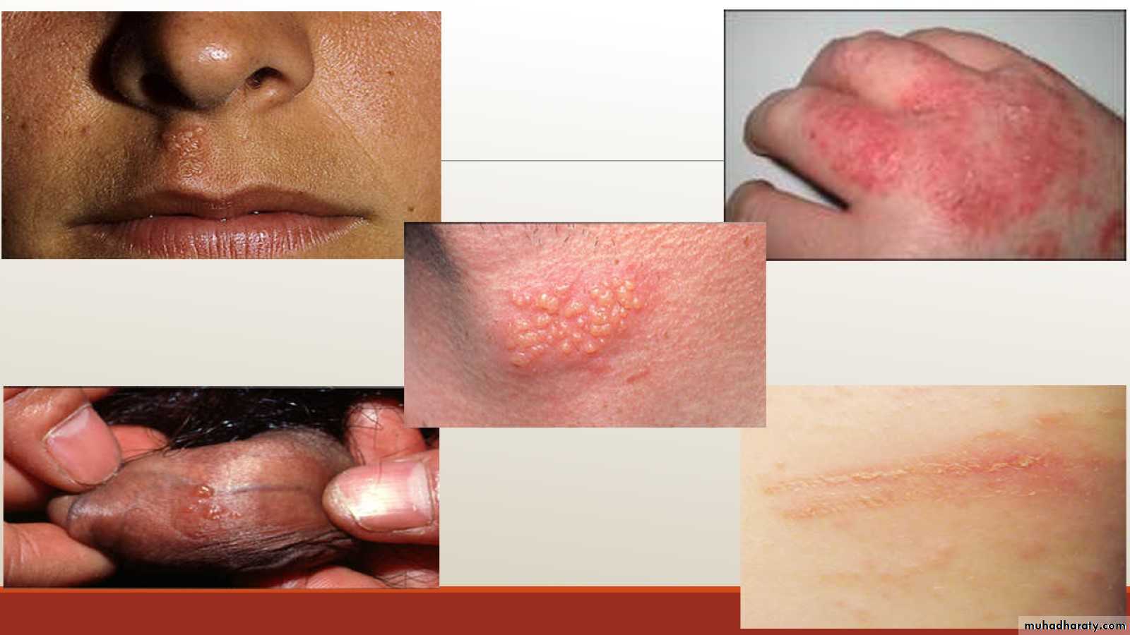
Eg of vesicles
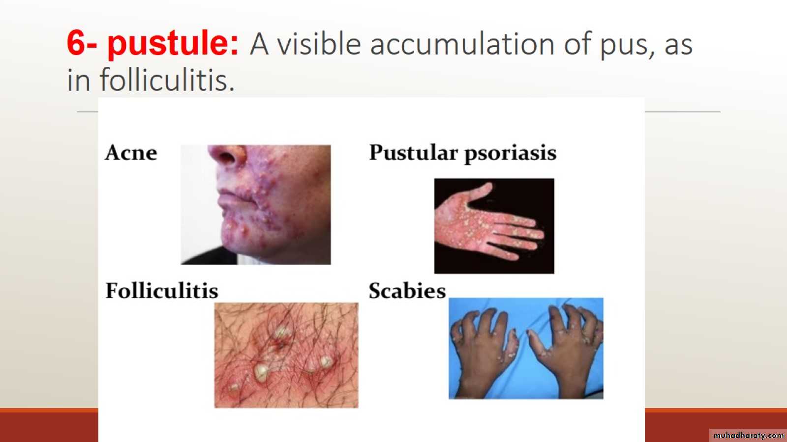
6- pustule: A visible accumulation of pus, as in folliculitis.
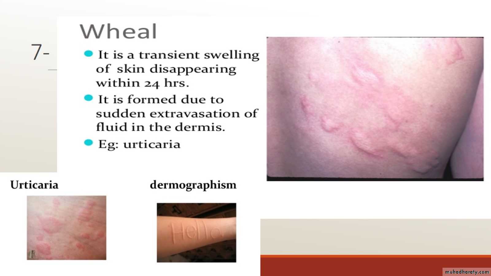
7-
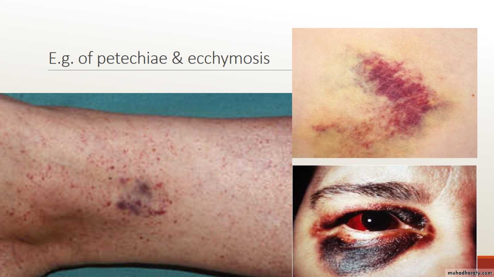
8- Purpura
E.g. of petechiae & ecchymosis
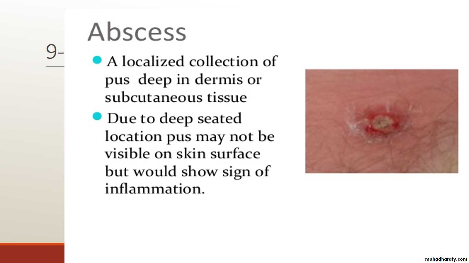
9-
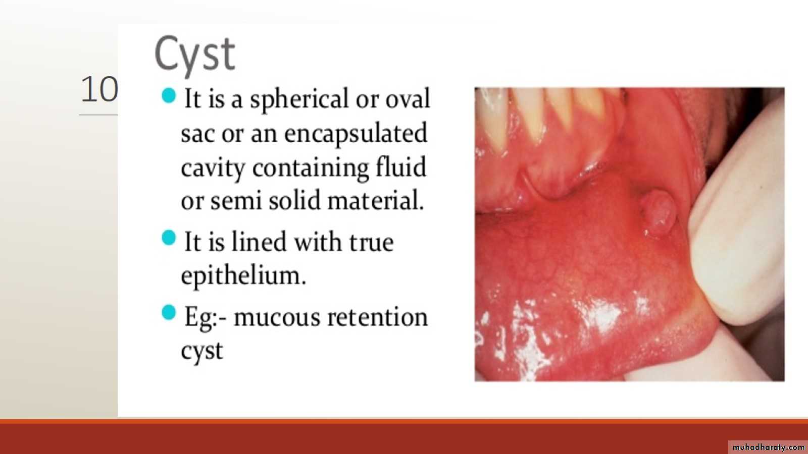
10-
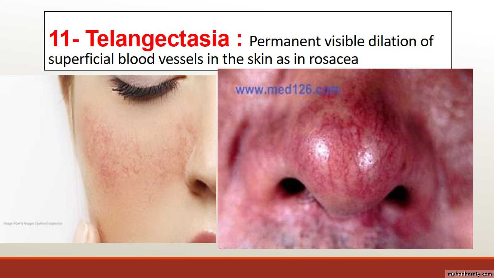
11- Telangectasia : Permanent visible dilation of superficial blood vessels in the skin as in rosacea
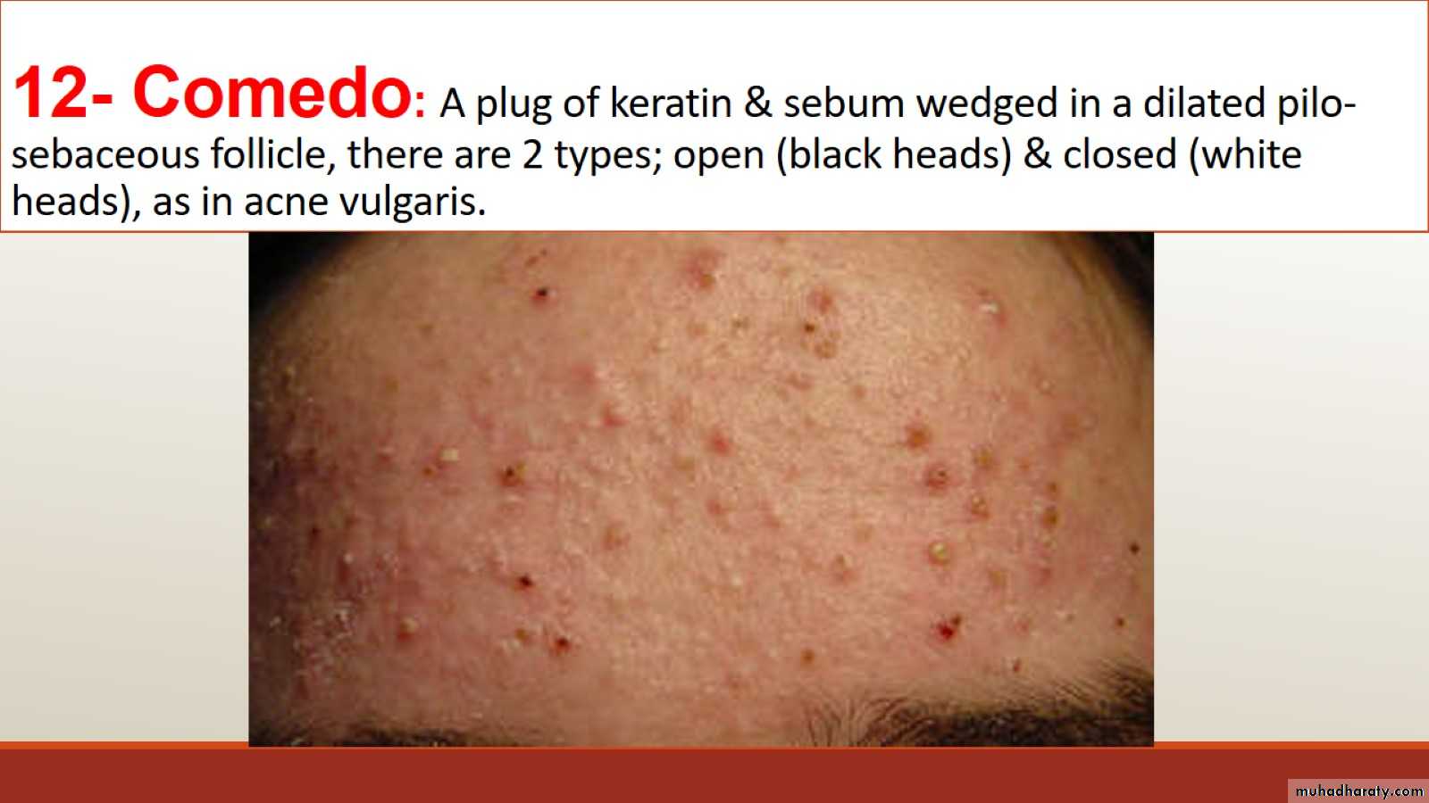
12- Comedo: A plug of keratin & sebum wedged in a dilated pilo-sebaceous follicle, there are 2 types; open (black heads) & closed (white heads), as in acne vulgaris.
Secondary skin lesions
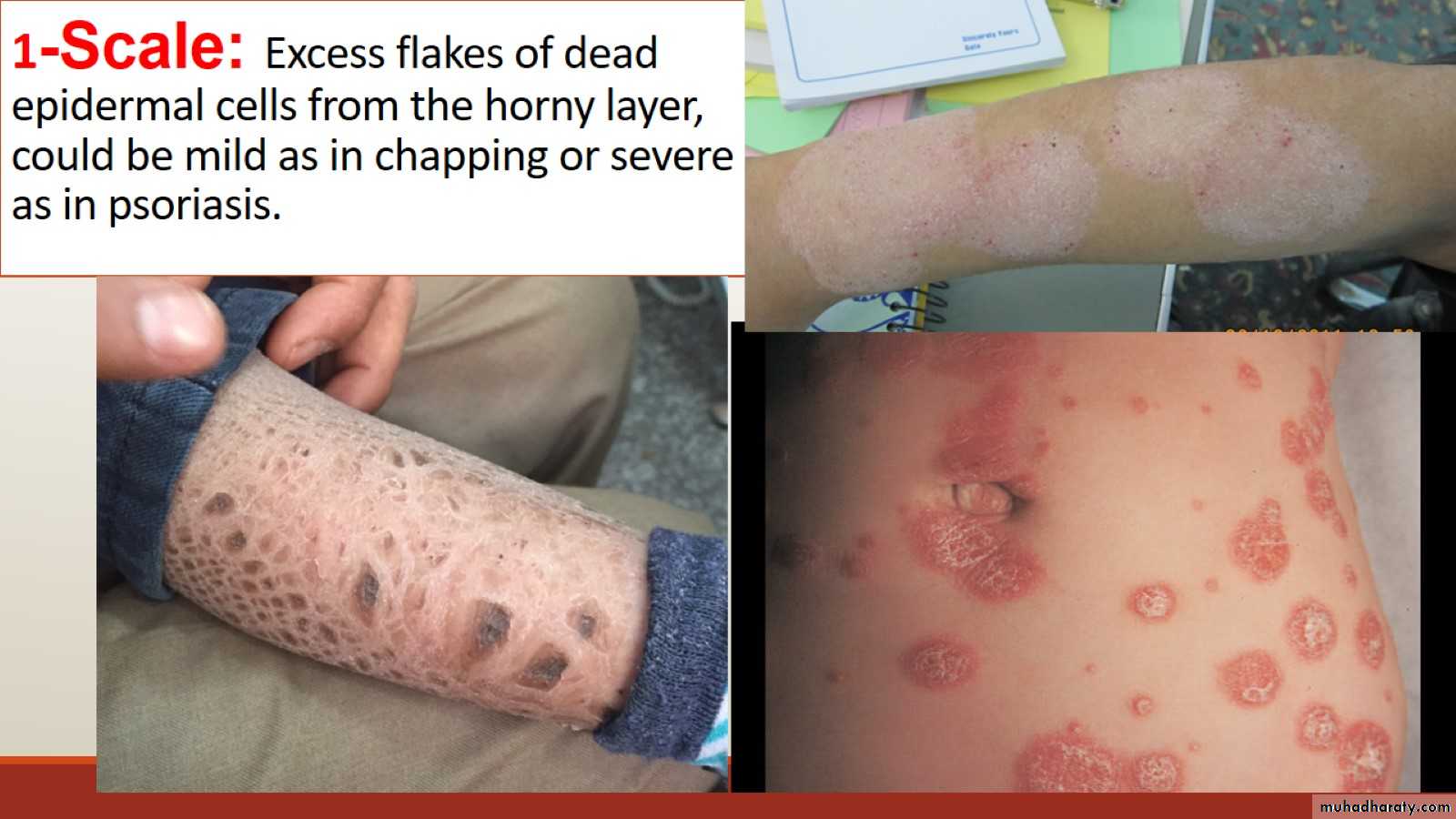
1-Scale: Excess flakes of dead epidermal cells from the horny layer, could be mild as in chapping or severe as in psoriasis.
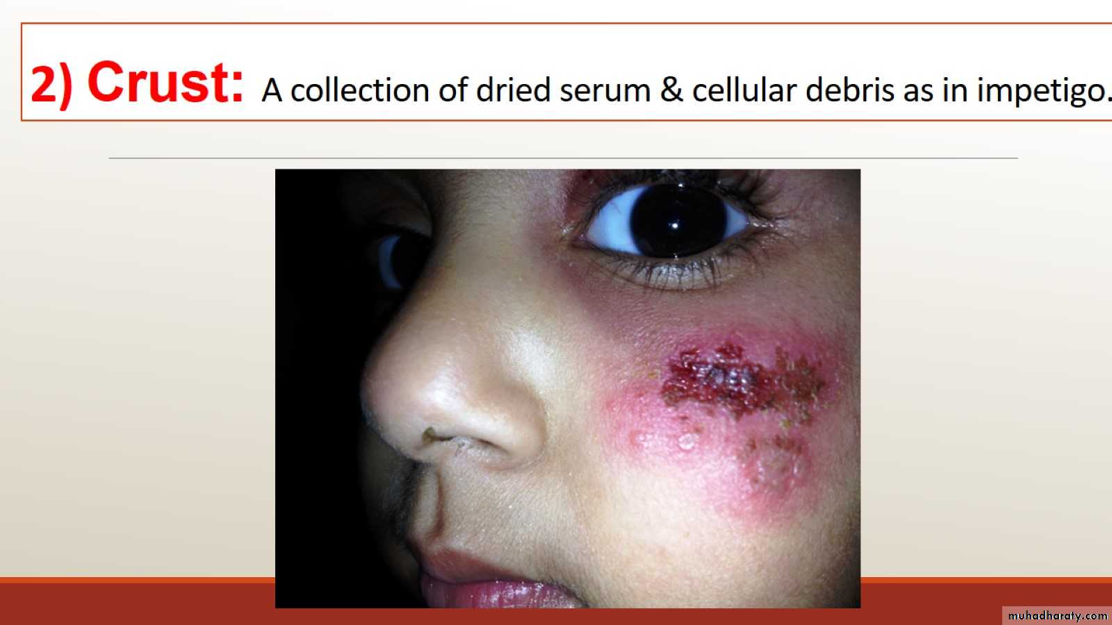
2) Crust: A collection of dried serum & cellular debris as in impetigo.
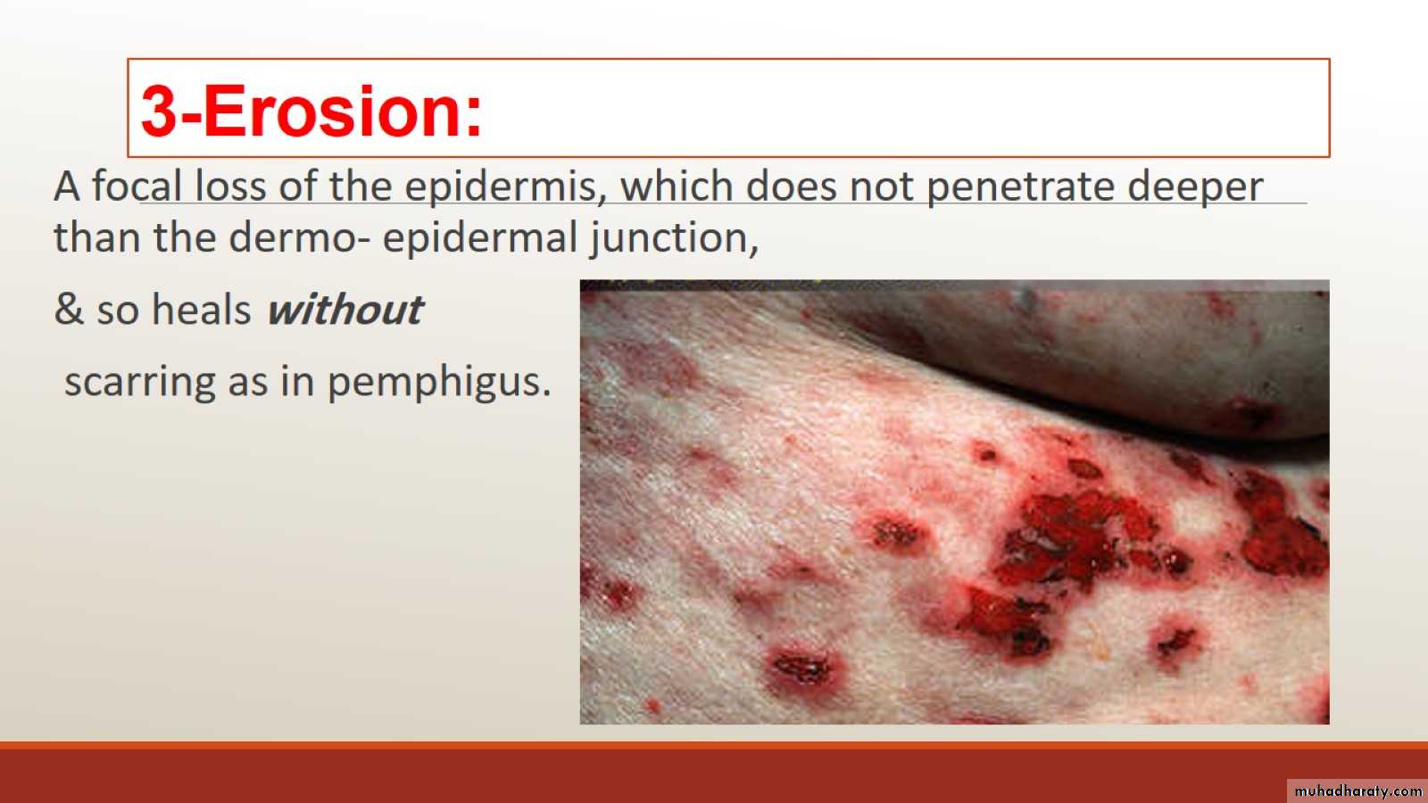
3-Erosion:
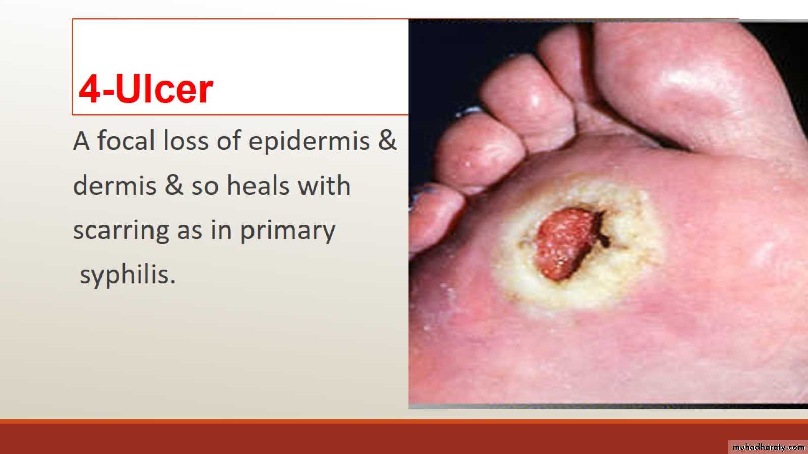
4-Ulcer
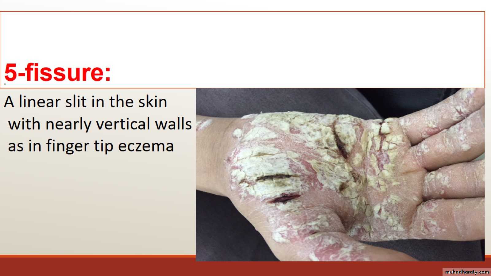
5-fissure:
6-Sinus :

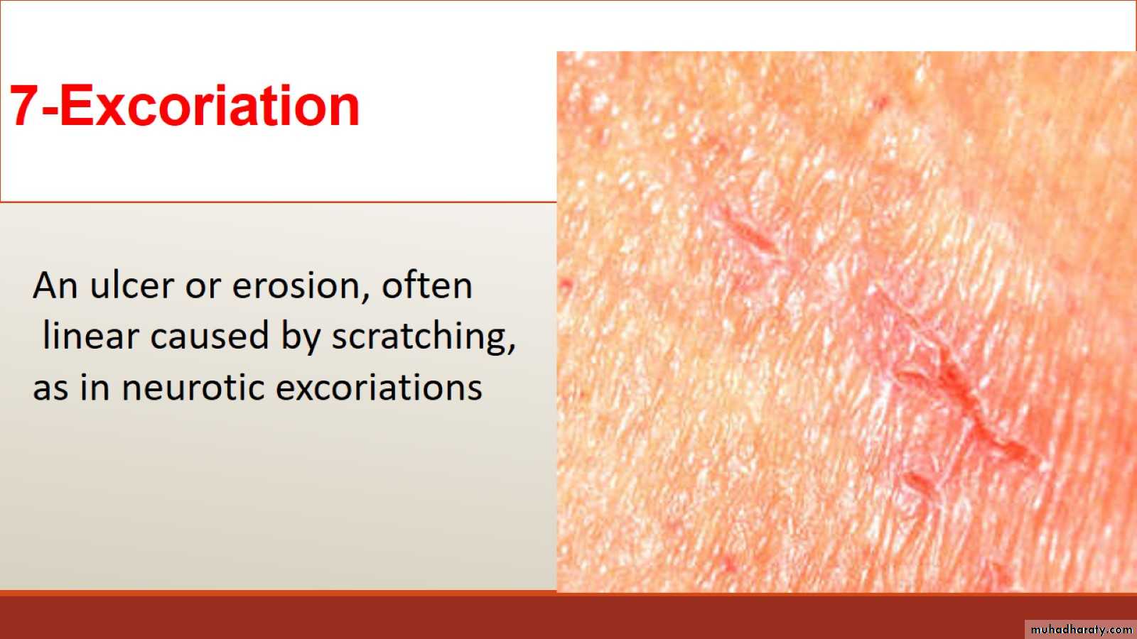
7-Excoriation
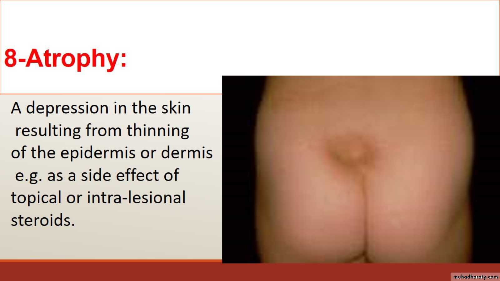
8-Atrophy:
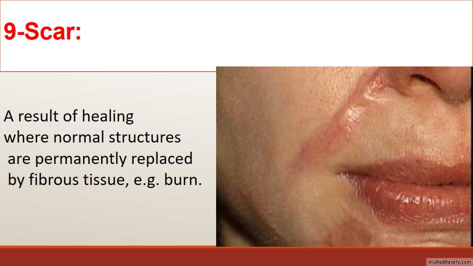
9-Scar:
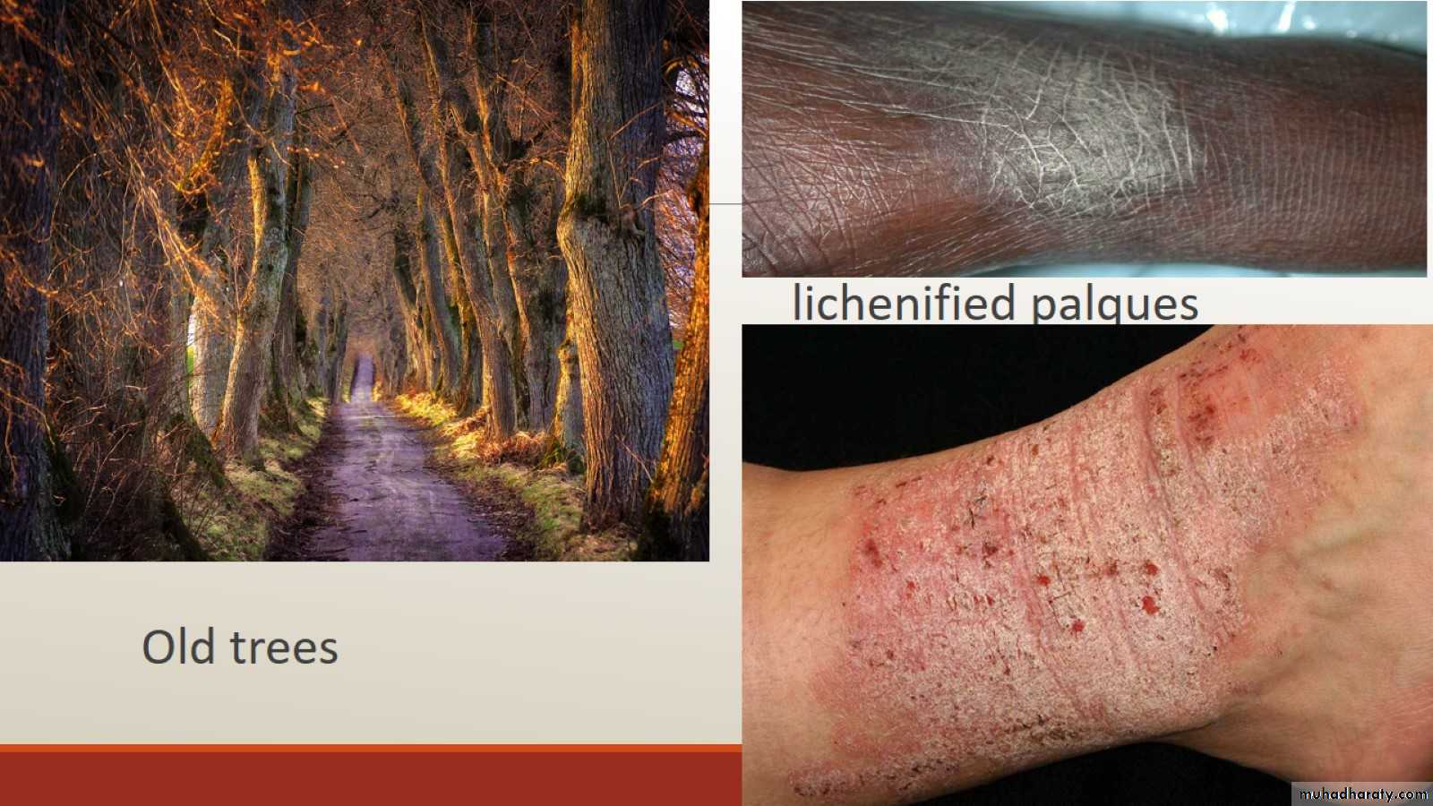
10-Lichenification
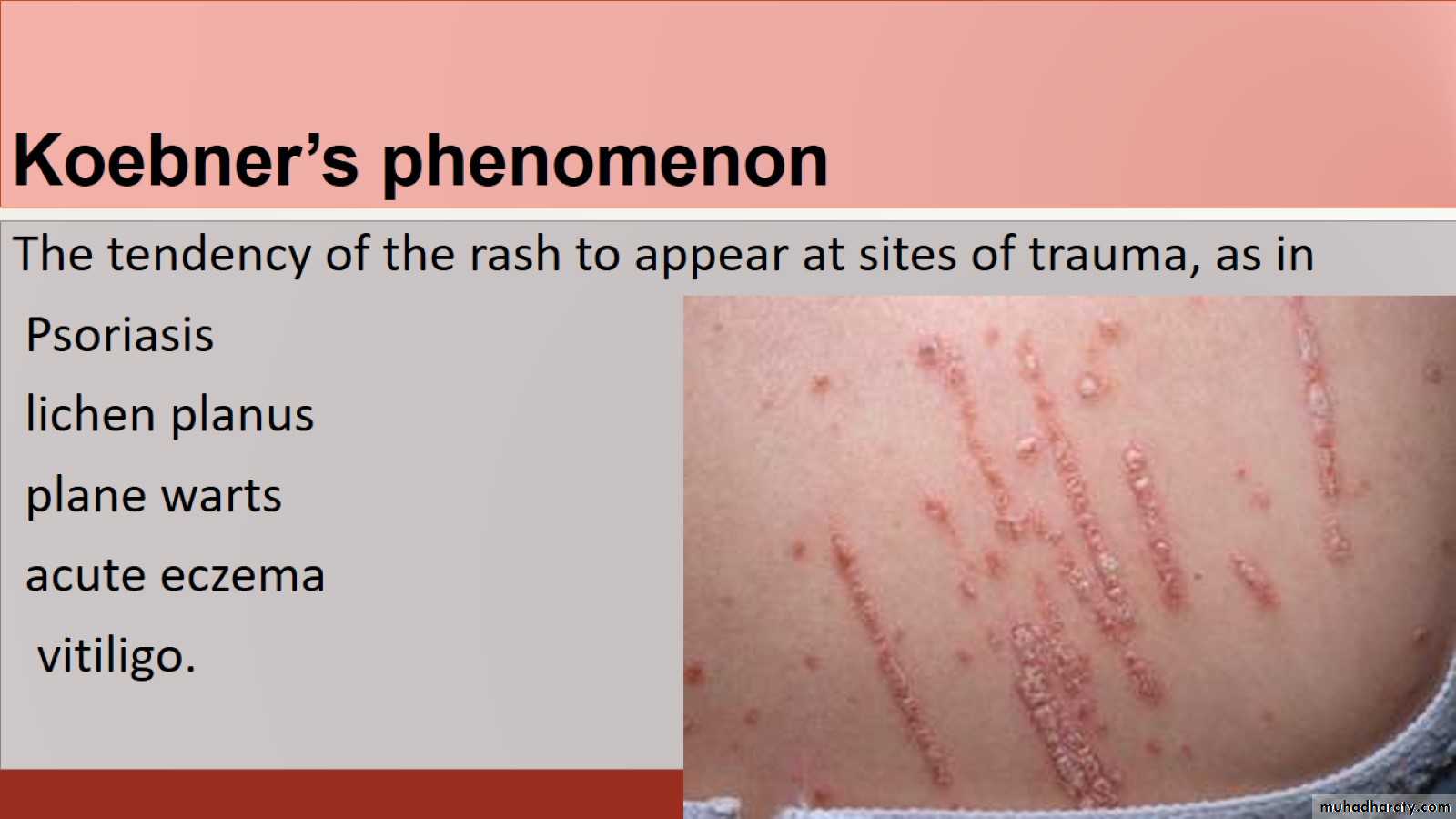
Koebner’s phenomenon
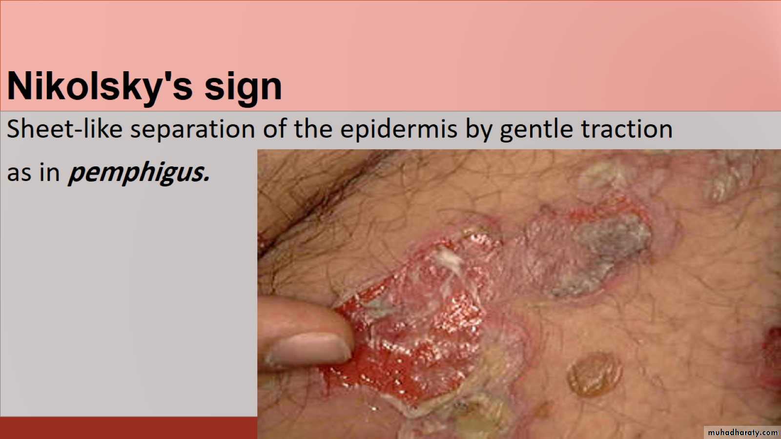
Nikolsky's sign
Nikolsky's sign
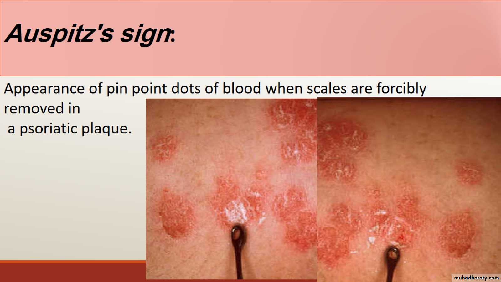
Auspitz's sign:
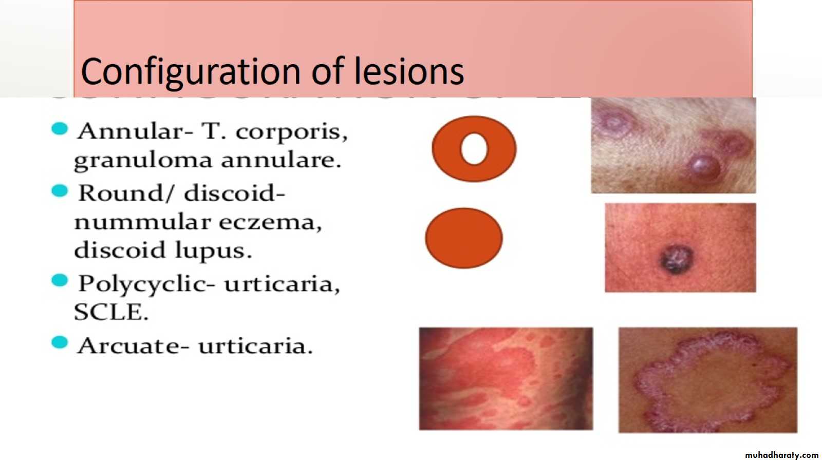
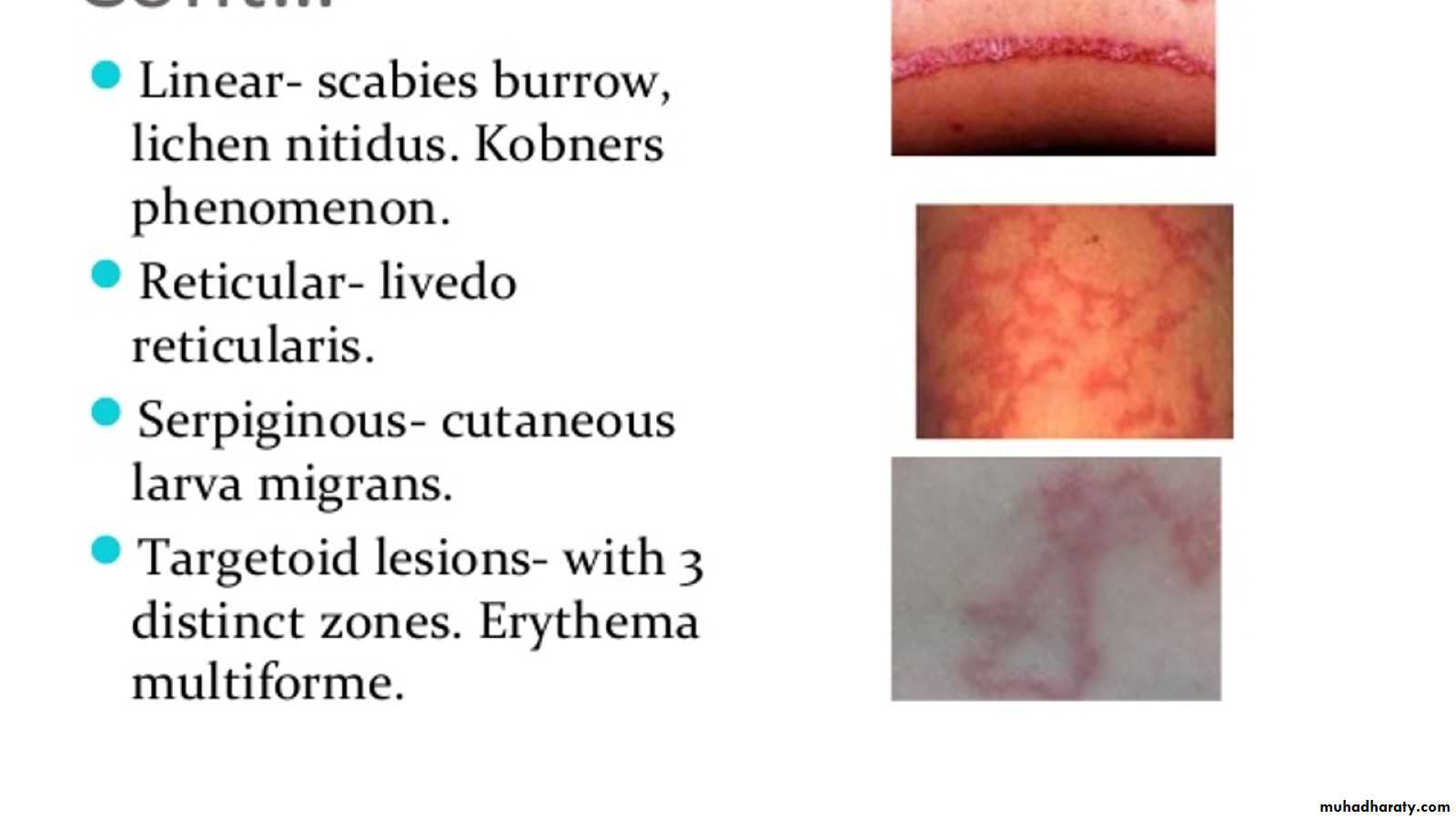
Configuration of lesions
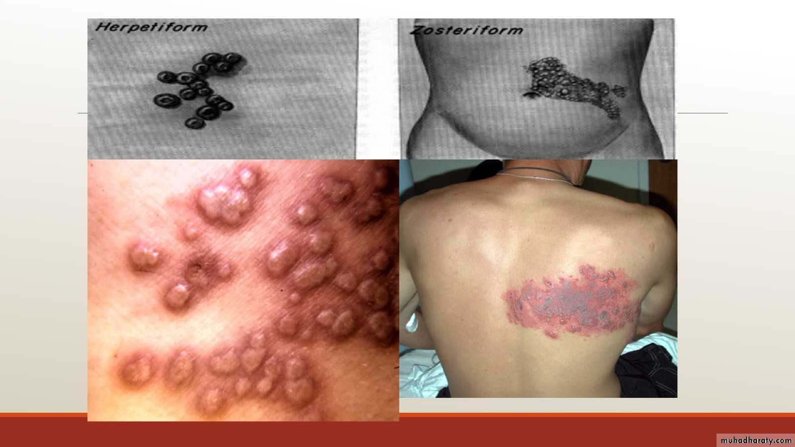
Arrangement of lesions

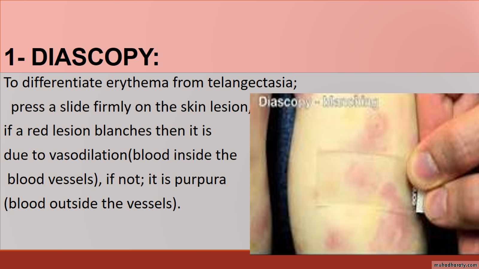
1- DIASCOPY:
In TB of the skin diascopy reveals an appearance called apple- jelly nodules.
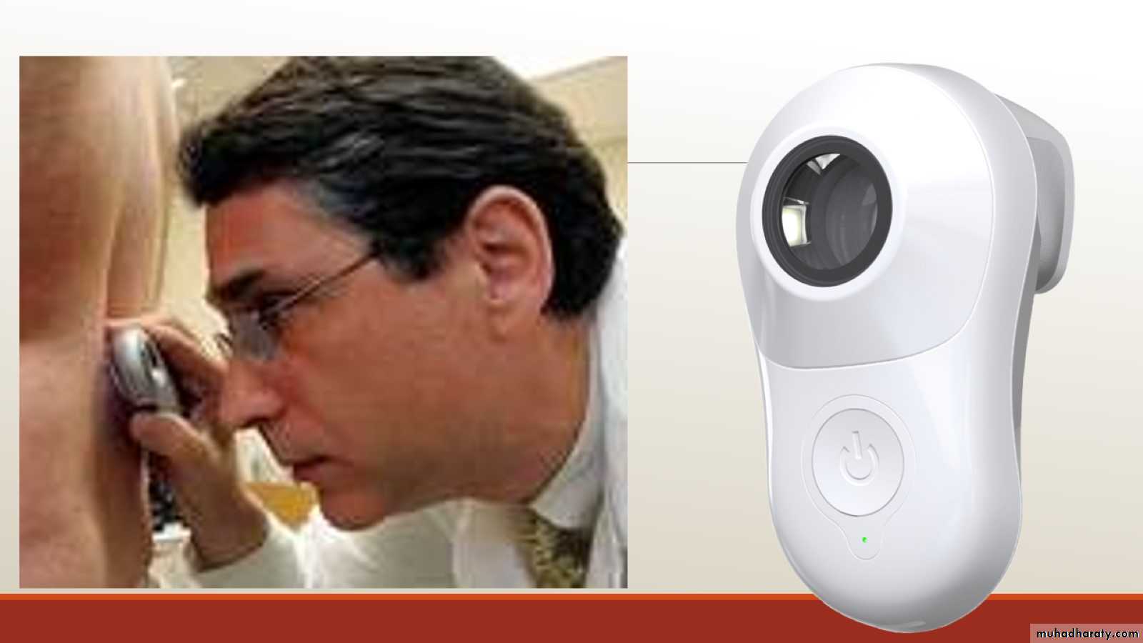
2- Dermoscopy
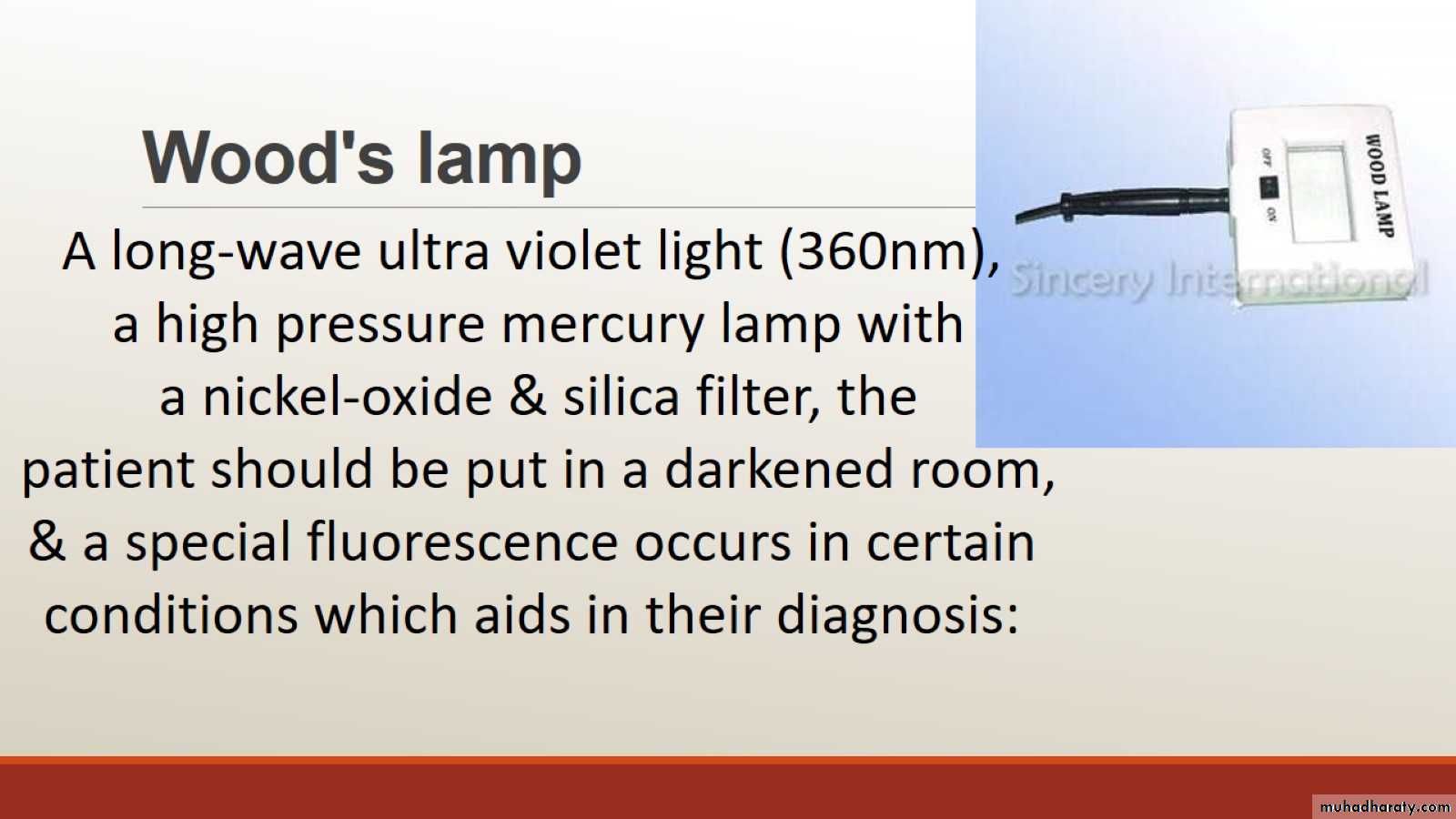
Wood's lamp
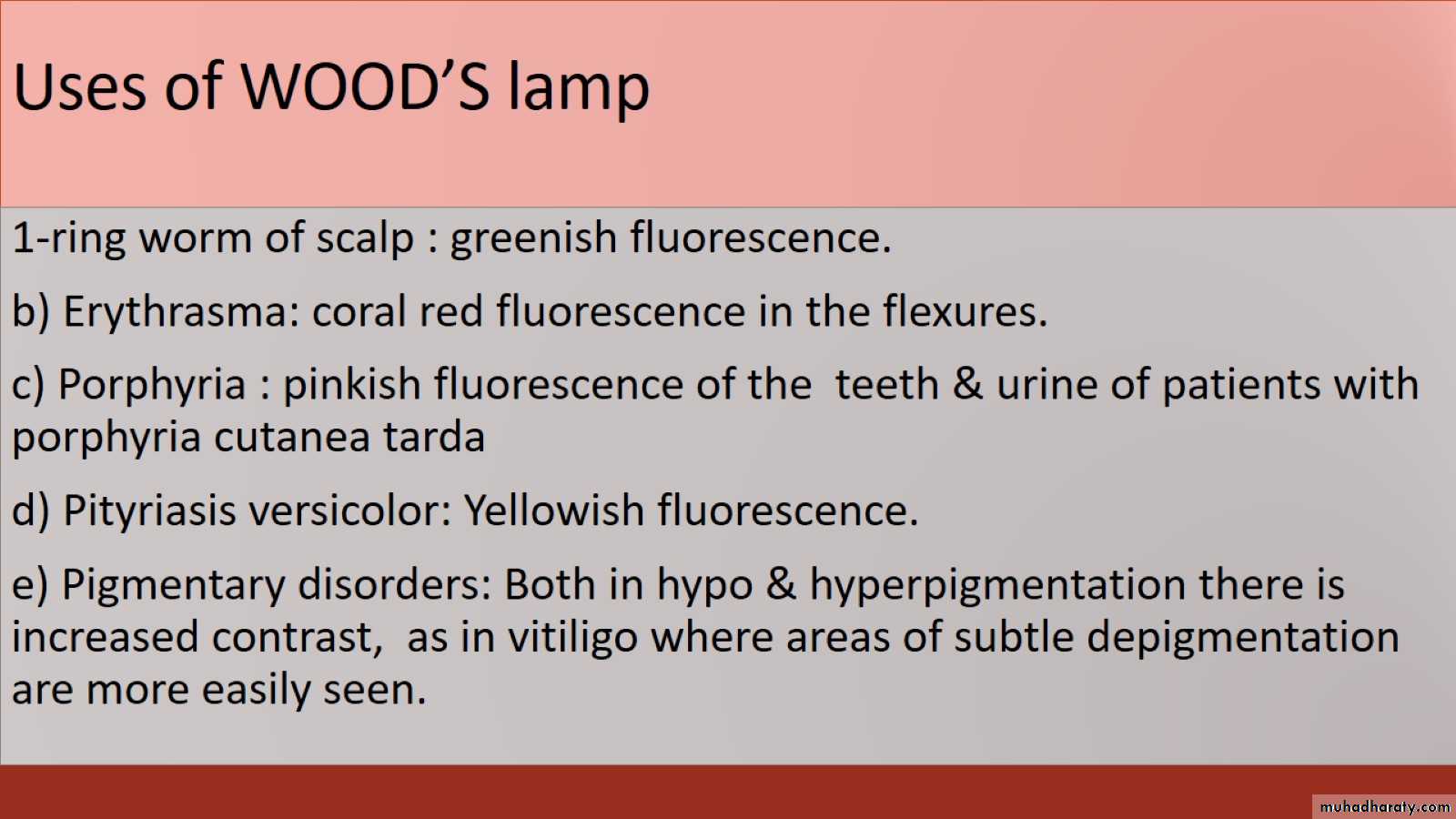
Uses of WOOD’S lamp
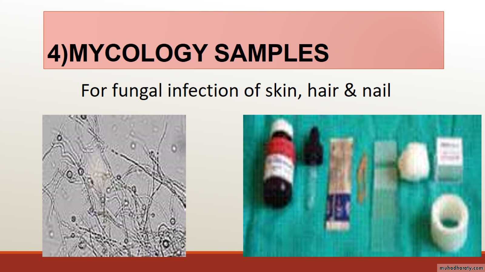
4)MYCOLOGY SAMPLES
5)LAB. INVESTIGATIONS :
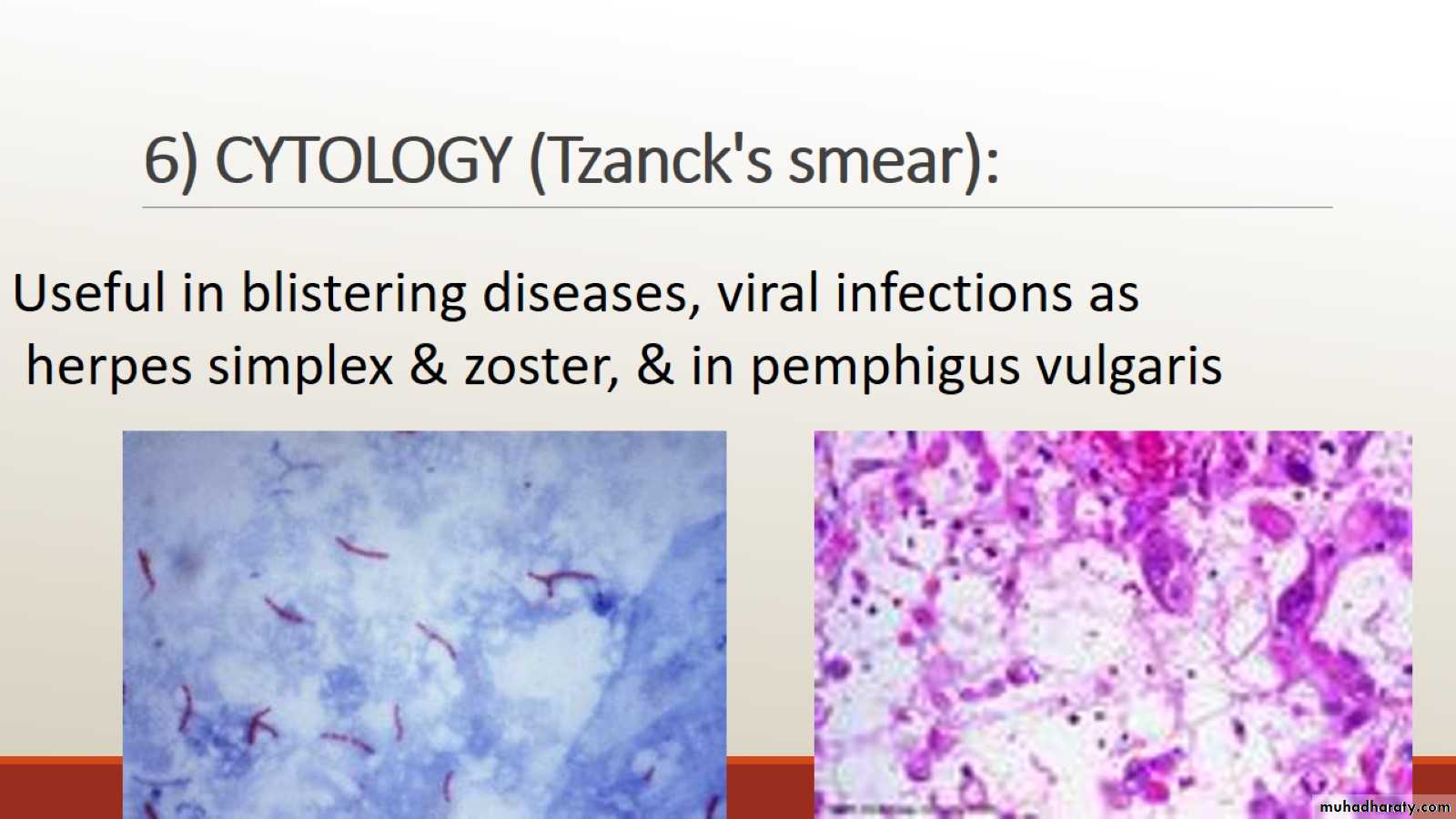
6) CYTOLOGY (Tzanck's smear):
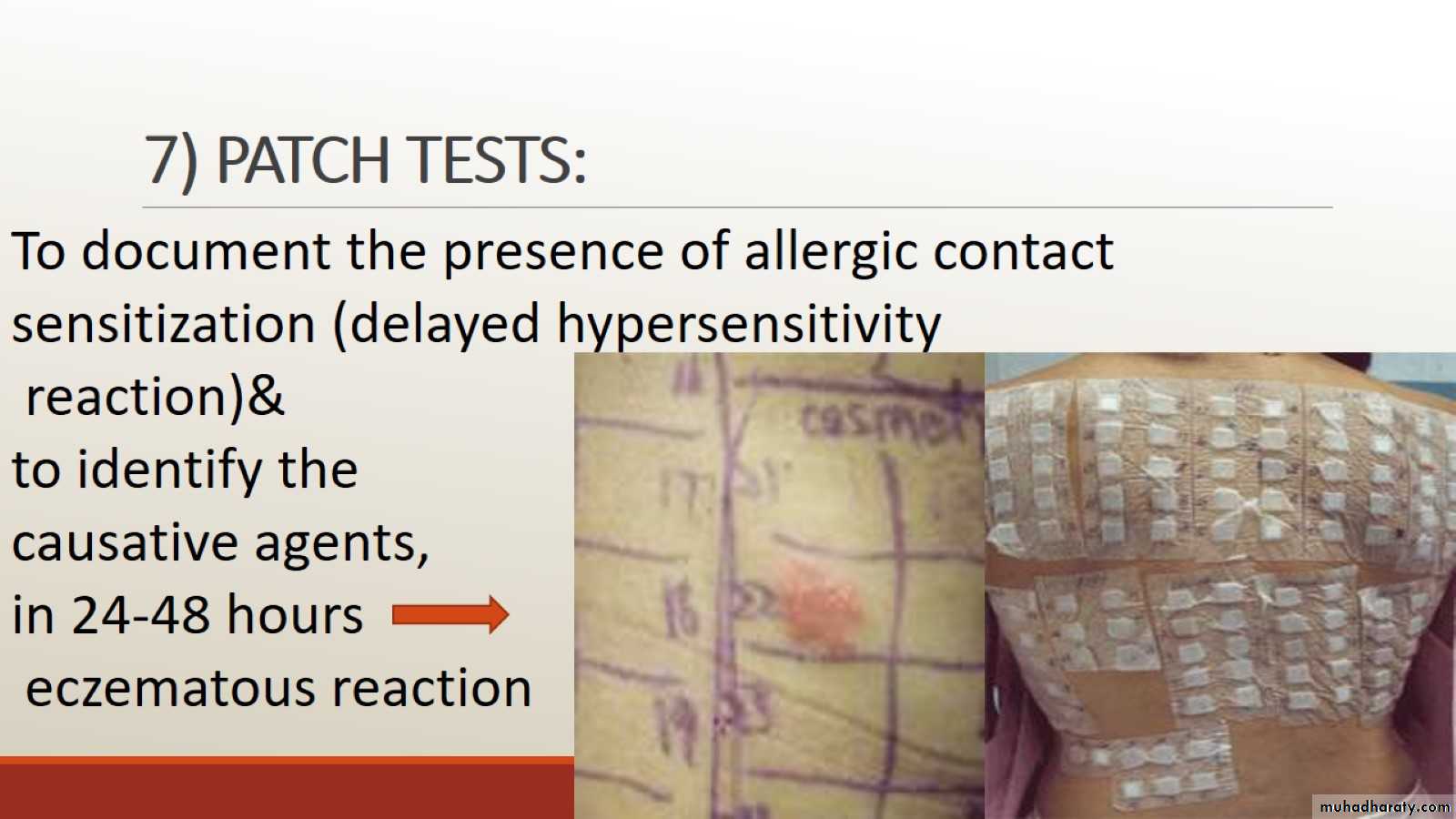
7) PATCH TESTS:
8) PRICK TESTS:

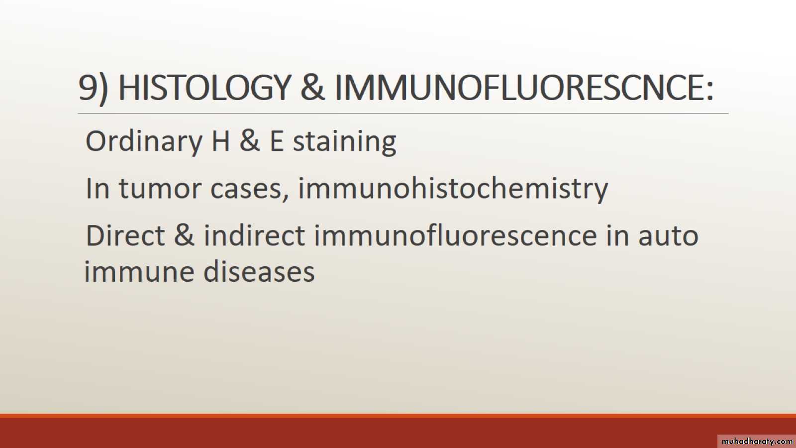
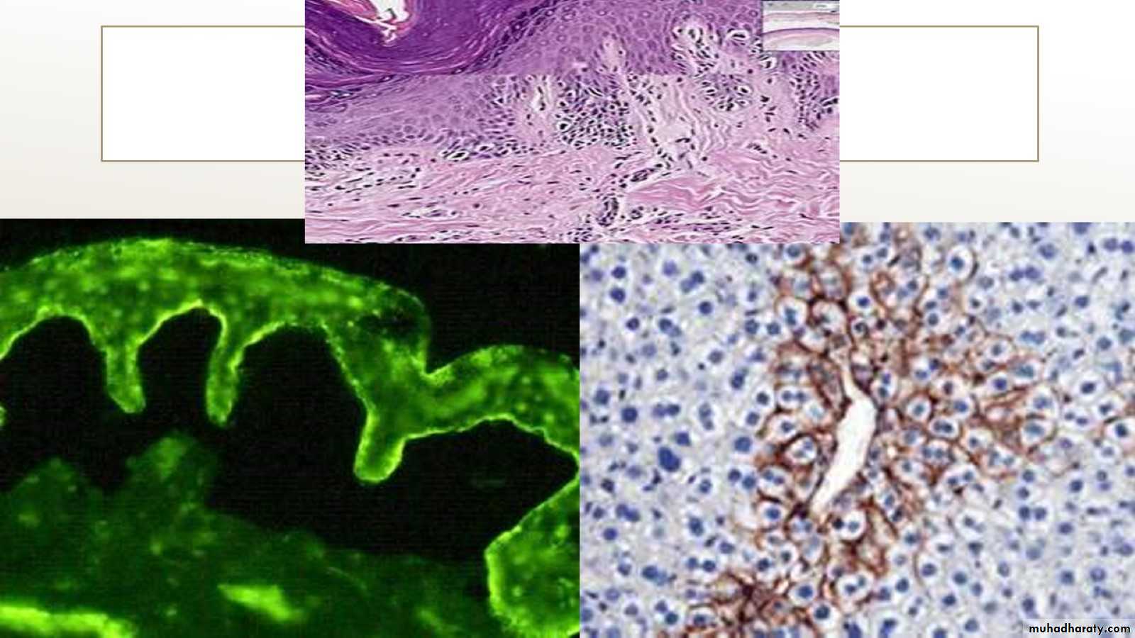
9) HISTOLOGY & IMMUNOFLUORESCNCE: