Renal Diseases 3
Renal Diseases 3
Third Year Class
By Dr.Riyadh A. Ali
Department of Pathology
TUCOM
Third Year Class
By Dr.Riyadh A. Ali
Department of Pathology
TUCOM
Titles
•
Dominant Polycystic Kidney Disease
•
Renal cell carcinoma
•
Wilm's tumor
•
Acute cystitis
•
Transitional cell carcinoma.
•
Dominant Polycystic Kidney Disease
•
Renal cell carcinoma
•
Wilm's tumor
•
Acute cystitis
•
Transitional cell carcinoma.

Dominant
Polycystic
Kidney Disease
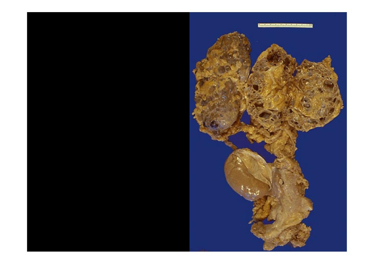
The cut surfaces of
these kidneys in a
patient with
DPKD
(
Dominant Polycystic
Kidney Disease)
reveal that the
parenchyma is
replaced by large
cysts. Note how large
these kidneys are in
relation to the normal
sized transplanted
kidney
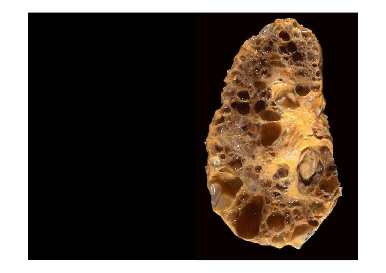
This kidney in a patient
with
DPKD
Dominant
Polycystic Kidney
Disease
weighed 3
kilograms cut section
reveal that the
parenchyma is replaced
by large cysts

Renal Cell
Carcinoma
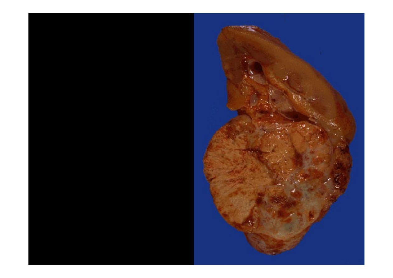
This is a
renal cell
carcinoma
arising in the
lower pole of the kidney.
It is fairly circumscribed.
The
cut
surface
demonstrates
a
variegated
appearance
with
yellowish
areas,
white
areas,
brown
areas, and hemorrhagic
red areas
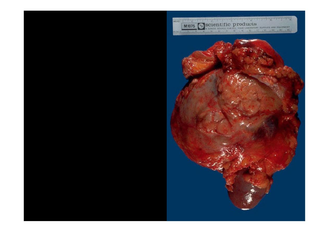
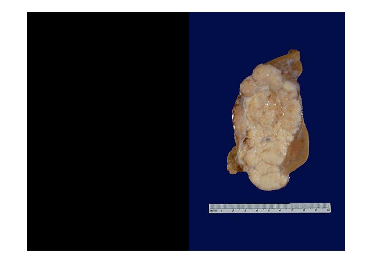
This
renal cell
carcinoma
is very
large, as indicated by
the 15 cm ruler. A
portion of normal
kidney protrudes at the
lower center. This
patient was a
physician himself and
just didn't have any
early symptoms.
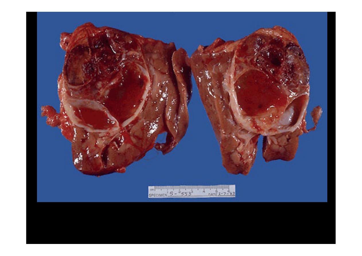
Here is a
renal cell carcinoma
that on sectioning is mainly cystic with
extensive hemorrhage
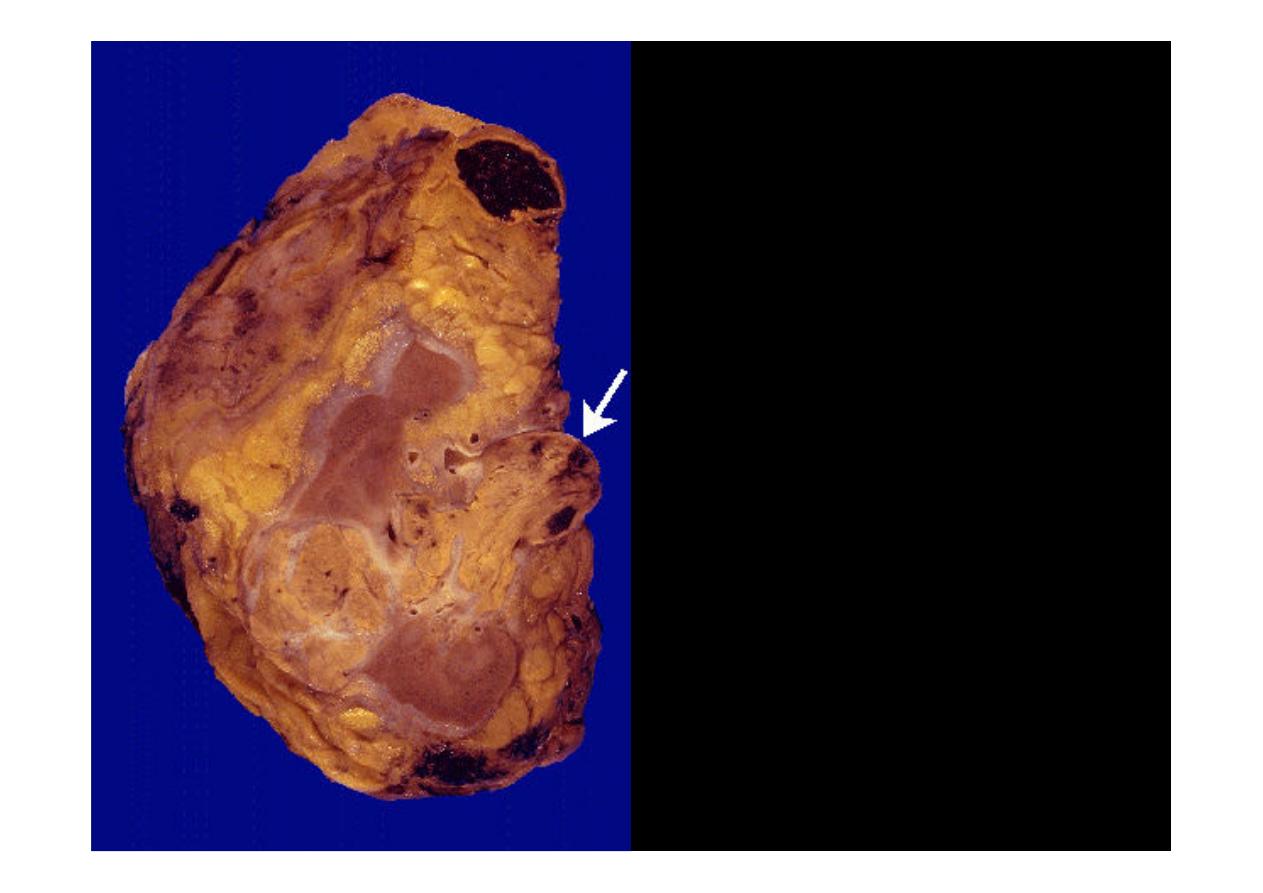
Renal cell
carcinomas
have a
tendency to invade
into the renal vein, as
shown here at the
white arrow in a
resected kidney
surrounded by
adipose tissue
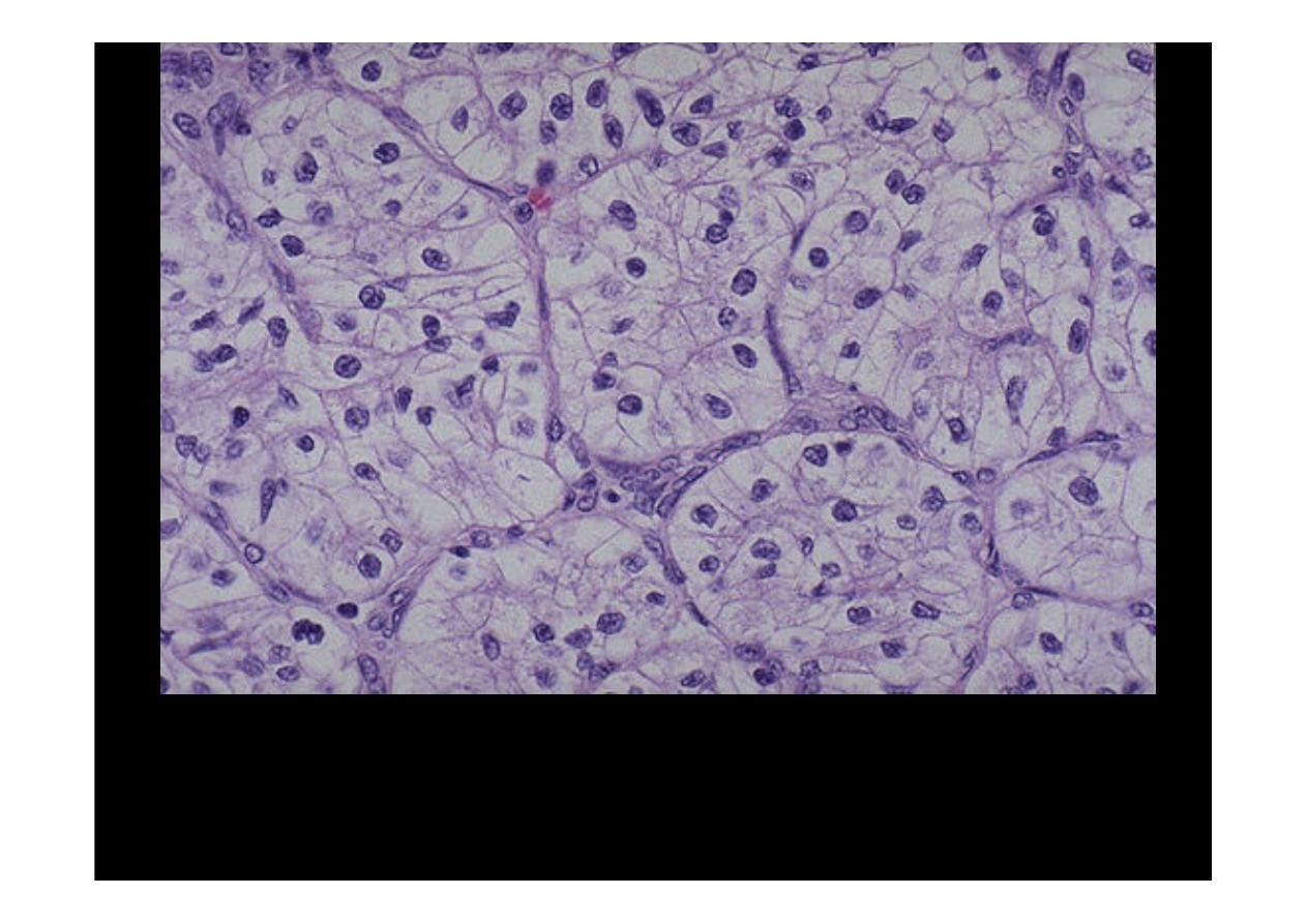
This is the classic histologic appearance of a
renal cell carcinoma
: the
neoplastic cells have clear cytoplasm and are arranged in nests with
intervening blood vessels. This appearance is why they are often called "clear
cell carcinomas".
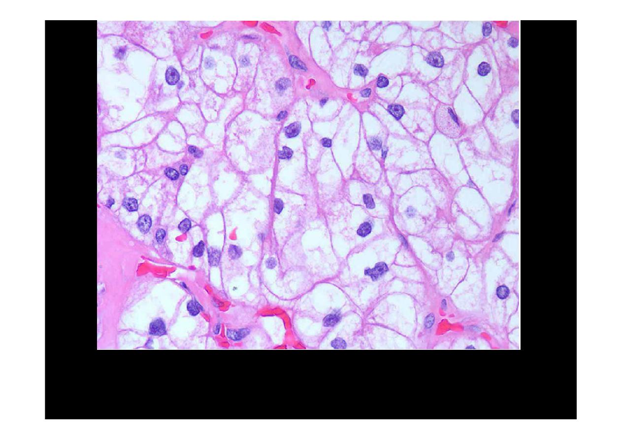
This is the classic histologic appearance of a
renal cell carcinoma
: the neoplastic
cells have clear cytoplasm and are arranged in nests with intervening blood
vessels. This appearance is why they are often called "clear cell carcinomas
Wilm's Tumor
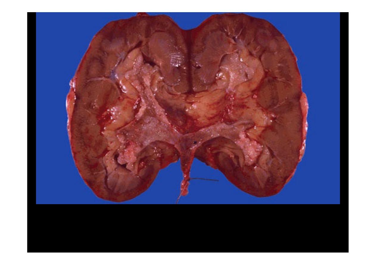
This small kidney from
a 4 year old child
contains a lobulated
tan-white mass. This
is
Wilm's tumor
of
the kidney
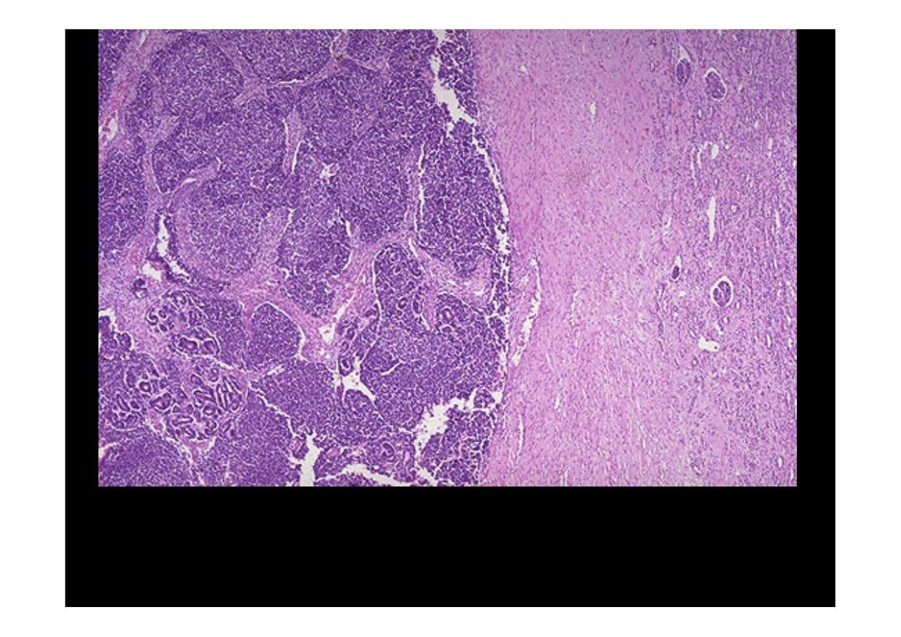
This is a
Wilm's tumor
that is composed microscopically of nests and
sheets of dark blue cells at the left with compressed normal renal
parenchyma at the right
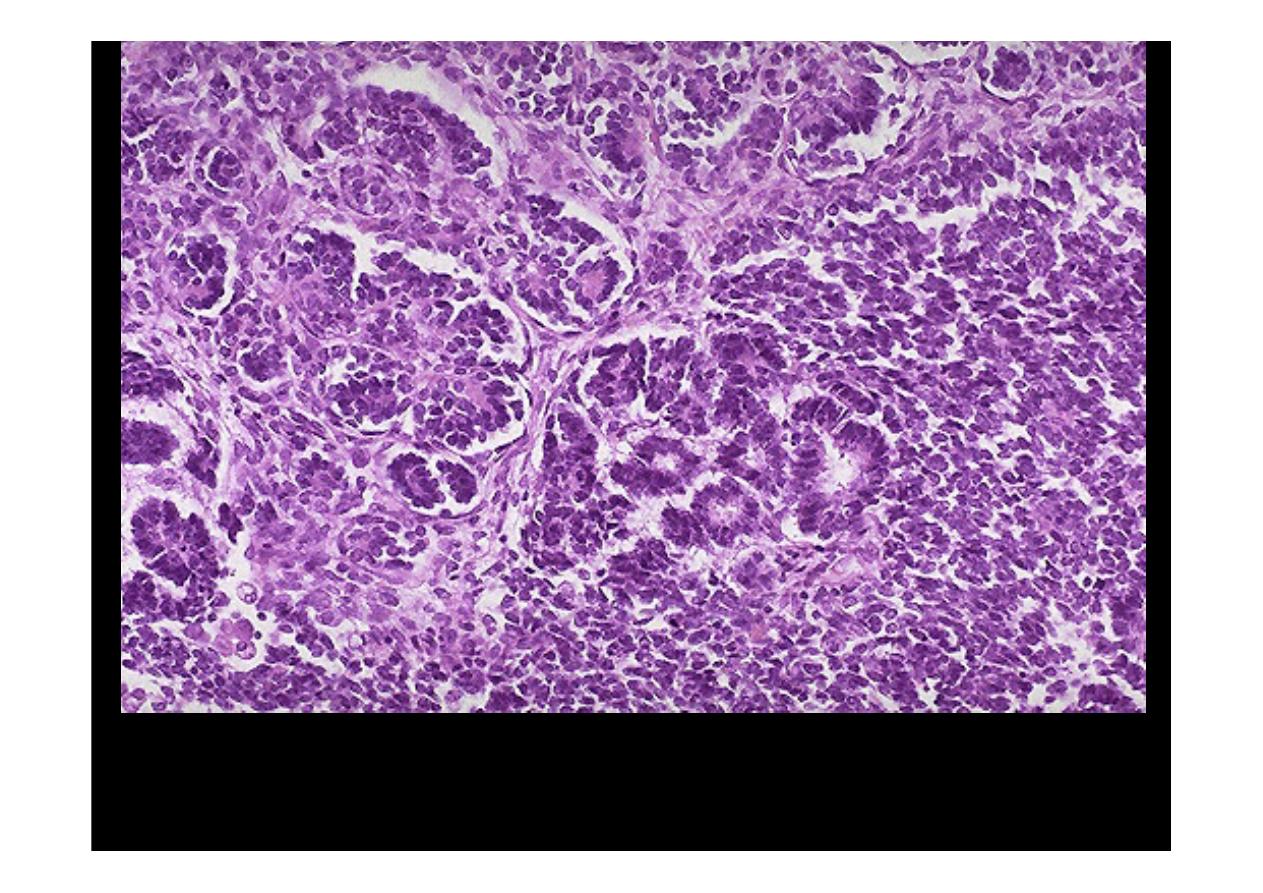
Wilm's tumor
resembles the fetal nephrogenic zone of the kidney. The tumor
shows attempts to form primitive glomerular and tubular structures.

Acute Cystitis
This bladder at autopsy has been opened to reveal areas of
hyperemia of the mucosa.
This is acute cystitis.

Transitional Cell
Carcinoma (TCC)

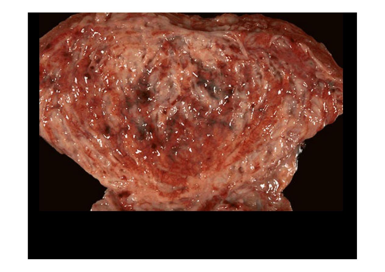
The opened bladder reveals masses of a neoplasm that histologically proved
to be
transitional cell carcinoma (TCC).
There is thicking of the wall and
mass found to occupy the bladder space with irregular ploypoidal projections
of red tan color.
The cut surfaces of the kidney removed surgically here demonstrate normal
cortex and medulla, but the calyces show focal papillary tumor masses of
transitional cell carcinoma.
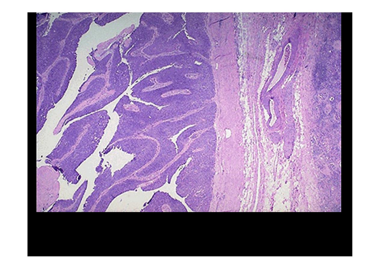
Transitional cell carcinoma of the urothelium
is shown here at low power to
reveal the frond-like papillary projections of the tumor above the surface to the left. It
is differentiated enough to resemble urothelium, but is a mass. No invasion to the
right is seen at this point.
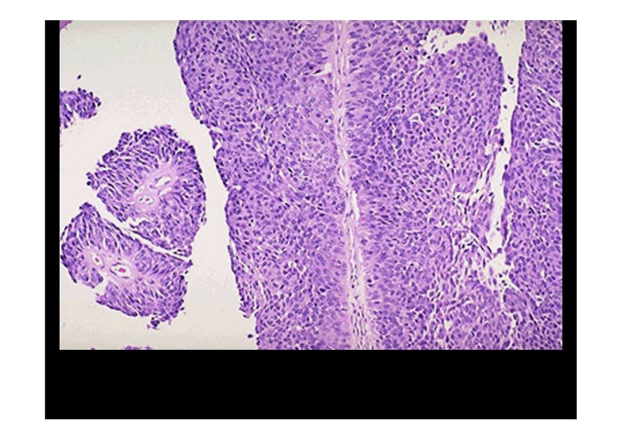
At high power, the
transitional cell carcinoma
does resemble urothelium, but the
thickness is much greater than normal and the cells show more pleomorphism