
Renal System 1
Third Year Class
By Dr.Riyadh A. Ali
Department of Pathology
TUCOM

Normal Kidney
Normal Kidney
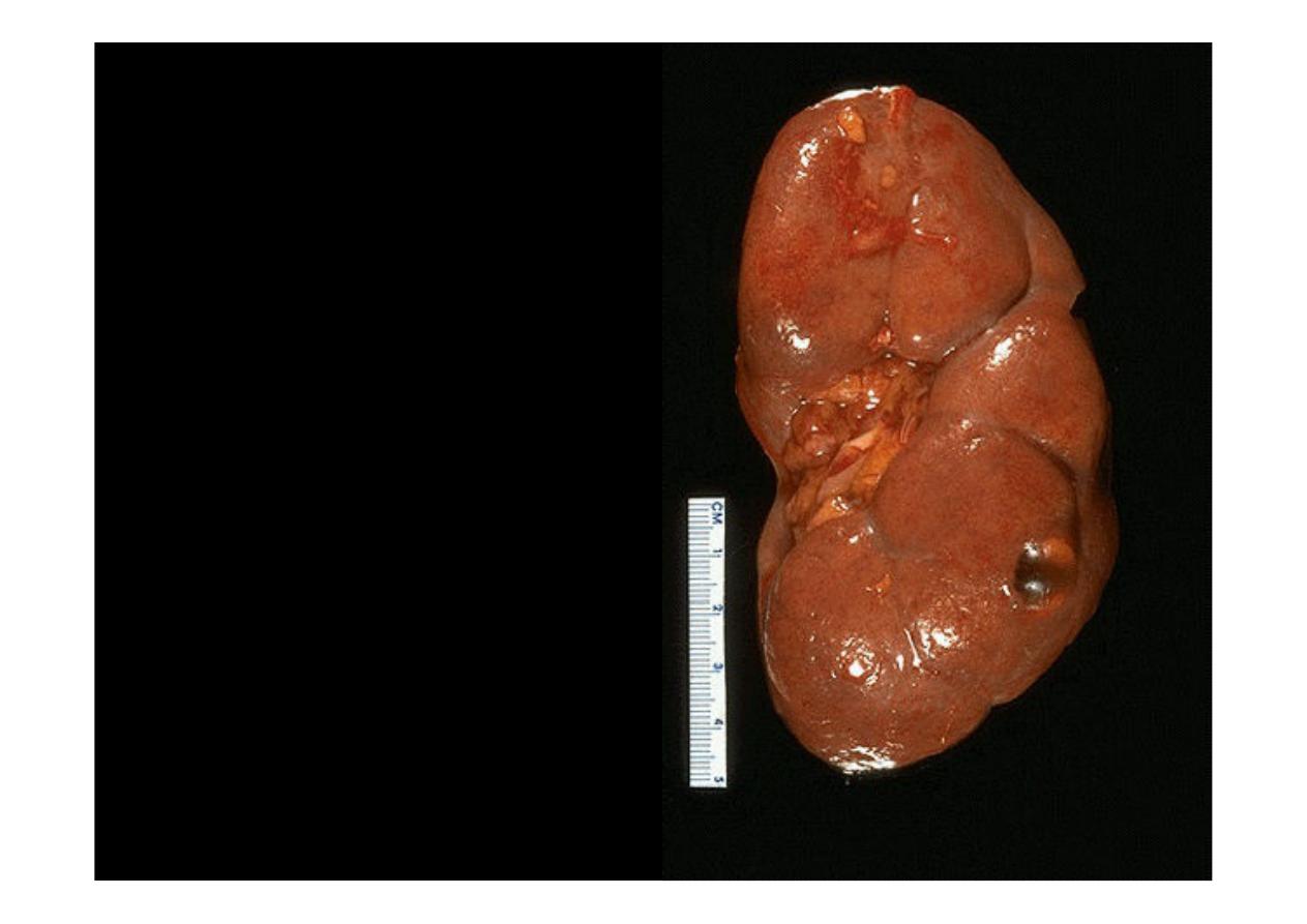
Here is a
normal adult
kidney
. The capsule has
been
removed
and
a
pattern of fetal lobulations
still
persists,
as
it
sometimes
does.
The
hilum
at
the
mid
left
contains
some
adipose
tissue. At the lower right is
a smooth-surfaced, small,
clear
fluid-filled
simple
renal
cyst.
Such
cysts
occur
either
singly
or
scattered around the renal
parenchyma and are not
uncommon in adults.
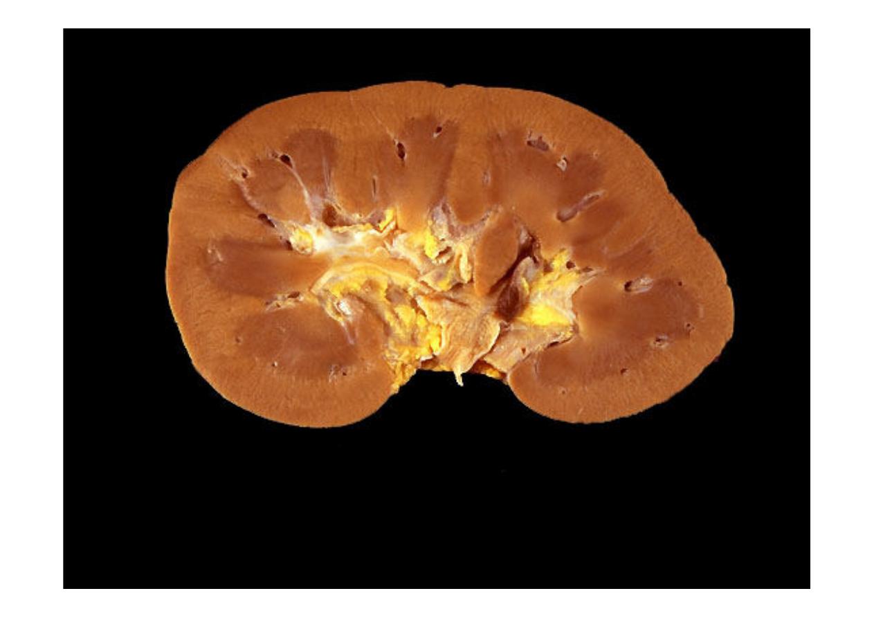
In cross section, this
normal adult kidney
demonstrates the
lighter outer cortex and darker medulla with central pelvis.

Kidney Diseases
(Grosses)
Simple renal cyst
Horseshoe kidney
Atrophic kidney
Hydronephrosis
Renal infarction
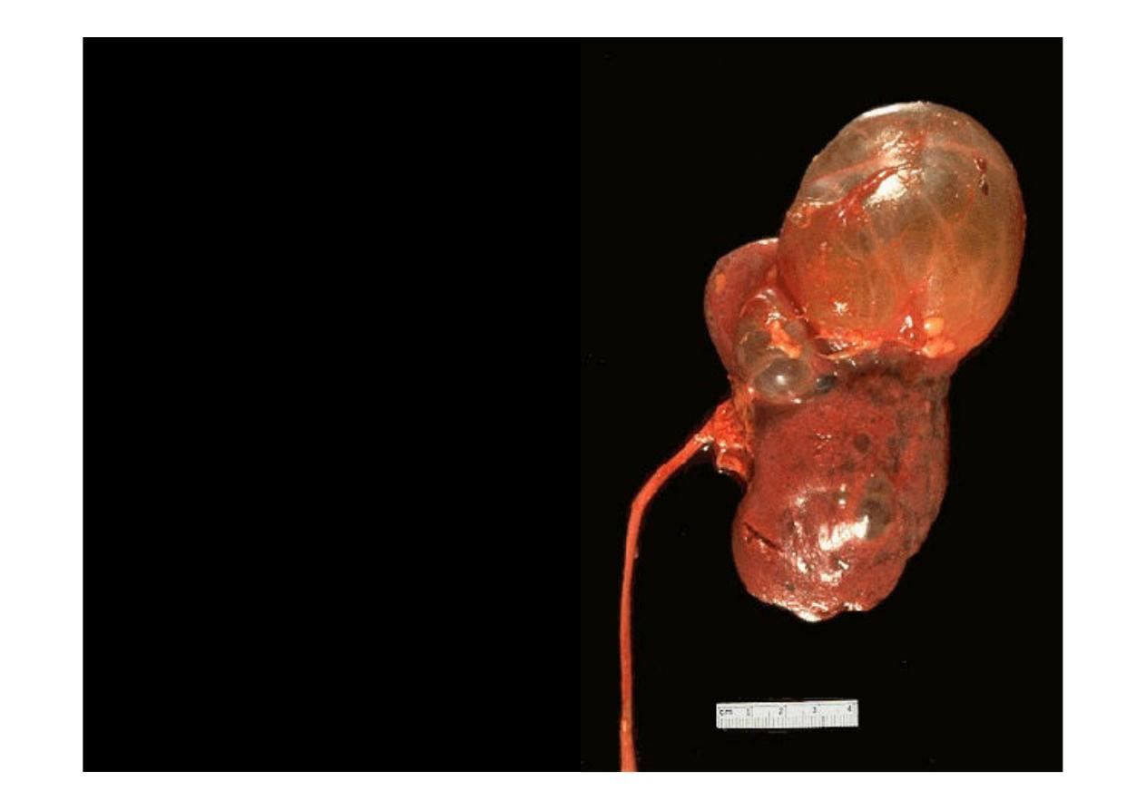
Here is a much larger
simple renal cyst
of the
upper pole. Other
smaller cysts are also
scattered around the
kidney. The ureter exits
south on the left.
Here is a "
horseshoe" kidney
. This is a congenital
anomaly
There is a relatively normal kidney at the left with only a few scattered,
shallow cortical scars and one fairly large pale tan-yellow scar in the
upper pole.
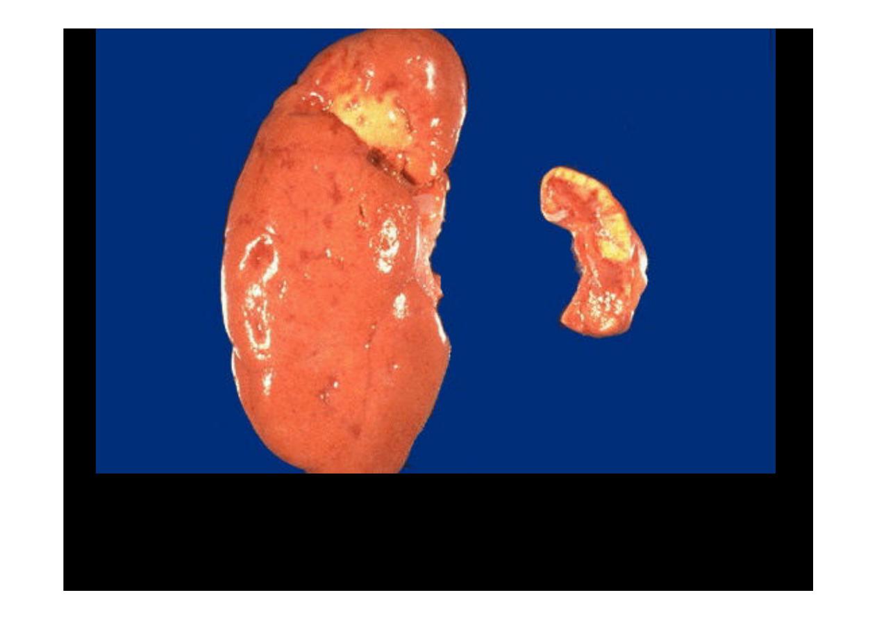
The left kidney is atrophic
because of renal arterial
occlusion
.
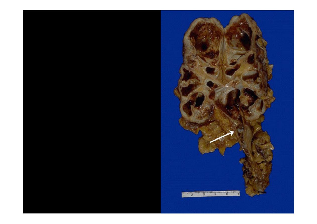
The arrow points to the
culprit in this case of
hydronephrosis
--a
ureteral calculus
at the
ureteropelvic
junction.
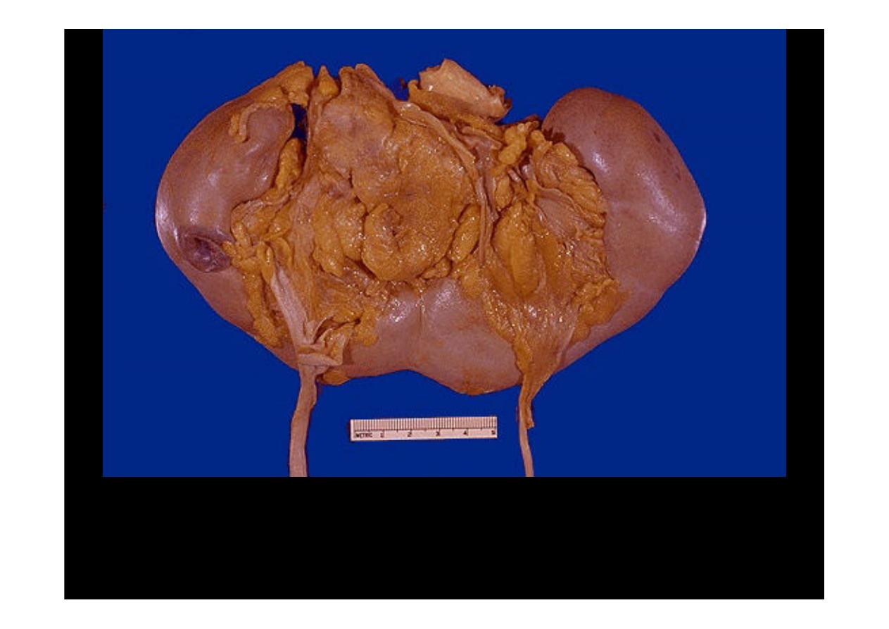
This kidney demonstrates
marked
hydronephrosis
with nearly complete loss
of cortex
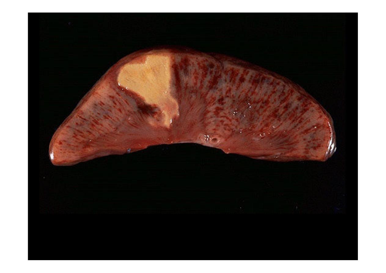
This is an
acute renal infarction
. Note the wedge shape of this zone of
coagulative necrosis resulting from loss of blood supply with resultant
tissue ischemia that produces the pale infarct. The remaining cortex is
congested, as is the medulla.

Pyelonephritis
Acute
Chronic
Xanthogranulomatous
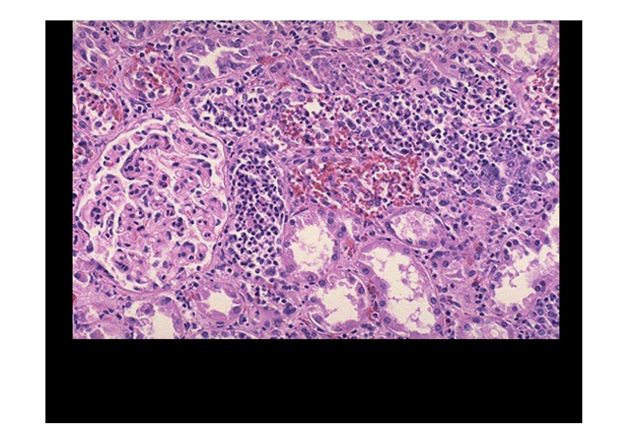
This is an ascending bacterial infection leading to
acute
pyelonephritis
. Numerous PMN's are seen filling renal
tubules across the center and right of this picture.
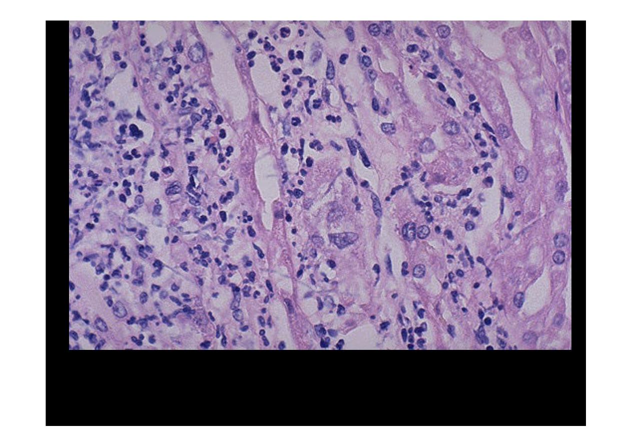
At high magnification, many neutrophils are seen in the
tubules and interstitium in a case of
acute pyelonephritis
.
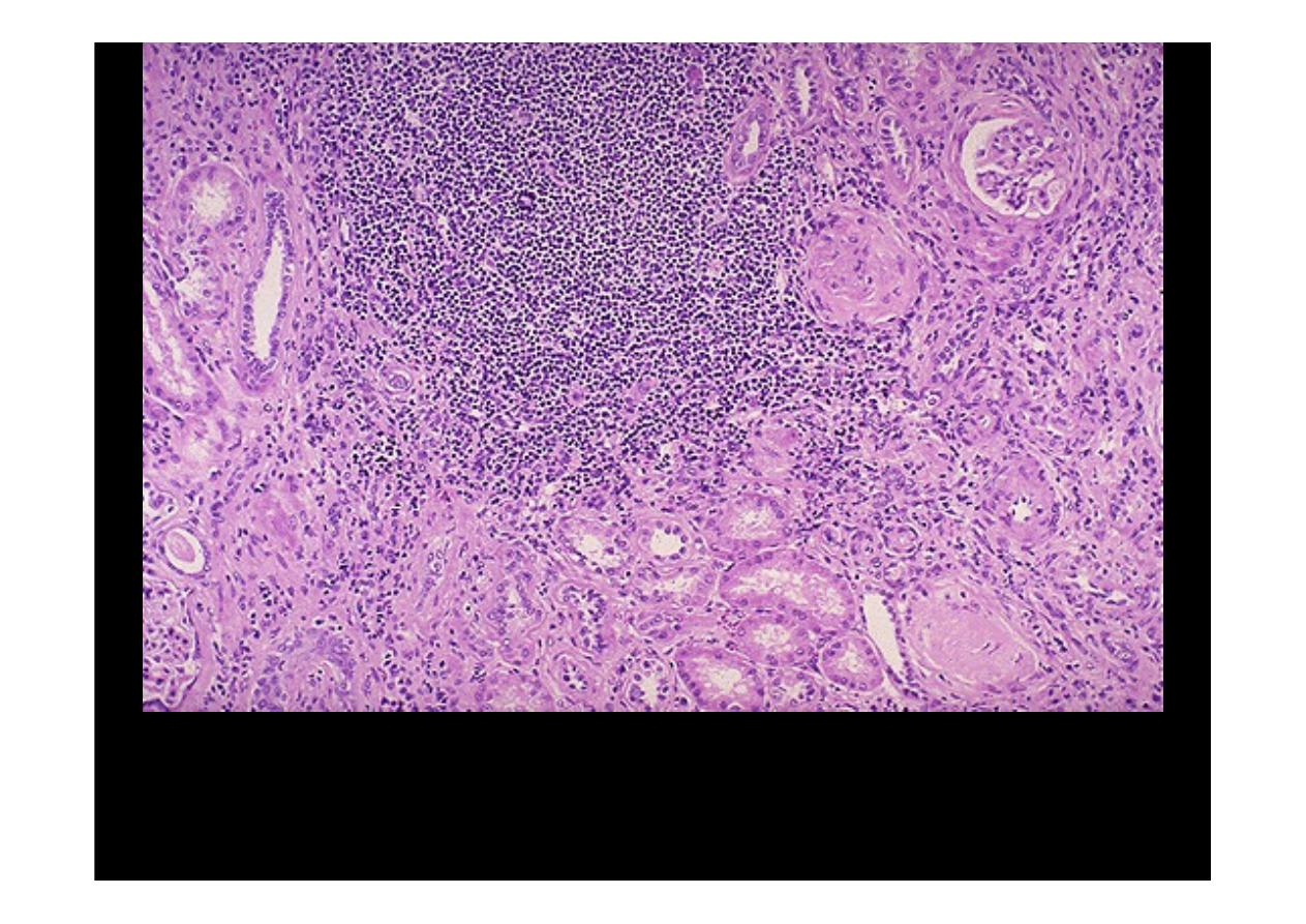
The large collection of chronic inflammatory cells here is in a
patient with a history of multiple recurrent urinary tract
infections. This is
chronic pyelonephritis
.
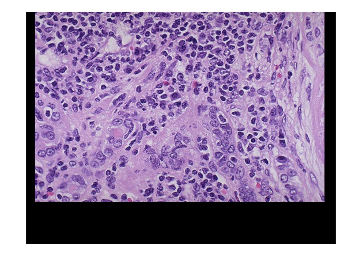
Both lymphocytes and plasma cells are seen at high magnification in this case of
chronic pyelonephritis.
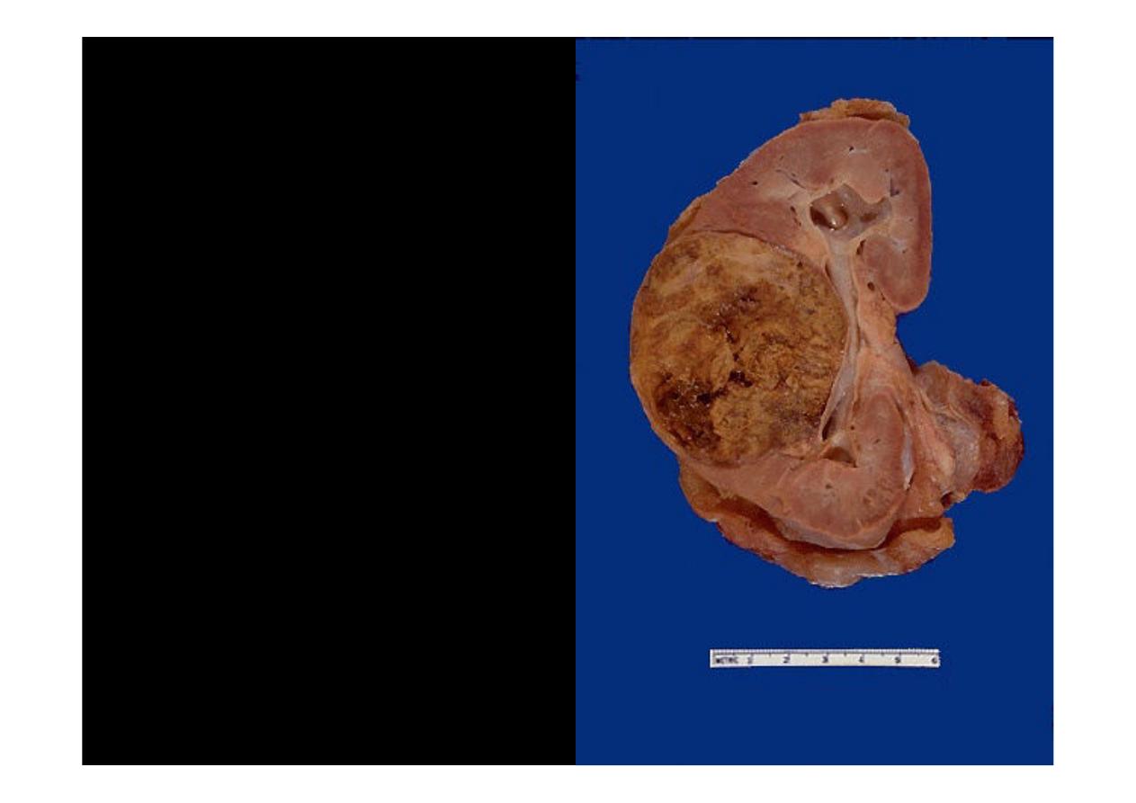
Sometimes
long-standing
infection may be localized
and form a mass-like lesion.
This is a disease known as
xanthogranulomatous
pyelonephritis
.
It is uncommon, but may
mimic a neoplasm.
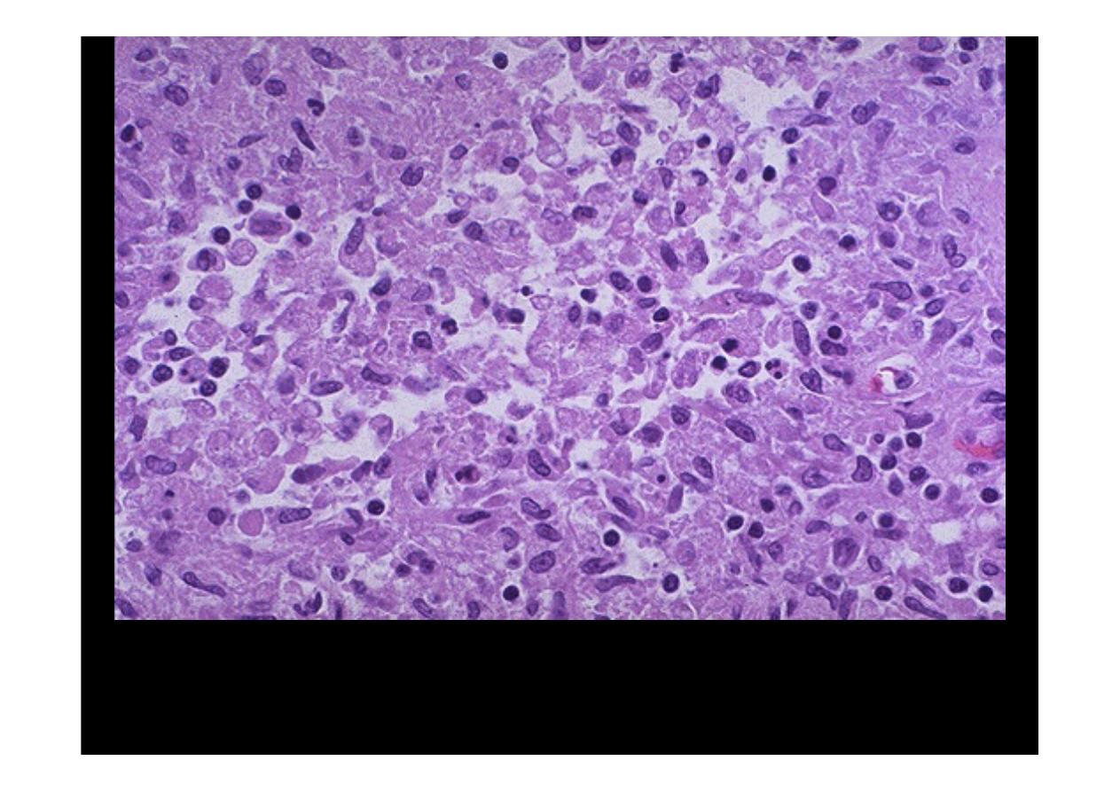
The microscopic appearance of
xanthogranulomatous pyelonephritis
shows many pale to foamy macrophages from breakdown of renal
parenchyma with ongoing inflammation
.