BONE PathologybyDr. Zahraa Marwan
- skeletal system is composed of 206 bones, it serves the following functions: -supports the body mechanically -keeps mineral homeostasis -houses the hematopoietic elements -protects viscera -determines body size and shape.bone is a connective tissue containing admixture of inorganic elements (calcium hydroxyapatite 65%), and organic matrix (osteoblasts and osteocytes 35%).osteoclasts are responsible for bone resorption.osteoblasts deposit collagen -random weave (woven bone) - orderly layered manner (lamellar bone).lamellar bone replaces woven bone during growth of the person
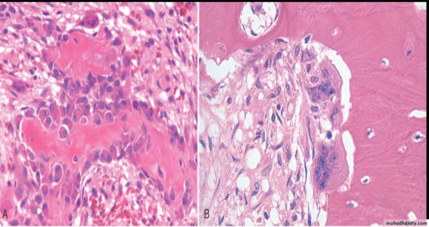
Osteoblasts synthesizing bone
Osteoclasts resorbingboneWoven bone (top) deposited on surface of lamellar bone
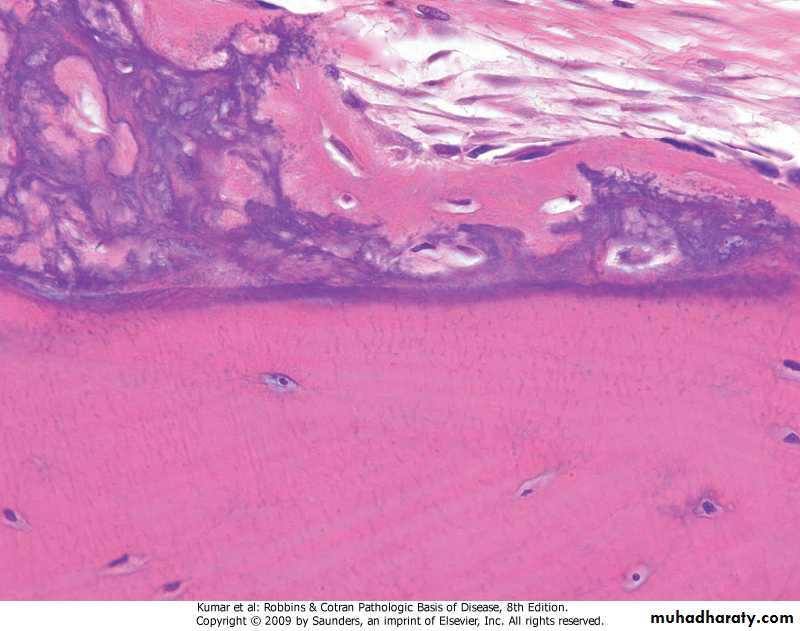
Woven bone (top) deposited on surface of lamellar bone
Bone Modeling And Remodeling .osteoblasts and osteoclasts act in coordination and are considered the functional unit of bone known as the basis multicellular unit (BMU) .modeling (growing and enlargement of skeleton) .remodeling (breakdown and renewal of bone)Diseases associated with deranged metabolism of collagen: -Osteogenesis imperfecta (OI), deficiency of type 1 collagen. - The fundamental abnormality in OI is too little bone, resulting in extreme skeletal fragility.- AD type of inheritance.- Blue sclera, hearing loss ; and dental imperfections (small, malformed, and blue-yellow teeth) secondary to a deficiency in dentin.
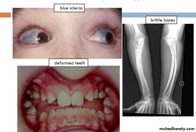
OI
OSTEOPOROSIS.reduction of bone mass, localized or generalized (primary or secondary).pathogenesis: -age-related changes -reduced physical activity -genetic factors -body’s calcium nutritional status -hormonal influences -prolonged glucocorticoid therapy
Morphology: -thinned trabeculae>microfractures .Diagnosis: -estimation of bone amount by biopsy -radiographic imaging techniques.Prevention and treatment: -exercise -calcium and vitamin D supplement -pharmacologic agents
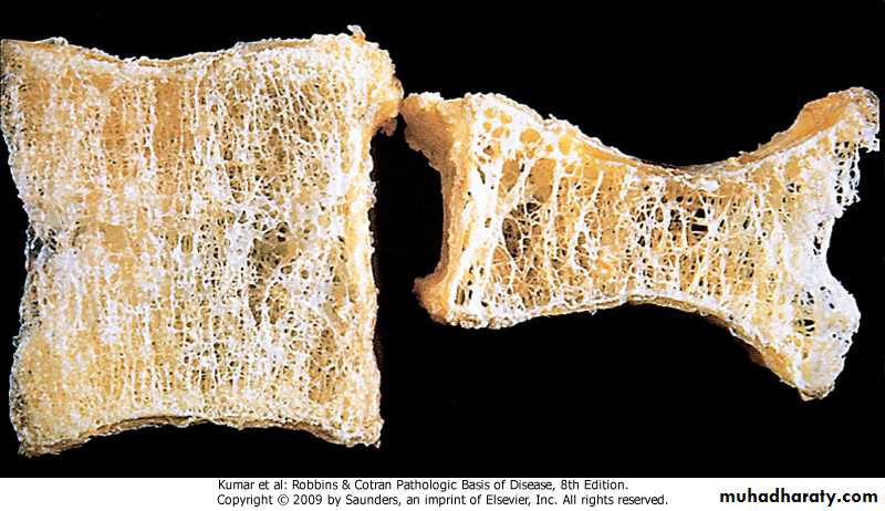
Osteoporosis, vertebral body with compression fractures (right)
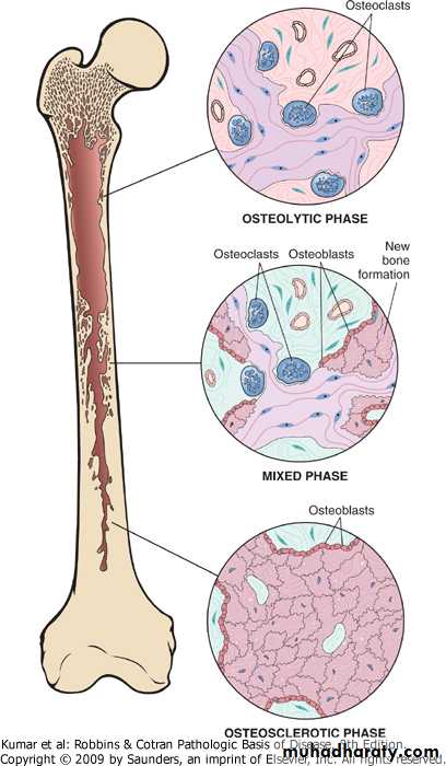
RICKETS AND OSTEOMALACIA.Rickets in children, and Osteomalacia in adults, are characterized by a defect in mineralization of matrix>osteopenia .causes -inadequate synthesis or dietary deficiency of vitamin D -decreased absorption of vitamin D -derangement in vitamin D metabolism -phosphate depletionPAGET DISEASE (OSTEITIS DEFORMANS).gain of disordered bone mass, divided into: -initial osteolytic stage -mixed osteoclastic-osteoblastic stage -osteosclerotic stage.pathogenesis -infection by paramyxovirus -abnormal osteoclasts -hereditary component
Morphology: -mosaic pattern of lamellar bone .clinical course: -usually discovered incidentally by radiology -can be monostotic or polystotic -most frequent features .pain, localized to the affected bone .benign tumors and tumor-like lesions .sarcomas
Paget disease of bone

Paget disease of bone, mosaic pattern
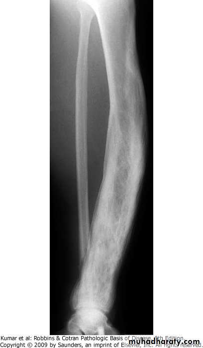
Paget disease of bone, tibia is sclerotic with thickened cortical and cancellous bone
FRACTURES: fracture is defined as loss of bone integrity resulting from mechanical injury and/or diminished bone strength. either complete or incomplete; simple or compound; comminuted (splintered bone); or displaced .complications: -delayed union -nonunion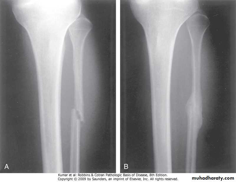
Fibula, fracture &callus formation
OSTEOMYELITIS: Osteomyelitis denotes inflammation of bone and marrow, virtually always secondary to infection..primary or follow systemic infection - Pyogenic Osteomyelitis: .almost always bacterial, reaching bone via: (1) hematogenous spread (majority) (2) extension from a contiguous site (3) direct implantation after compound fractures or orthopedic procedures. -Changes associated with osteomyelitis depend on: -The stage (acute, sub acute, or chronic) - Location of the infection
In the acute phase, bacteria proliferate and induce a neutrophilic inflammatory reaction. Necrosis of bone cells and marrow arises within the first 48 hours. The bacteria and inflammation spread longitudinally and radially to reach the periosteum. Lifting of the periosteum further impairs the blood supply to the affected region, contributing to the necrosis. The dead bone is known as a sequestrum. Rupture of the periosteum leads to a soft tissue abscess Brodie abscess (intraosseous abscess), which can channel to the skin, creating a draining sinus.
After the first week, chronic inflammatory cells release cytokines that stimulate osteoclastic bone resorption, ingrowth of fibrous tissue, and the deposition of reactive bone at the periphery.
The newly deposited bone can form a shell of living tissue, known as an involucrum, around the segment of devitalized infected bone.
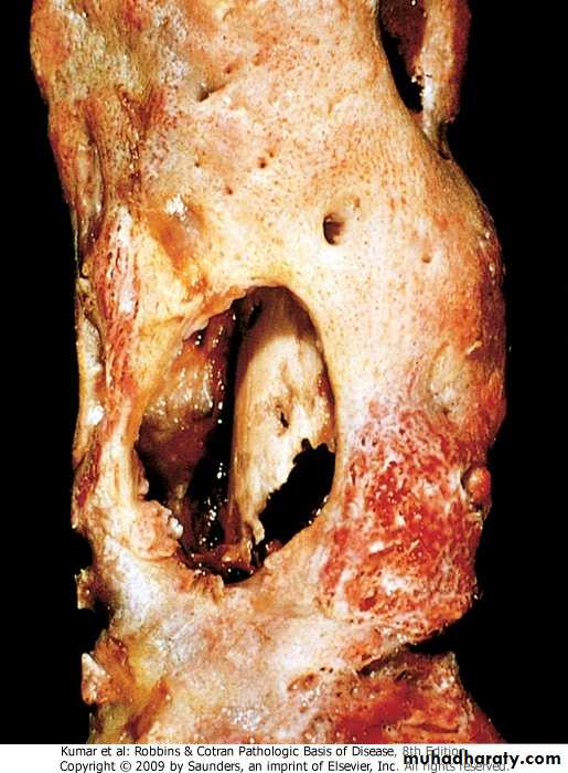
Femur, outer (involucrum), inner (squestrum)
• The histologic findings of chronic osteomyelitis are more variable but typically involve marrow fibrosis, sequestrum, and an inflammatory infiltrate of lyhocytes and plasma cells.Tuberculous Osteomyelitis .organisms are usually blood-borne .the lesion is usually solitary, most commonly involved is the spine (Pott disease)
BONE TUMORS AND TUMOR-Like LESIONS.most are classified according to cell of origin .benign tumors outnumber their malignant counterparts .benign tumors have their peak incidence within the 1st 3 decades of life.in the elderly a bone tumor is more likely to be malignant
.Etiology of bone tumors -the cause of most bone tumors is unknown, associated factors include .genetic .bone infarcts .chronic osteomyelitis .Paget disease .radiation .metal prosthesis
Osteochondromas (Exostoses).benign cartilage-capped outgrowths with a bony stalk .can be solitary (adolescents, young people) or multiple (children), . with M:F ratio of 3:1 .present as slowly growing masses of long bones, especially abound the knee, sometimes painful.rarely (<1%) give rise to sarcoma
Osteochondroma
BONE-FORMING TUMORSOsteomas.solitary projection from subperiosteal or endosteal surfaces of the cortex, in middle-aged adults .slow-growing asymptomatic, unless -obstructing a sinus cavity -invading on the brain or eye -interfering with function of oral cavity -producing cosmetic problemsOsteoid Osteomas And Osteoblastomas.both have identical histologic features, but differ in size, sites, and symptomsOsteoid Osteomas:.are less than 2 cm in maximum diameter occur in young patients, arise in any bone painful (nocturnal), relieved by aspirinOsteoblastomas:.are larger, frequently involve the spine, and the pain is not responsive to aspirin
.both have round to oval shape with hemorrhagic gritty tan tissue .histology, woven bone trabeculae rimmed by osteoblasts, stroma consists of loose fibrous tissue
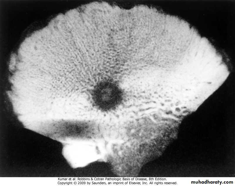
Osteoid osteoma, specimen radiogram (translucent nidus surrounded by sclerotic bone)
Osteoid osteoma
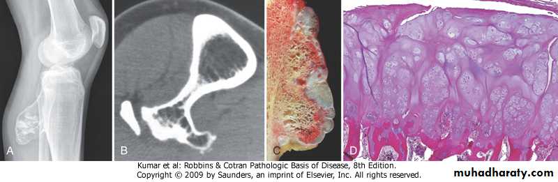
Osteosarcomas:.most common primary malignant tumors of bone, with bimodal age incidence; 1st 2 decades of life, and in elderly secondary to, (Paget disease, bone infarcts, prior radiation).more common in males, arising from the metaphysis of long bones, especially around the knee, presenting as painful mass, or as bone fracture .Codman triangle is a characteristic but not a diagnostic x-ray finding
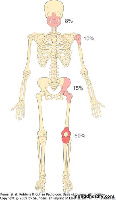
Osteosarcoma, major sites of origin
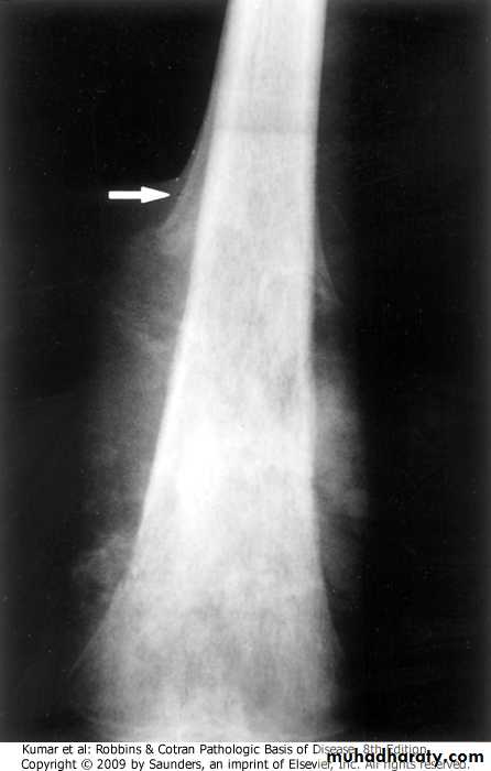
Femur, osteosarcoma, Codman triangle
FIBROUS CORTICAL DEFECTS:.affect up to 50% of children older than 2y.developmental eccentric defects at metaphysis of distal femur and proximal tibia.about one half are bilateral or multiple .about 0.5 cm in diameter, and when they grow to about 6 cm>nonossifying fibromas>pathologic fracture .majority are asymptomatic and undergo spontaneous resolution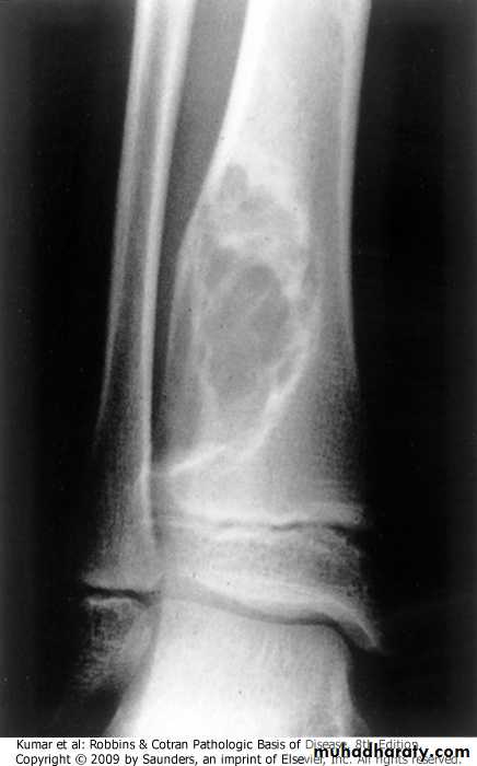
Tibia, fibrous cortical defect
FIBROUS DYSPLASIA:benign tumor that has been related to a localized developmental arrest of bone constituents..there are 3 clinical patterns -monostotic (70%), .minimal symptoms -polyostotic (27%), without endocrine abnormalities .recurring fractures .deformities of long & craniofacial bones .sarcoma-polyostotic (3%) associated with .café au lait skin pigmentations .endocrine abnormalities, especially precocious puberty.medullary in location consisting of woven bone trabeculae without osteo- blastic rimming, embedded in fibrous stroma
FD showing irregular trabecullae (C-shaped or Chinese characters shaped trabaculae, (described as "Chinese letters“) which are usually coarse woven bone, are seen instead of well-organized lamellar bone. with compact stroma of interlacing collagen fibers
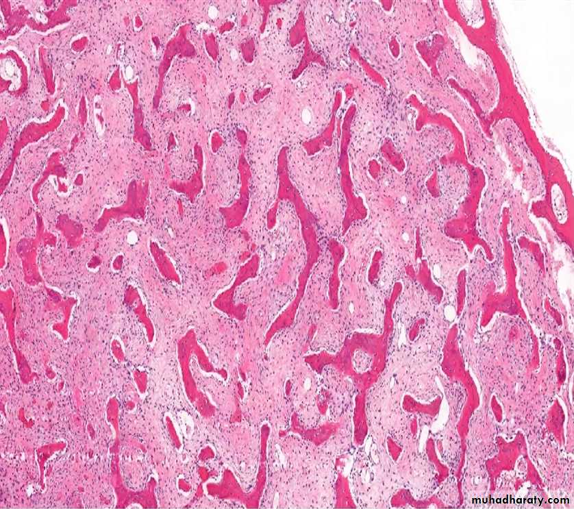
FD. Composed of curvilinear trabeculae of woven bone that lack conspicuous osteoblastic rimming and arise in a background of fibrous tissue.
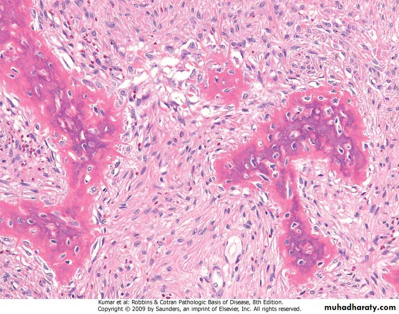
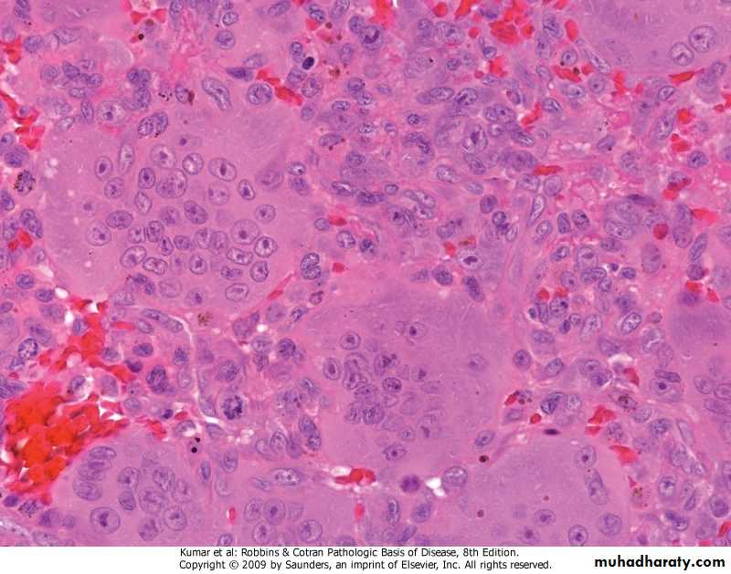
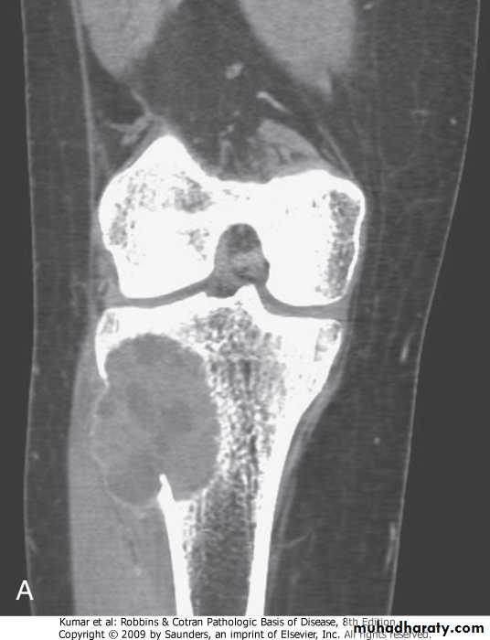
GIANT CELL TUMOR.uncommon locally aggressive tumor .affects people between 20 and 40 years.usuaIly solitary, in adults involves both epiphysis and metaphysis, in adolescents involves metaphysis only, around knee.histology, consists of oval mononuclear and multi-nucleated giant cells .curettage is associated with recurrence
Benign giant cell tumor
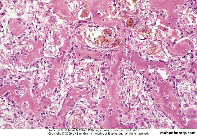
ANURYSMAL BONE CYST:.benign tumor of bone characterized by multiloculated blood-filled cystic spaces.mostly occurs during 1st 2 decades of life, and involves metaphyses of long bones and vertebral bodies.causes pain and swelling, rarely fractures.most lesions are lytic with shell of reactive bone at the periphery
Tibia, aneurysmal bone cyst
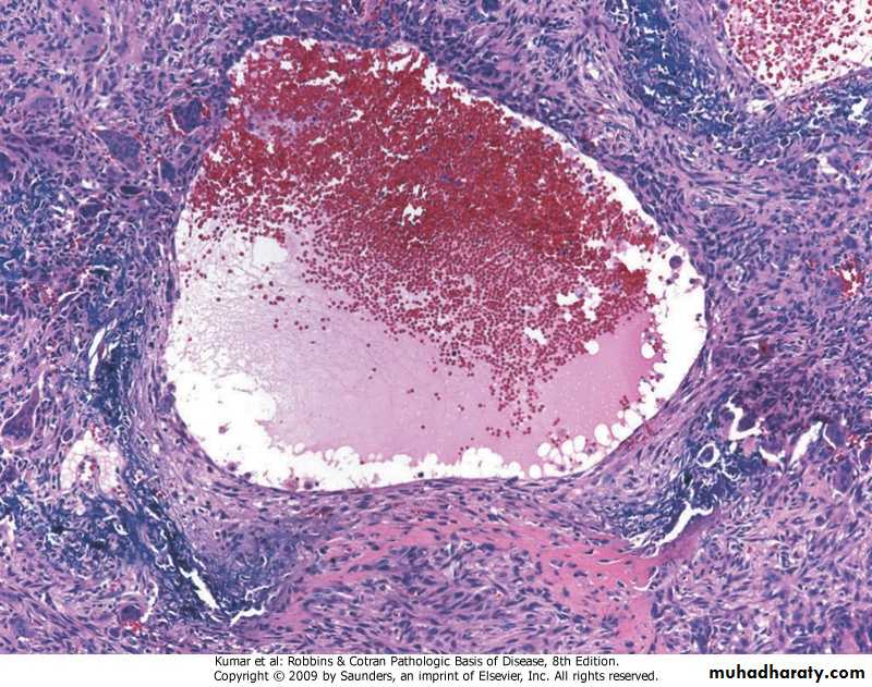
Aneurysmal bone cyst with blood-filled cystic space surrounded by a fibrous wall containing proliferating fibroblasts, reactive woven bone, and osteoclast-type giant cells
METASTATIC TUMORS.far more common than primaries .pathways of spread include -direct extension -lymphatic or hematogenous -intraspinal seeding