1
L3 Gynecology D. Huda Adnan
Congenital malformation of FGT
Normal genetic differentiation
Following fertilization the normal embryo, contain 46 chromosomes including 22 autosomes and
1 chromosome derived from each parent.
46 xx develop as a female and 46 xy develop as a male.
Y chromosome contain a region known as SRY region (sex determining region of the chr. Y), in
males this gene triggers testis formation.
Ovarian differentiation determined by the presence of two x chromosomes and ovarian determinant
gene located on short arm of X chromosome.
Normal anatomical differentiation
Embryologically mesonephric (wollfian) develop into male genital structures
The two-paramesonephric (mullarian) ducts develop as upper female genital structures
including fallopian tubes and after fusion of both tubes develop into uterus, upper third of
vagina.
Urogenital sinus develop into lower two thirds of vagina.
Ovaries differentiated from coelomic epithelium.
Disorders of sexual development
Hearmophroditisim
In which ovarian snd testicular tissue present at the same time ina various degree result in
ambigious genitalia.
Most common karyotyping 46xx but may be 46xx/xy or 46 xy.
Management
The sex will determined at puberty by gender role or functional capability of the external genitalia
and removal of inappropriate organ.
Congenital adrenal hyperplasia
Most common disorder.
Deficiency of 21-hydroxylase enzyme lead to increase 17 hydroxyprogesteron.
Karyotyping 46xx
Autosomal recessive disease.
Present at birth as ambiguous external genitalia like labial adhesion and clitoral
hypertrophy and salts losing syndrome, normal internal organs

2
Management by replacement of mineralocorticoid at birth and cortisol administration
long life.
Turner syndrome
Karyotyping 45O
Phenotype female with features of short stature, webbed neck, and shield shape thorax,
elbow deformity, primary amenorrhea due to gonadal dysgenesis, cardiac and renal
diseases.
Management by hormone replacement therapy by estrogen and growth hormone to
achieve height of 150 cm and breast development and after age of 11 years by estrogen
and progesterone .
Pregnancy can be obtained by ova donation.
5 alpha reductase deficiency
Karyotyping 46 xy
The defect in 5 alpha reductase which responsible for conversion of testosterone into
dihydrotestosterone which is important in development of male external genitalia.
Presented as ambiguous genitalia at birth and some degree of virilization at puberty.
Sex determined by gender role
Androgen insensitivity syndrome
Karyotyping 46 xy
Receptors not respond to testosterone hormone .
Presented with female external genitalia at birth and absence of menstruation (no uterus)
and spare axillary and pubic hair at puberty, blind end vagina, with presence of testis in
the abdomen or inguinal region.
Management by gonadectomy after puberty because of risk gonadoblastoma and creation
of vagina for sexual satisfaction.
Mayar-Rokatinsky-Kuster-Hauser syndrome
Karyotyping 46 xx
Mullarian agenesis result in absent uterus and tube and upper vagina.
Female present with normal external genitalia ,blind end vagina and normal secondary
sexual charechteristices with primary amenorrhea.
Associated with urinary tract anomalies in 40% and skelatal abnormality in 25%.
Management by psychological counselling,vaginal cration and pregnancy by surrogate
uterus.
Other structural malformation
Imperforated hymen due to incomplete canalization of vagina, managed by crciate
incision of hymen.
3
Septate uterus due to incomplete failure of fusion of two mullarian ducts may present
with recurrent miscarriage and malpresentatin during labor and management by removal
of septum
Uterine didyphylus dut to complete failure of fusion of two mullarian ducts (pregnancy
occur in one cavity)
Bicornuate uterus when there is deep indentation of fundus present with recurrent
miscarriage and malpresentation.
Unicornuate uterus when there is failure of development of one mullarian duct
associated with recurrent miscarriage and ectopic pregnancy.
Longitudinal vaginal septum presented with dyspareunia managed by removal of
septum.
Transverse vaginal septum presented with primary amenorrhea managed by surgical
removal of septum.
Slide
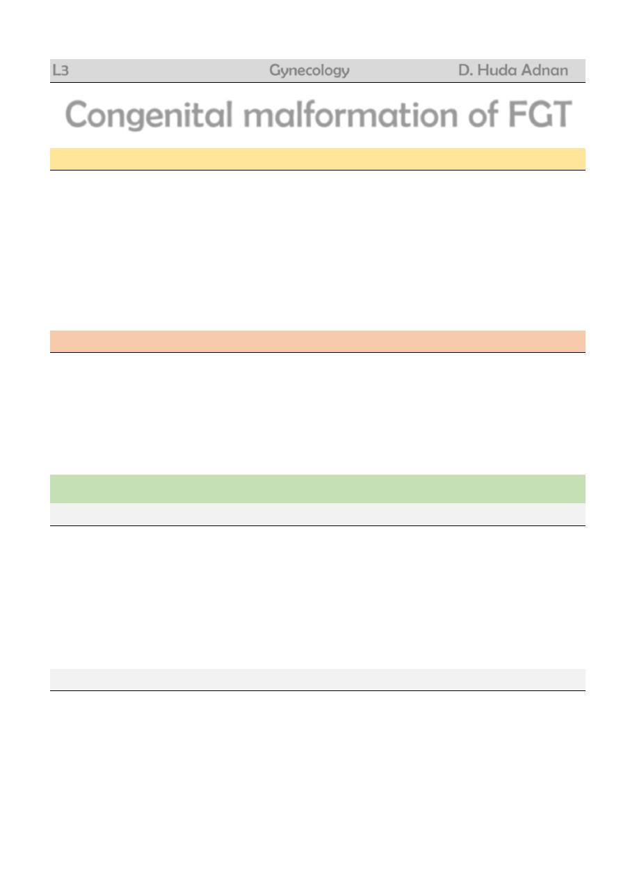
The girl shown in the picture of 16 yers and 140 cm
length old age present with delayed onset of
menarche
A. What is the most likely diagnosis?
B. What do you suspect the result of karyotyping?
C. Give 4 main other clinical features of the
condition.
D. How can this patient get pregnant in the
future?
Slide
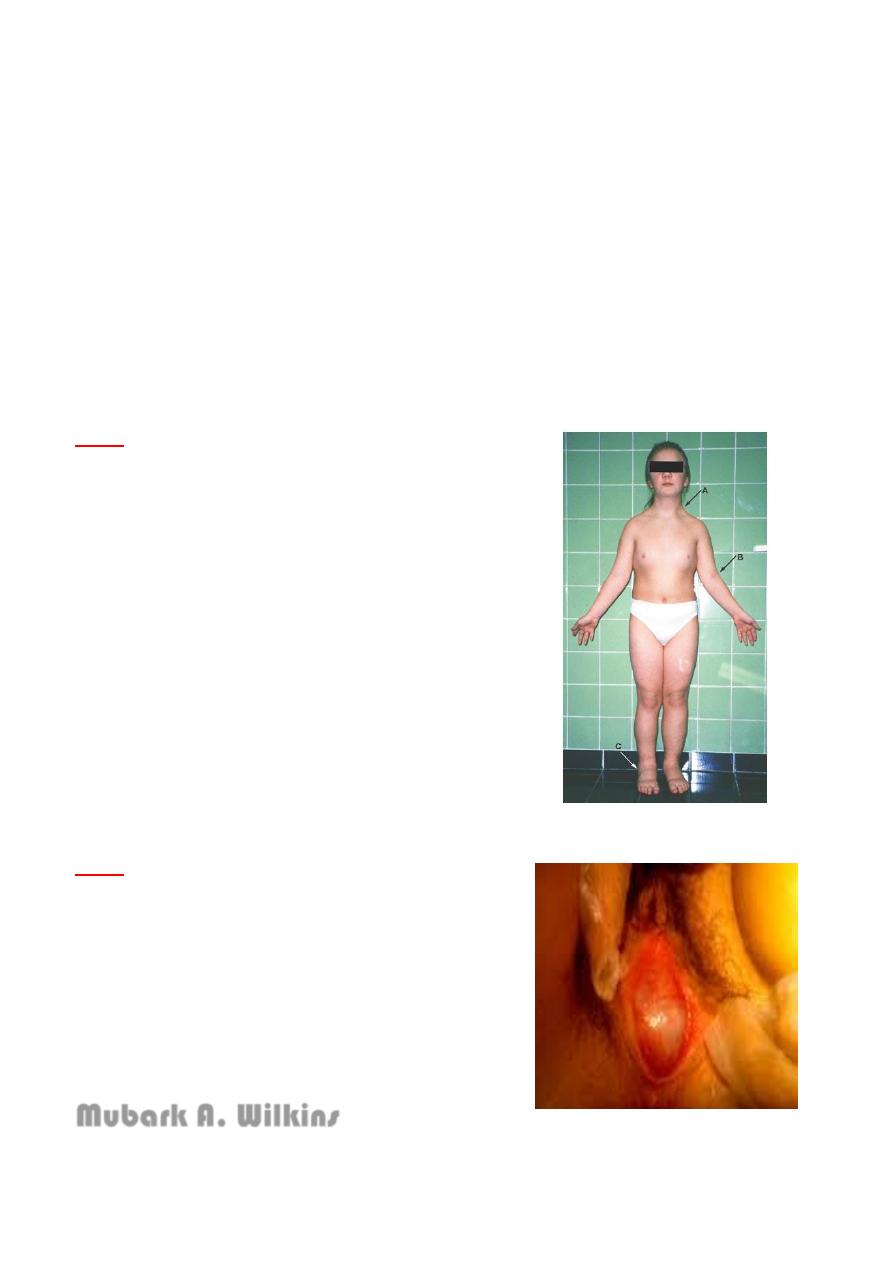
A 14 years old girl present with periodic abdominal
pain and delayed onset of puberty, perineal
examination showing in this picture.
A) What is the diagnosis?
B) What are other clinical findings you suspect?
C) What are the ultrasonic findings you suspect?
D) What is the management?
Mubark A. Wilkins