HEMORRHAGIC CEREBROVASCULAR DISEASE
15% of stroke are caused by hemorrhage10% are intra-cerebral hemorrhage
5% are subarachnoid hemorrhage
Intracerebral hemorrhage
Causes1-Hypertension
2-Amyloid (congophilic) angiopathy
3-Arteriovenous malformation
4-Bleeding diathesis
5-Drugs (amphetamines, cocaine, anticoagulants, thrombolytics)
6-head injury
7-tumour
In hypertensive ICH
Rupture of microaneurysms (Charcot-Bouchard aneurysms, 0.8-1.0 mm diameter) and degeneration of small deep penetrating arteries are the principal pathology.Such hemorrhage is usually massive, often fatal and occurs in chronic hypertension and at well-defined sites - basal ganglia, pons, cerebellum and subcortical white matter .
In normotensive patients
particularly over 60 years, lobar intracerebral hemorrhage occurs - in the frontal, temporal, parietal or occipital cortex.
Cerebral amyloid angiopathy (rare) is the cause in some of these hemorrhages, and the tendency to re-bleed is associated with particular apolipoprotein E genotypes
Clinical Manifestations
Although not particularly associated with exertion, intracerebral hemorrhages almost always occur while the patient is awake and sometimes when stressed.The hemorrhage generally presents as the abrupt onset of focal neurologic deficit. Seizures are uncommon. The focal deficit typically worsens steadily over 30–90 min and is associated with a diminishing level of consciousness and signs of increased ICP, such as headache and vomiting
Putamen hemorrhage
The putamen is the most common site for hypertensive hemorrhage, and the adjacent internal capsule is usually damaged Contralateral hemiparesis is therefore the sentinel sign..When hemorrhages are large, drowsiness gives way to stupor as signs of upper brainstem compression appear. Coma ensues, accompanied by deep, irregular, or
intermittent respiration, a dilated and fixed -pupil, and decerebrate rigidity. In milder cases, edema in adjacent brain tissue may cause progressive deterioration over 12–72 h.
Pontine hemorrhage
In pontine hemorrhages, deep coma with quadriplegia usually occurs over a few minutes.There is often prominent decerebrate rigidity and "pin-point" (1 mm) pupils that react to light., severe hypertension, and hyperhidrosis are common.
Death often occurs within a few hours, but small hemorrhages are compatible with survival
Cerebellar haemorrhage
There is headache and rapid reduction of consciousness with signs of brainstem origin (e.g. nystagmus, ocular palsies). Gaze deviates towards the haemorrhage. Skew deviation may develop.There are unilateral or bilateral cerebellar signs, if the patient is awake.
Cerebellar haemorrhage sometimes causes acute hydrocephalus. Emergency surgical clot evacuation is often necessary after imaging
Diagnosis
Intracerebral hemorrhage often cannot be distinguished from other types of strokes based on clinical findings alone.
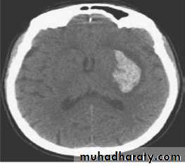
The test of choice for making the diagnosis is a non– contrast-enhanced CT scan that shows areas of hemorrhage as zones of increased density, which may or may not have associated regions of decreased density indicating infarction.
Treatment
General MeasuresWith initial care directed at maintenance of the airway, oxygenation, nutrition, and prevention and treatment of secondary complications.
Optimal blood pressure treatment is uncertain, although the general guidelines for excessive hypertension and reduction of cerebral perfusion apply as for ischemic strokes
There is no accepted protocol for the management of increased intracranial pressure; osmotherapy, hyperventilation, and neuromuscular paralysis rarely are beneficial.
Fluid management should maintain euvolemia;
fluid
restriction or volume expansion is not of proven value.
Seizures are particularly harmful in critically ill patients and are treated despite lack of data from randomized trials.
Maintenance of normal body temperature is theoretically desirable because fever may accelerate tissue destruction
Medical Therapy
Intravenous administration of recombinant factor VIIa within 4 hours after onset reduces the volume of hemorrhage and surrounding cerebral edema, as measured by CT, and improves neurologic outcomes at 90 days despite an increase in ischemic stroke and myocardial infarction [. 2]
Corticosteroids may increase the risk for infectious complication
Surgical Therapy
The goal of surgical treatment of intracerebral hemorrhage is to remove as much blood clot as possible as quickly as possible. Ideally, surgery should remove the underlying cause, such as an AVM, and prevent hydrocephalus. Early surgical intervention to evacuate intracerebral hematomas within 24 hours is no better than medical therapy .However, patients who have cerebellar hemorrhages and are deteriorating because of brain stem compression and hydrocephalus caused by ventricular obstruction are still recommended by some for removal of the clot or amputation of part of the cerebellum, although no proof exists to support this approach