Endocrine System 1
Third Year Class
By Dr. Riyadh A. Ali
Department of Pathology
TUCOM
NORMAL THYROID
NORMAL THYROID
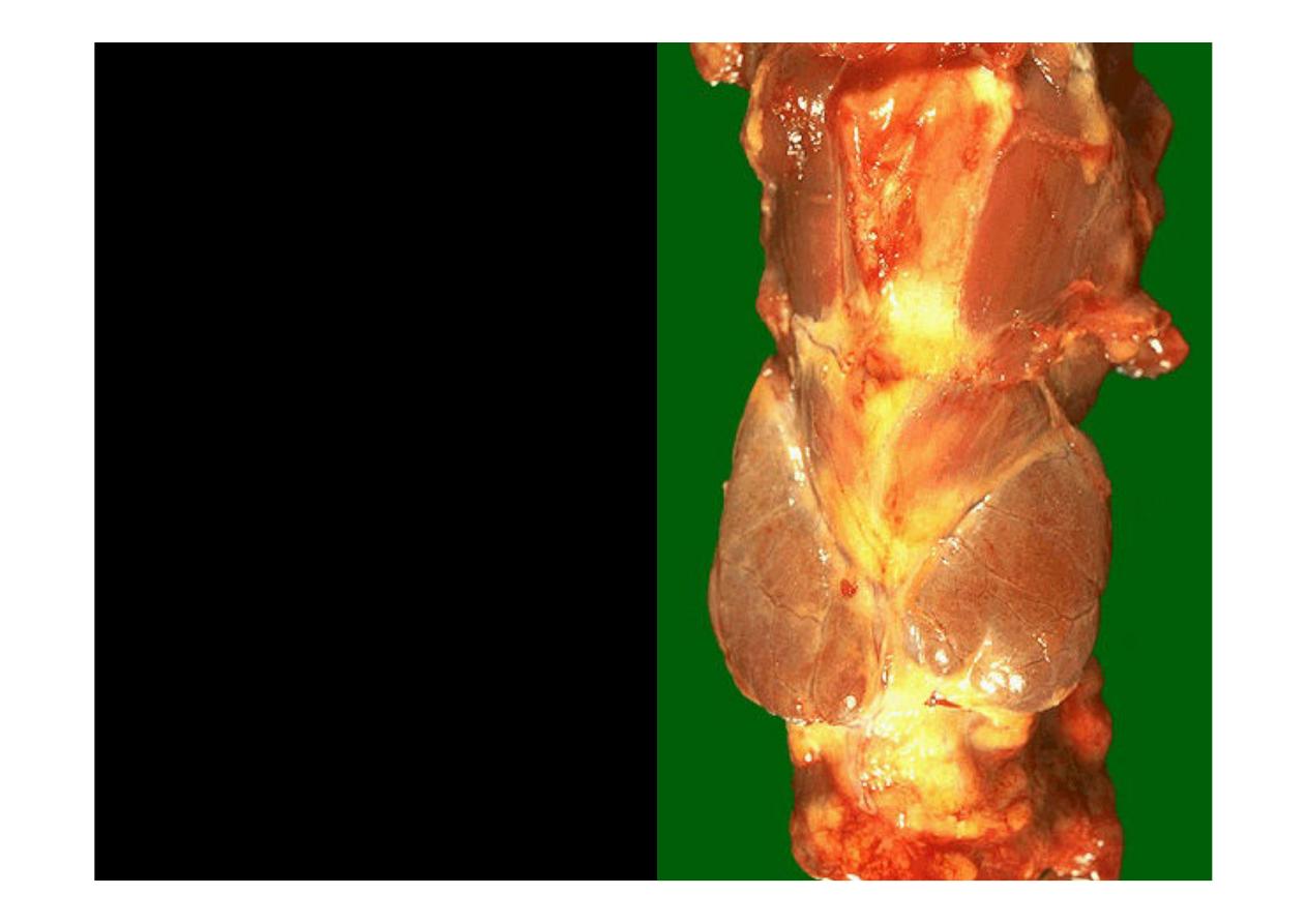
This is the normal
appearance of the
thyroid
gland on the anterior trachea
of the neck. The thyroid
gland has a right lobe and a
left lobe connected by a
narrow isthmus. The normal
weight of the thyroid is 10 to
30 grams
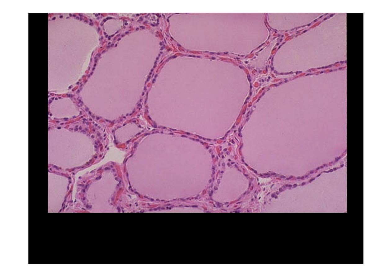
Normal thyroid
seen microscopically consists of follicles lined by a an
epithelium and filled with colloid. The follicles vary somewhat in size. The
interstitium, which may contain "C" cells, is not prominent.
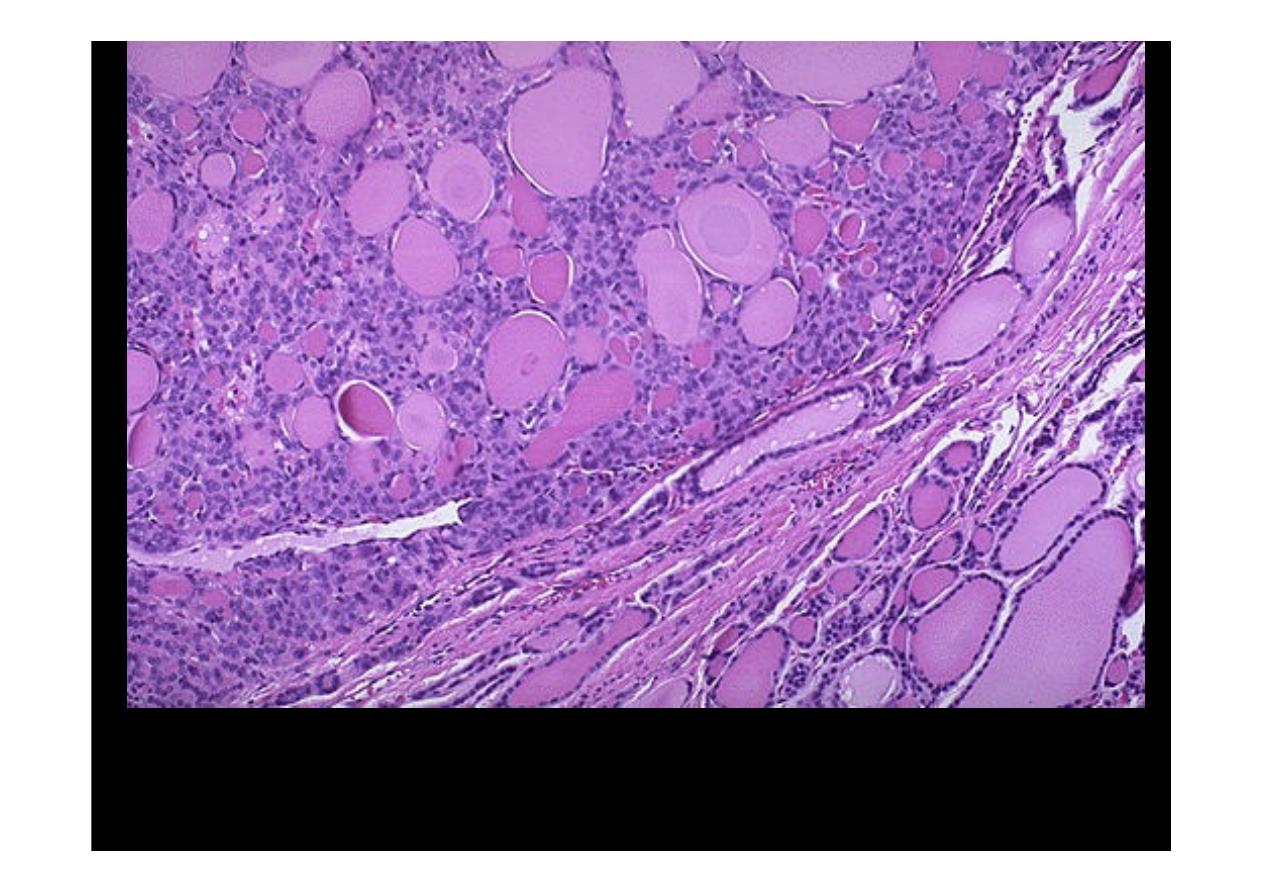
Hashimoto's Thyroiditis
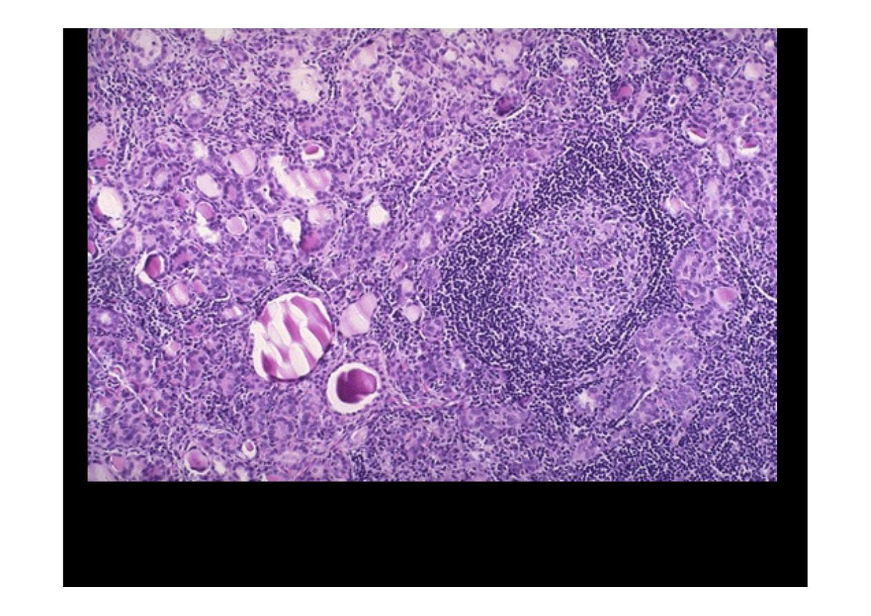
Low power microscopic view of a thyroid with
Hashimoto's
thyroiditis
. Note the lymphoid follicle at the right center
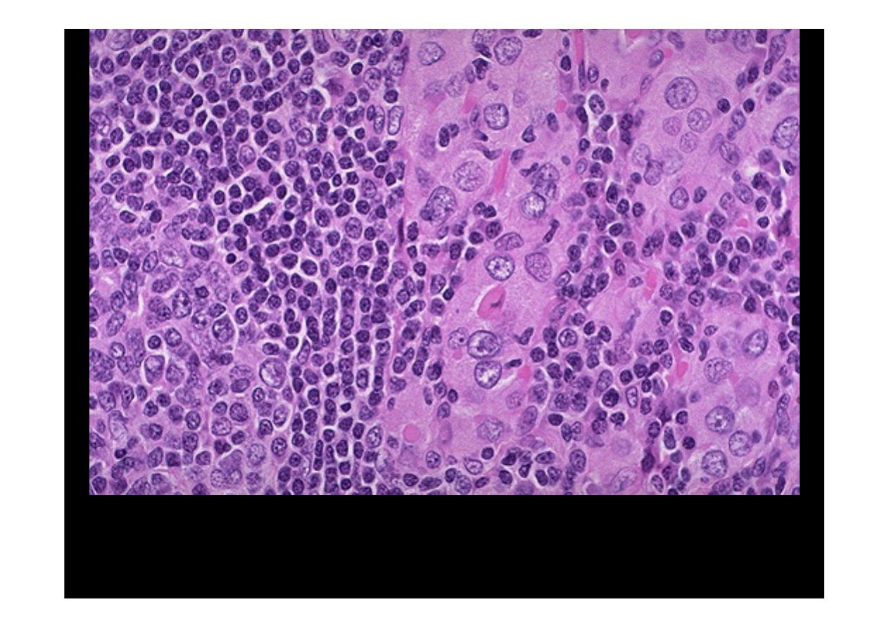
Hashimoto's thyroiditis
demonstrates the pink Hurthle cells
at the center and right. The lymphoid follicle is at the left.

Nodular Goiter
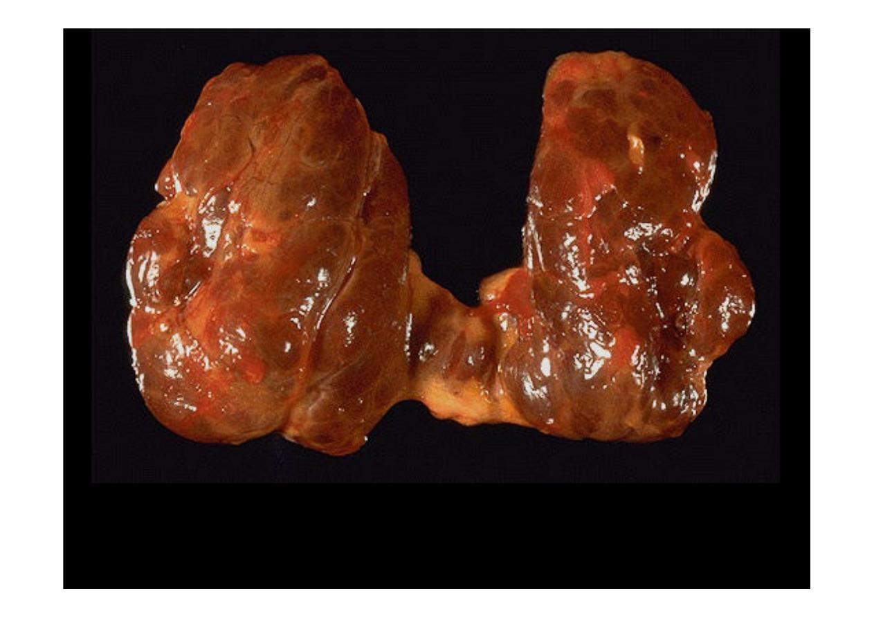
This diffusely enlarged thyroid gland is somewhat nodular,
thyroid--a nodular goiter
Grave's
Disease
A diffusely enlarged thyroid gland associated with hyperthyroidism is
known as
Grave's disease
. At low power here, note the prominent
infoldings of the hyperplastic epithelium
At high power, the tall columnar thyroid epithelium with
Grave's disease
lines the
hyperplastic infoldings into the colloid. Note the clear vacuoles in the colloid next to the
epithelium where the increased activity of the epithelium to produce increased thyroid
hormone has led to scalloping out of the colloid
Follicular Neoplasm
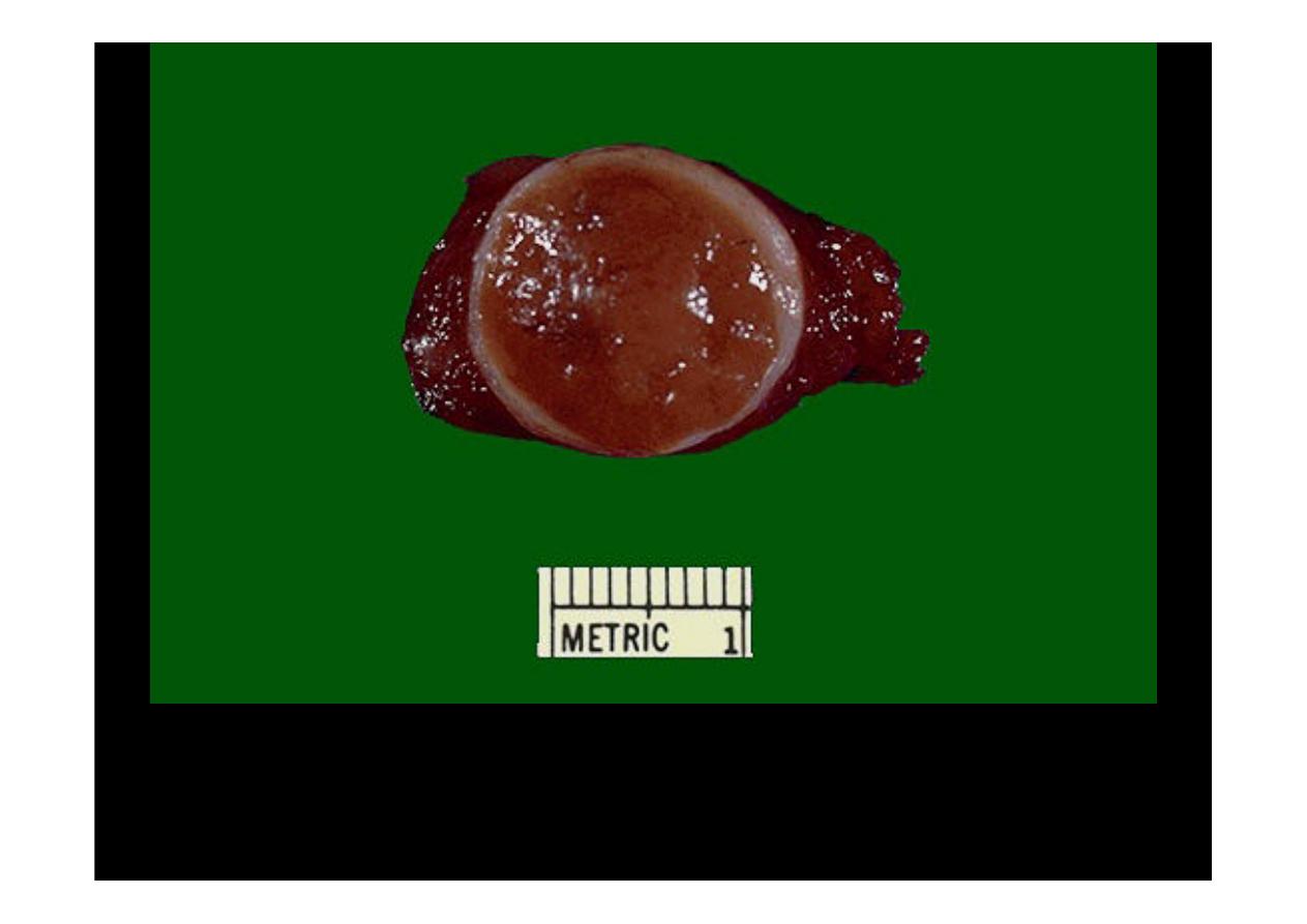
This is
follicular neoplasm
(a follicular adenoma histologically)
that is surrounded by a thin white capsule.
Normal thyroid follicles appear at the lower right. The
follicular adenoma
is at the center to
upper left. This adenoma is a well- differentiated neoplasm because it closely resemble normal
tissue. The follicles of the adenoma contain colloid, but there is greater variability in size than
normal.
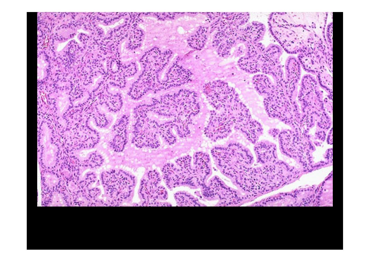
Papillary Carcinoma
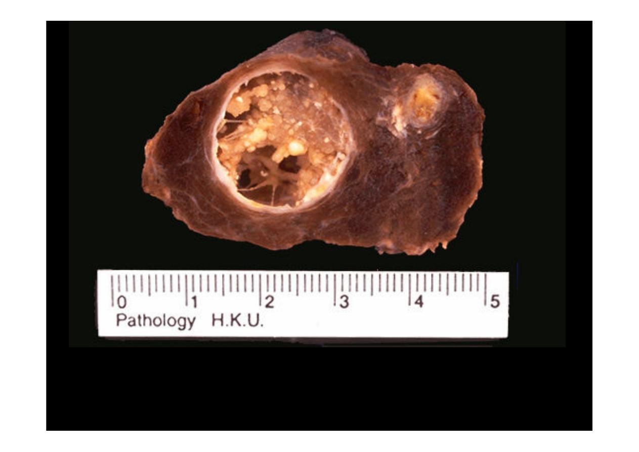
Papillary Carcinoma
. The larger mass is cystic and contains
papillary excresences.
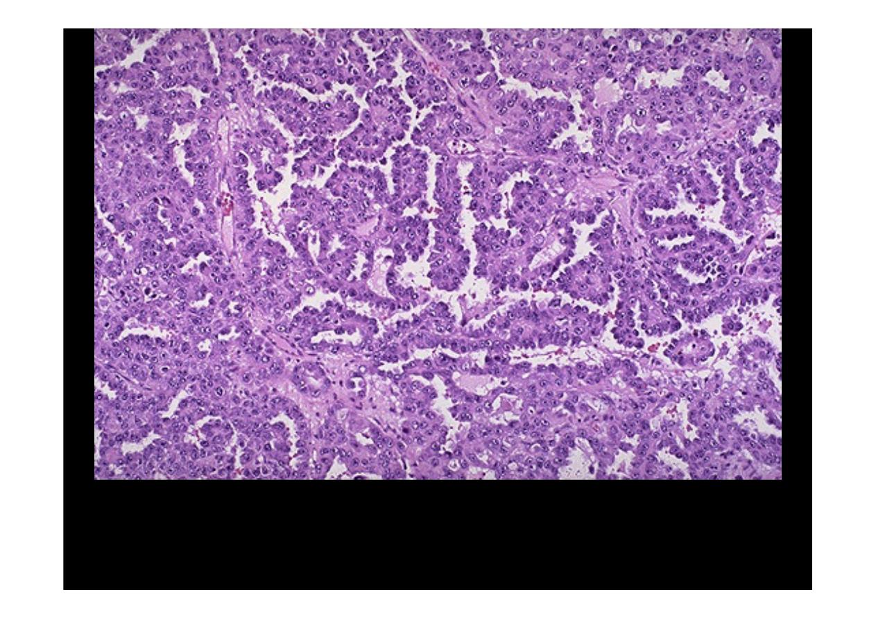
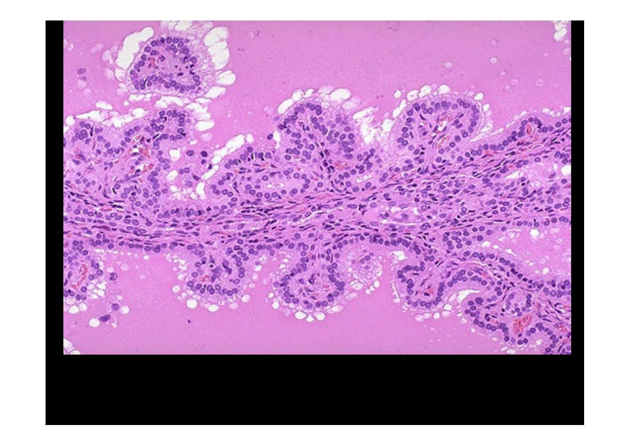
A
papillary carcinoma
of the thyroid. The fronds of tissue have
thin fibrovascular cores. The fronds have an overal papillary
pattern.
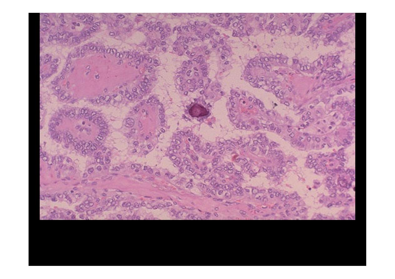
This is
papillary carcinoma
of thyroid. small psammoma body in the center.
The cells of the neoplasm have clear nuclei.