Gastrointestinal Radiology
Dr wasan Ali(C.A.H.S)
Department of Radiology
Medical collegue of MosulSmall& large bowel Radiology
Learning objectivesRevision of their radiological anatomy.
To be aware of the common imaging modalities available to evaluate them.
Recognize radiological features of common disorders related to small& large bowel .
Anatomy
Large bowelperipheral
haustra
Small bowel
central
valvulae conniventes
Stomach
Liver
Pancreas
Ascending colon
Transverse colonStomach
Descending colon
Sigmoid colon
RectumHaustra
Valvulae conniventes:
mucosal folds thatgo right across small bowel
peripheral
haustra± faeces
larger calibre (>5cm<9cm)
central
valvulae conniventes
no faeces
smaller calibre
(2-2.5cm)
Large versus small bowel on AXR
Large bowel Small bowel
What are the common imaging modalities available to evaluate the bowel?
Plain film (AXR)
Barium and contrast studiesUltrasound
Computed tomography
Angiography
Imaging modalities
Limitations:
cannot evaluate bowel mucosacannot assess bowel motility
cannot determine level of obstruction accurately
cannot demonstrate the site of perforation/leaks
Plain AXR
Can provide radiological signs for pneumoperitonuem
Barium and water-soluble contrast media (contains Iodine) are dense elements which appear white on X-rays
Coat and show bowel mucosa
Demonstrate bowel motility
Demonstrate site of obstruction and leaks
Barium and contrast studies
Barium studies
Ba swallowBa meal
Ba follow through
Small bowel enema
Barium enema
Water soluble contrast GIT studies
Used when barium is contraindicated:Suspected perforation
Check anastomosis post-surgery
What imaging studies can be performed to image the small bowel?
Barium follow-through: performed at the end of either barium meal or swallow examination
Small bowel enema: barium is passed directly into the small bowel via a catheter inserted via the nose/mouth
Small bowel evaluation
Indication of BA follow through examination :
• _1)Inflammatory bowel disease (CD & UC)• _2)Mal absorption syndromes
• 3)swelling and/or inflammation of the small intestine walls
• 4)tumors
• 5)Ulcer
• Contraindications for a barium follow through may include:
• 1)suspected bowel perforation
• 2)bowel obstruction
• 3)conditions where aspiration of barium is likely
Feathery appearance
Valvulae conniventes
Small bowel enema
Tube in 4th part of duodenum
Better distension and delineation of small bowelBarium enema
IndicationsAlteration in bowel habit
Chronic diarrhoea or constipation
Rectal bleeding
Abdominal pain
Suspected abdominal mass
Obstruction
How is it done?
Barium enema
The colon should be cleaned by laxative or enema.Patient lies on X-ray couch
Rectal tube is inserted
Barium ± air is then infused
X-rays are taken with patient in different positions
Barium enema
Rectal catheterB
A
C
D
E
HF
SF
F
B
A
C
D
E
HF
SF
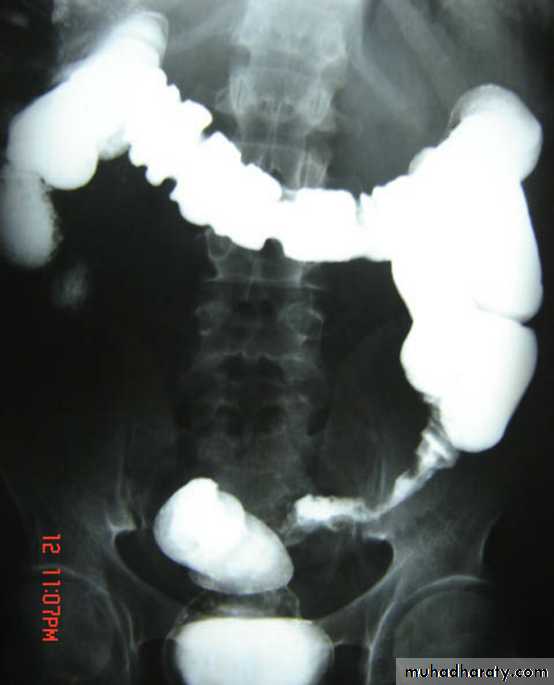
Barium enema
A: caecumB: ascending colon
C: transverse colon
D: descending colon
E: sigmoid colon
HF: hepatic flexure
SF: splenic flexure
F: terminal ileum
F
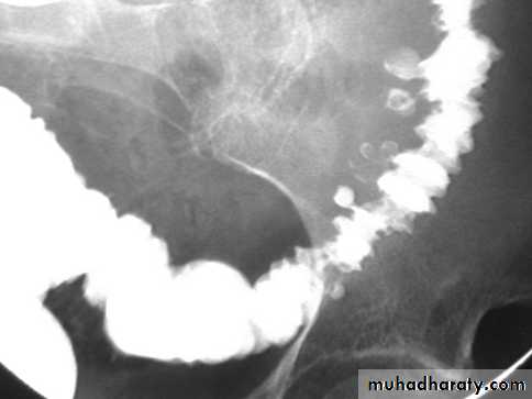
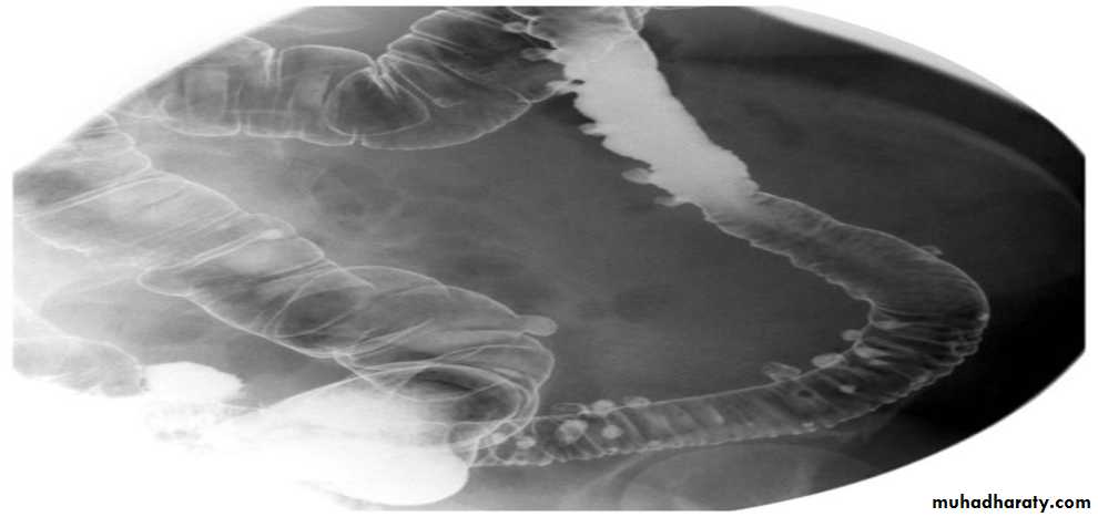
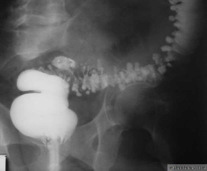
Crohns Disease :
• Crohn disease remains idiopathic• Radiographic features
• The characteristic of Crohns disease is the
• presence of :
• _skip lesions
• _multiple discrete ulcers.
• _The frequency with which various parts of the gastrointestinal tract are affected varies widely :
• *small bowel: 70-80%
• *small and large bowel: 50%
• *large bowel only: 15-20%
Radiological finding of CD in BA follow through :
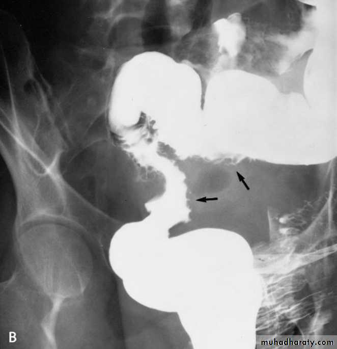
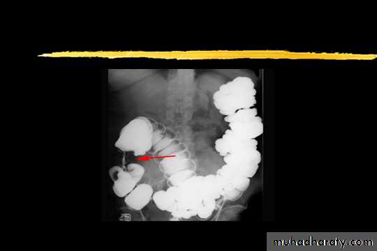
• _Multiple mucosal ulcers aphthous ulcers.• _Transmural ulcer (Rose thorn appearance )
• _longitudinal fissures
• _Multiple skip lesions
• _when severe leads to cobblestone appearance
• _May lead to sinus tracts and fistulae
• _widely separated loops of bowel due to fibro-fatty proliferation
• _Thickened folds due to edema.
• _Pseudo diverticula formation: due to contraction at the site of ulcer with ballooning of the opposite site.
Crohns disease
CD
CD
CD
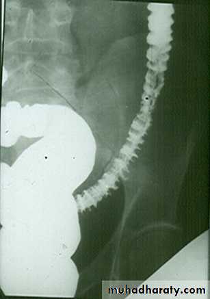
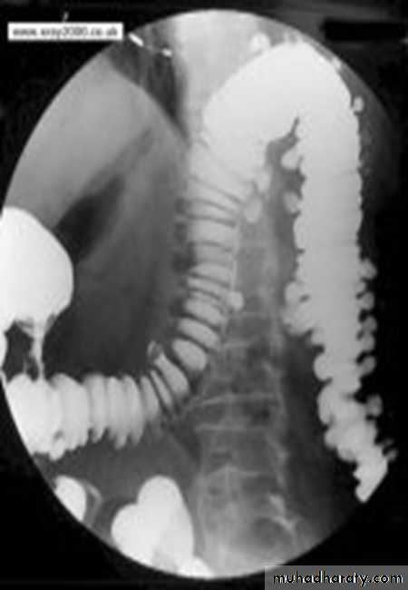
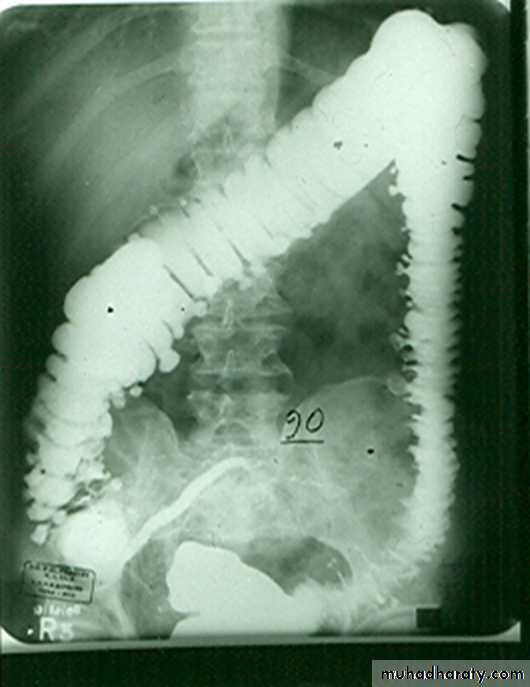
Ulcerative colitis(UC)
• _is an inflammatory bowel disease which predominantly affects the colon, but also has extra intestinal manifestation• _Involvement of the rectum is almost always present (95%) , with the disease involving variable amounts of the more proximal colon, in continuity. The entire colon may be involved, in which case edema of the terminal ileum may also be present (so-called back-wash ileitis).
• _The ulcer are huge in number and super fiscial and healed by diffuse fibrosis of the colon .
• _In very severe cases, the colon becomes atonic, with marked dilatation, worsened by bacterial overgrowth. This leads to toxic mega colon which although uncommon has a poor prognosis..
Ulcerative Colitis
• Plain film• Non specific but may show evidence of mural thickening (more common), with thumb printing also seen in more severe cases
• Ba enema
• Double contrast barium enema allows for detail of the colonic mucosa, and also allows bowel proximal to strictures to be assessed. It is however contraindicated if acute severe colitis is present due to the risk of perforation.
• Mucosal inflammation lends a granular appearance to the surface of the bowel. As inflammation increases , the bowel wall and haustra thicken.
• Mucosal ulcers are undermined (button-shaped ulcers). When most of the mucosa has been lost, islands of mucosa remain giving it a pseudo-polyp appearance.
• In chronic cases the bowel becomes featureless with loss of normal haustral markings, luminal narrowing and bowel shortening (lead pipe sign).
Radiological features of UC :
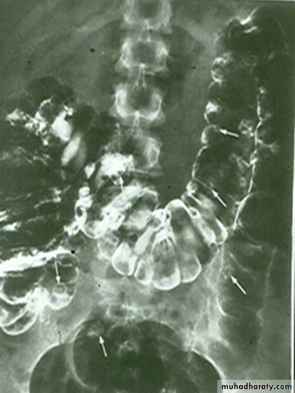
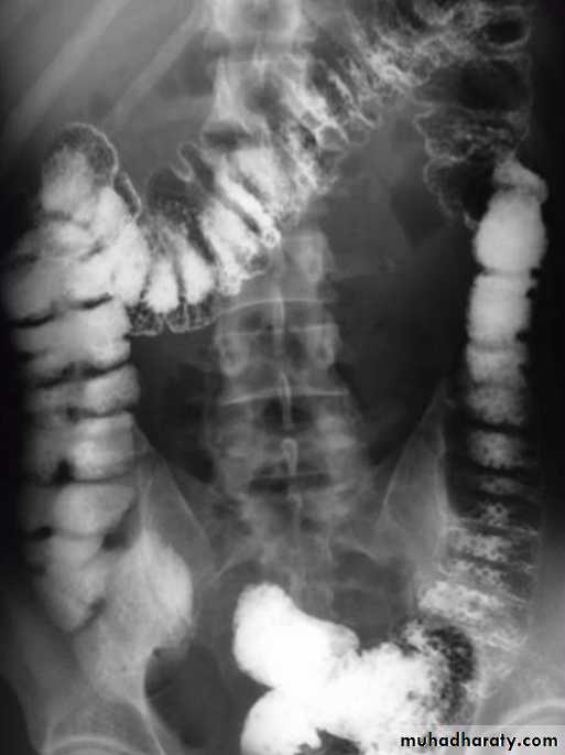
• Plain film : non specific ,may show marked dilatation of colon due to acute toxic dilatation ( fulminating case ), mural thickening with thumb printing in more sever cases Ba. Enema : usually double contrast allow for detail of colonic mucosa and contra-indicated in toxic dilatation due to the risk of perforation :1)- Widening of pre-sacral space ( Lat. View ).2)-The mucosal ulcers are undermined (Button _shaped ulcer) when most of the mucosa has been lost , island of mucosa remian giveing it ( a pseudo polyp) apperance .3)- Loss of haustration .4)- Narrowing of the colon , in early stage due to spasm , later on due to fibrosis .
• 5)- Shortening of the colon due to fibrosis 6)- In late stage combination of (3 ,4 ,and 5) give rise to Lead pipe appearance .7)- Cobble – stone mucosa due to pseudo-polyposis .8)- Constant narrowing not relieved by relaxant indicate either stricture or malignant changes.
UC
The colon is distended with air
The descending & sigmoid colon are featureless (no haustral pattern)
•
Thumb printing
Complication OF UC
• Colorectal carcinoma in the setting of ulcerative colitis is more frequently sessile and may appear to be a simple stricture.• toxic megacolon (TM) is complication that can be seen in both types of inflammatory bowel disease more in UC , in infectious colitis, as well as in some other types of colitis.
Radiological features of toxic megacolon :
• The colon (typically transverse colon) becomes dilated to at least 6 cm (usually greater). There is additional loss of haustral markings
• It is serious acute abdominal condition
• More in UC > CD
• Practical points
• barium studies and colonoscopy should be avoided, due to the risk of perforation
Toxic megacolon
toxic mega colon
• Crohn's disease vs. ulcerative colitis
• Due to the overlap in clinical presentation of Crohn's disease (CD) and ulcerative colitis (UC), imaging often has a role to play in distinguishing the two. Distinguishing features include:• bowel involved
• CD: small bowel 70-80%, only 15-20% have only colonic involvement
• UC: rectal involvement 95%, with terminal ileum only involved in pancolitis (backwash ileitis)
• distribution
• CD: skip lesions typical
• UC: continuous disease from rectum up
• gender
• CD: no gender preference
• UC: male predilection
• Terminal ileum involvement
• UC: involved ( terminal ileitis )
• CD: un common, backwash ileitis
Lymphoma of small bowel
• Splaying & separation of the bowel loops due to enlarged LN• Thickening of the mucosa , irregular in outline ( saw tooth pattern ) .
• LATER stage could be present as sign of Malabsorption syndrome ( flocculation & segmentation of the Ba ) .
Mal absorption syndrome
TUMOUR OF THE COLON
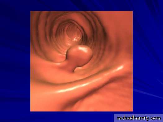
• Colonic Polyp:_Solitary or multiple._Produce small , well-defined rounded filling defect best seen in double contrast enema or post-evacuation enema ._ May be sessile or pedunculated ._ The pedicle appear as two parallel line of filling defect (stalk) and shows change of position with change of posture of patient .Familial adenomatosis polyposis syndrom (FAPS)
• The condition is familial .• Its a predesposition to colonic carcinoma .
• The colon shows multiple filling defects through out its length of different sizes.
• Imaging usually under estimates the number of polyps because most of them< 5mm in size .
• Signs of malignant changes can be seen.
colorectal CA
• Is the most common cancer of the gastrointestinal tract and the second most frequently diagnosed malignancy in adults.• Colorectal cancers can be found anywhere from the caecum to the rectum, in the following distribution :
• _recto-sigmoid: 55%
• _caecum and ascending colon: 20%
• _ileocaecal valve: 2%
• _transverse colon: 10%
• _descending colon: 5%
Ba enema
• Morphological types:• 1_ Ulcerative : give rise to irregularity of the colon with ulceration.
• 2_ Constrictive or infiltrative ( Annular ) type :
• a- Constant narrowing .
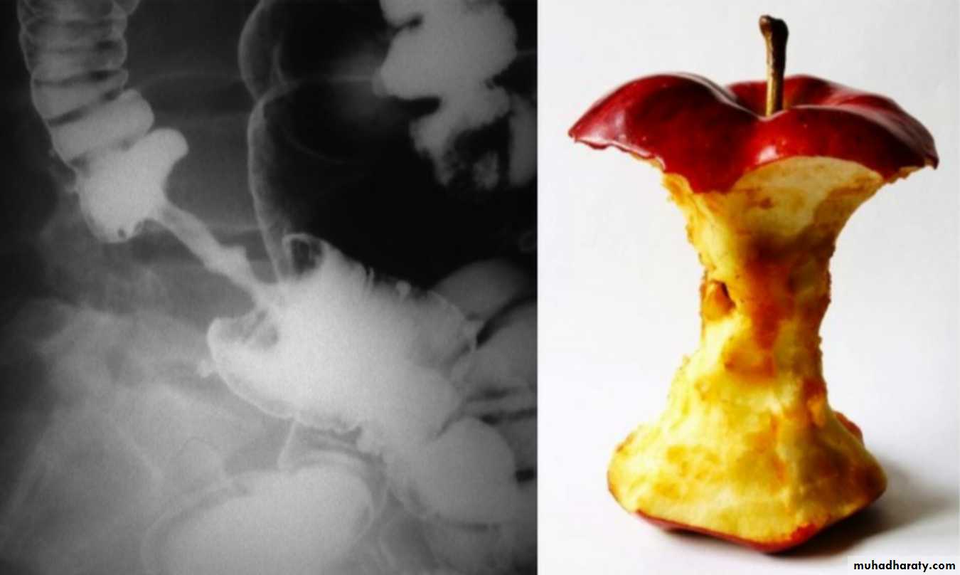
• b-Shouldering sign, apple core sign .
• c- Destruction of mucosa at narrow area .
• d- Double track due to fistula .
• e- In severe constriction ; stoppage of Ba. Flow with proximal dilatation .
3_Proliferative type : give rise to :
a- Large , constant filling defect with irregular margin .
b- Destruction of mucosa .
c- Intestinal obstruction
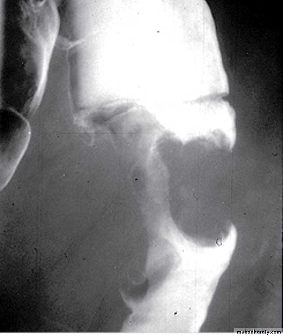
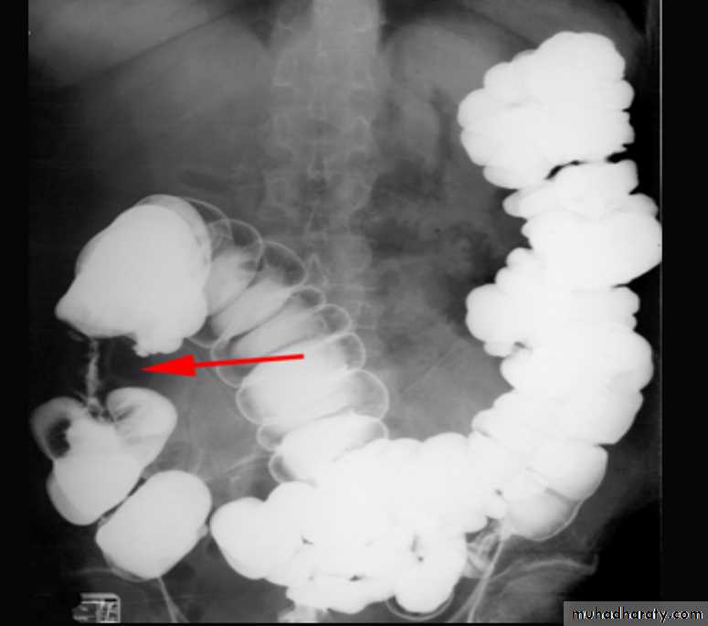
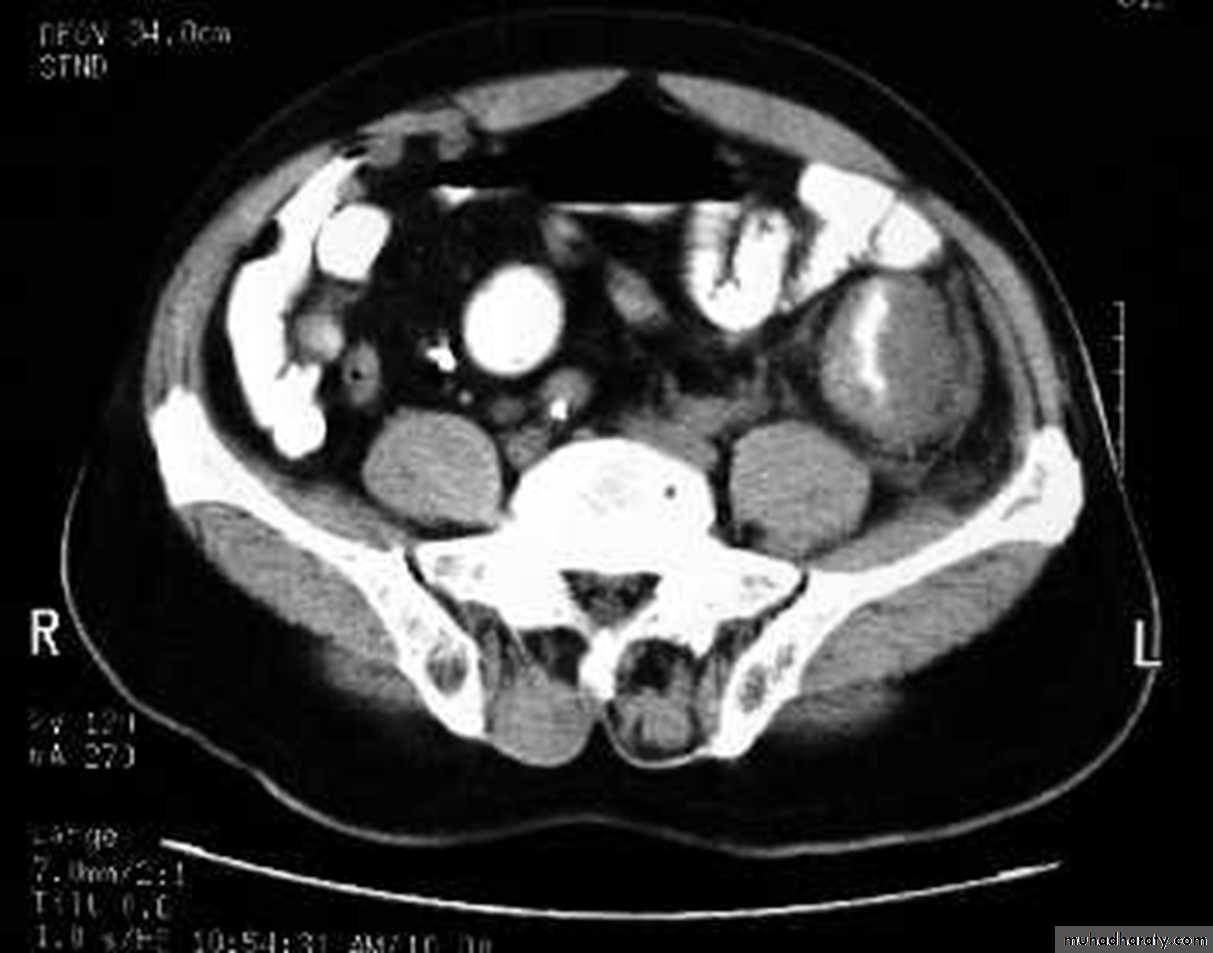
apple core lesion in the descending colon
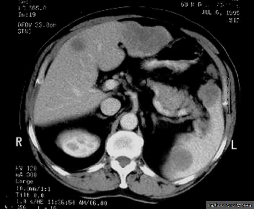
Multiple lesions are seen in the liver and spleen consistent with metastases
•Diverticular disease of the colon
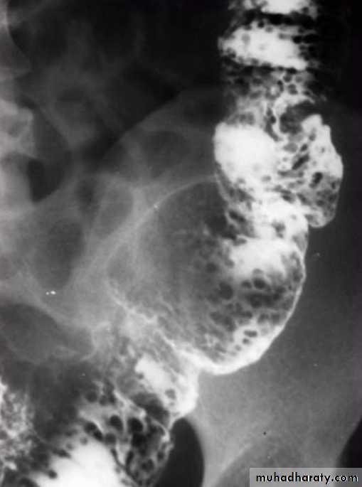
• Due to over activity and hypertrophy of transverse bands of smooth muscle which result in increased intra-luminal tension with herniation of mucosa through week point in the wall.The condition is commonly seen in left hemi-colon.Radiological appearance:Plain film: show air trapping .• Ba. Enema :
• *Early appearance show saw-teeth colon (pre-diverticular stage).• *The diverticulum is consist of body and neck , best seen in post evacuation film .
• *Faecal impaction may result in incomplete filling of the body or flask shape .
• Complication of diverticular disease
• 1- Diverticulitis(inflammation & infection). 2- Ulceration and haemorrhage . 3- Perforation …. Pneumo-peritonium . 4- Fistula … air fluid level in bladder , or sinus track . 5- Peri-colic abscess….localized narrowing . 6- Intestinal Obstruction …. Air fluid level . 7- Malignant change …. Irregular narrowingcongenital anomaly of the colon:
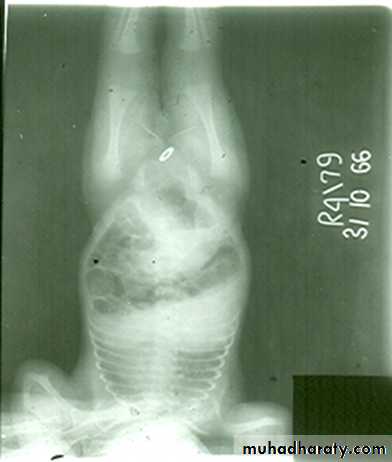
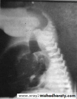
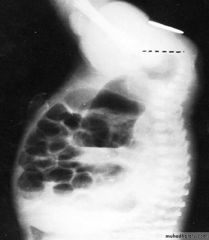
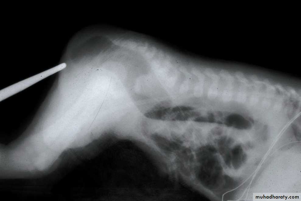
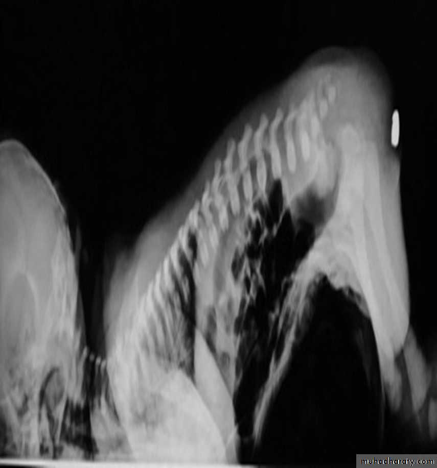
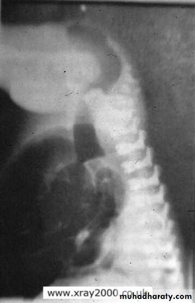
• 1- Anal Atresia ( imperforated Anus).2-Meconium ileus ( neonatal obstruction).3-Duplication.4-Malrotation .5-Mega colon ( Hirschsprung’s disease ,aganglionic colon ).Imperforated anus
• Clinically the newborn delivered with abdominal distension and failure to pass meconium .• The anomaly vary from thin mucosal membrane to severe developmental anomalies of rectum and according to that there are two types :-
• A- Low type ( perineal approach ).
• B- High type (Abdominal approach ).
• The value of radiological examination to assess these types prior to surgery .
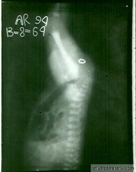
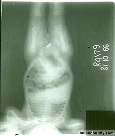
• Technique of Examination :• 1- The examination should be done at least 12-18 hours after delivery to allow time for swallowed air to reach the rectum .
• 2- The anal dimple is identified by metallic marker
• 3- AP and Lateral views are taken with inverted position(invertogram) .
• Signs of low atresia :
• 1- Multiple air fluid level due to obstruction .
• 2- In AP view the distance between the terminal rectal segment and the marker should not exceed 2 cm.
• 3- In lateral view the terminal air shadow is seen above the pubo-coccygeal line.
• Signs of high atresia
• 1- In AP view the distance more than 2 cm .• 2-In lateral view the terminal air is below the pubo-coccygeal line .
Low type
High type
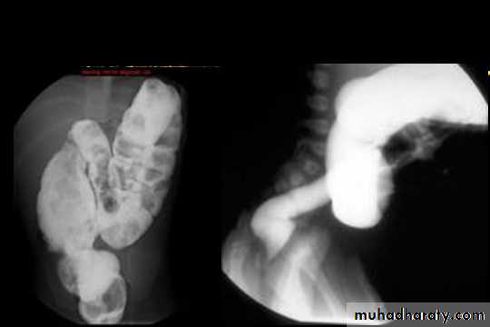
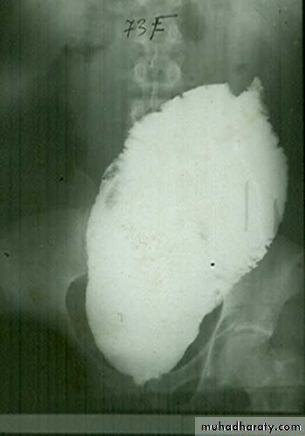
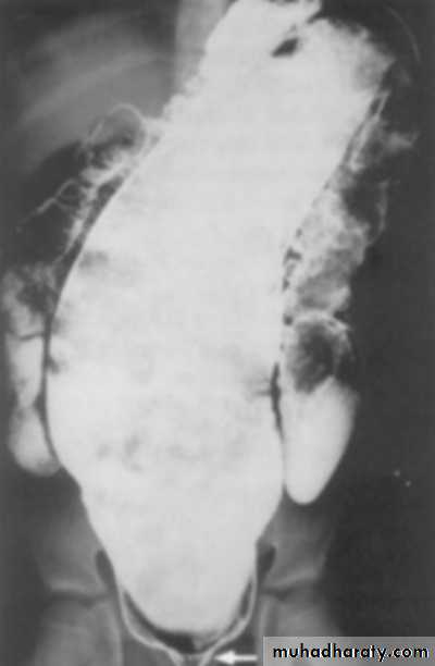
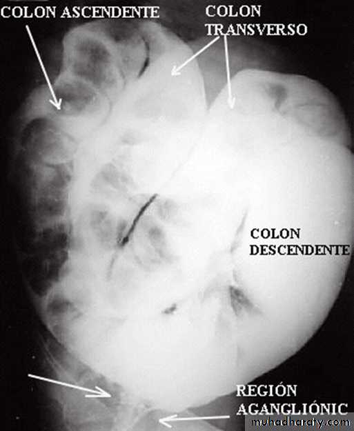
congenital megacolon (Hirschprung disease )
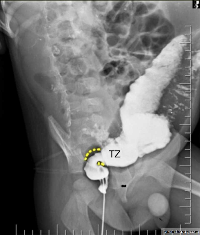
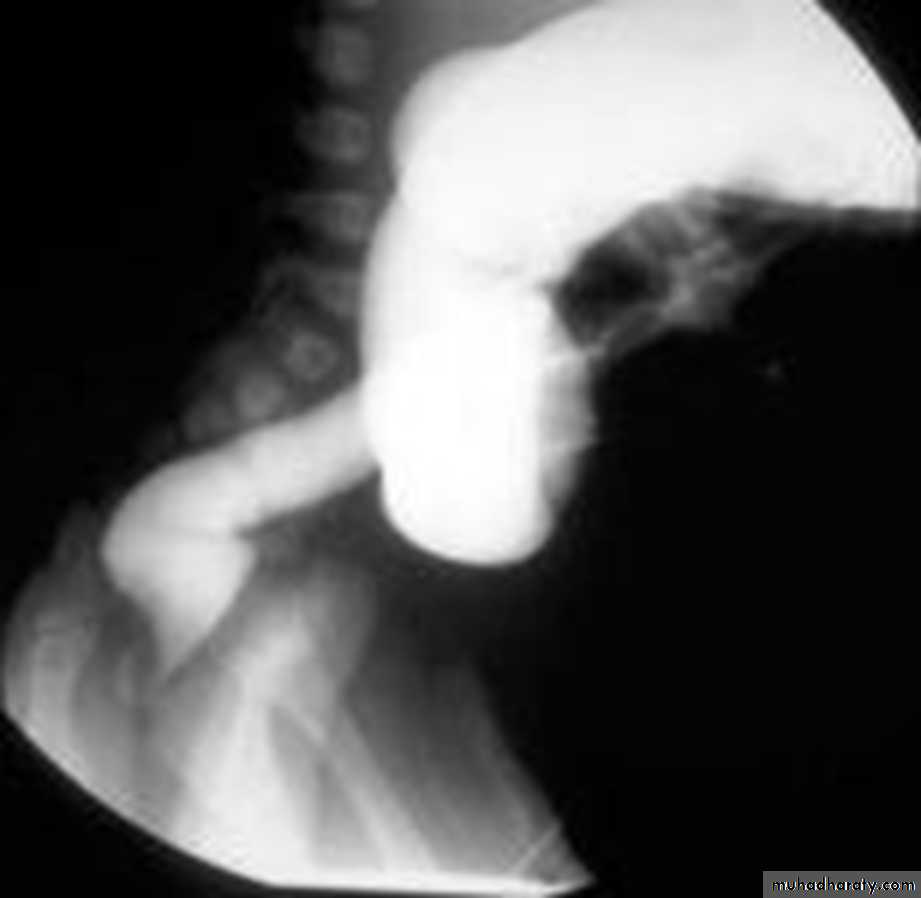
• There is one or more segment of colon devoided from innervation resulting in constriction with proximal dilatation of rest of colon due to weak innervation .• The agangloinic segment usually short , at or near the recto- sigmoid junction .
congenital mega colon
• Rarely the whole colon can be affected result in micro-colon .• 5. Clinically the patient suffering from constipation and abdominal distention since birth .
• 6. The value of Ba Enema is to spot the narrow segment especially prior to surgery .
• 7. Instant Ba Enema is usually done and the Barium used is usually hypertonic .
• Views of particular importance include:
• early filling views that include rectum and sigmoid colon allowing for rectosigmoid ratio to be determined.
• transition zone


