

Is a funnel-shaped fibromuscular tube,
10-12 cm in length in adults. It extends from
the base of the skull superiorly to the
oesophagus at level of C6. The
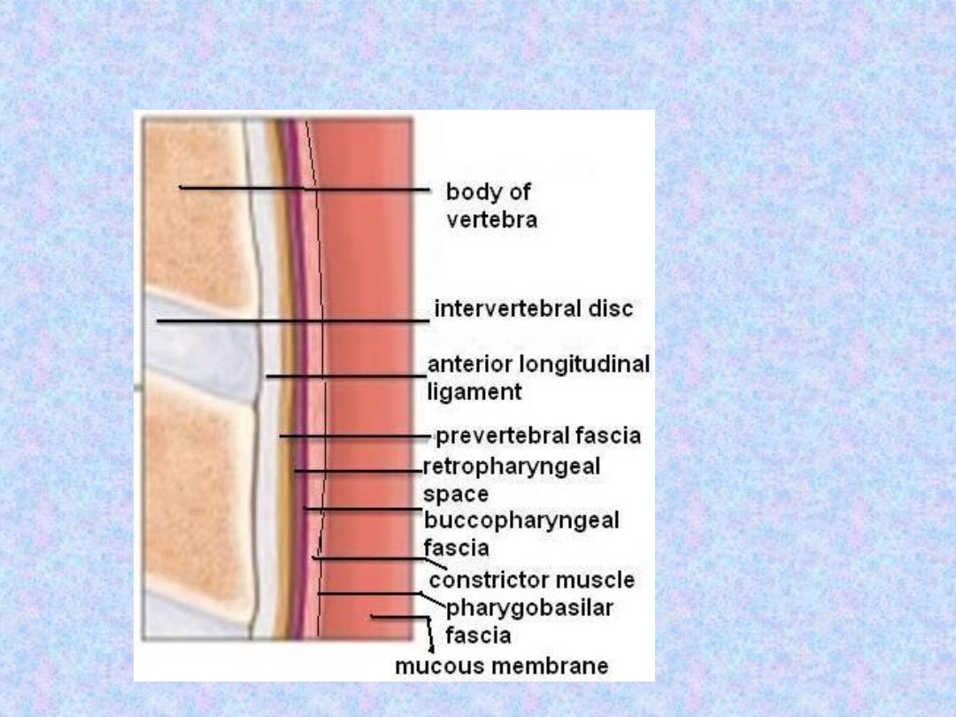
pharyngeal wall consists of 4 layers:
1. Mucous membrane.
2. Pharyngobasilar fascia.
3. Muscle layer.
4. Buccopharyngeal fascia
.

Pharyngeal Wall

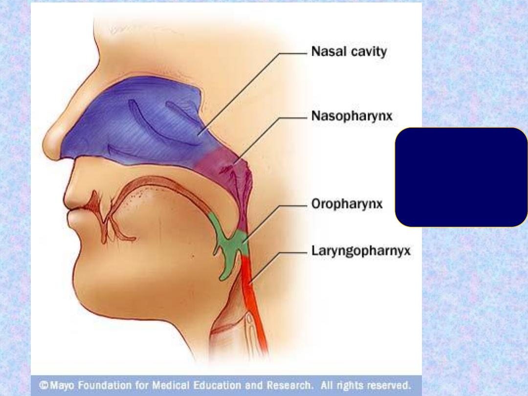
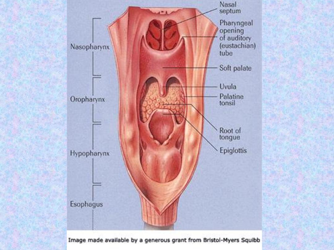
The pharynx is divided anatomically
into 3 parts defined by the openings
of its anterior surface; nasopharynx,
oropharynx and laryngopharynx
Behind
:
The Nose
The Mouth
The larynx
Anatomy of the pharynx
Compartments
•
Nasopharynx
•
Oropharynx
•
Laryngopharyn
x
•
(Hypopharynx
)
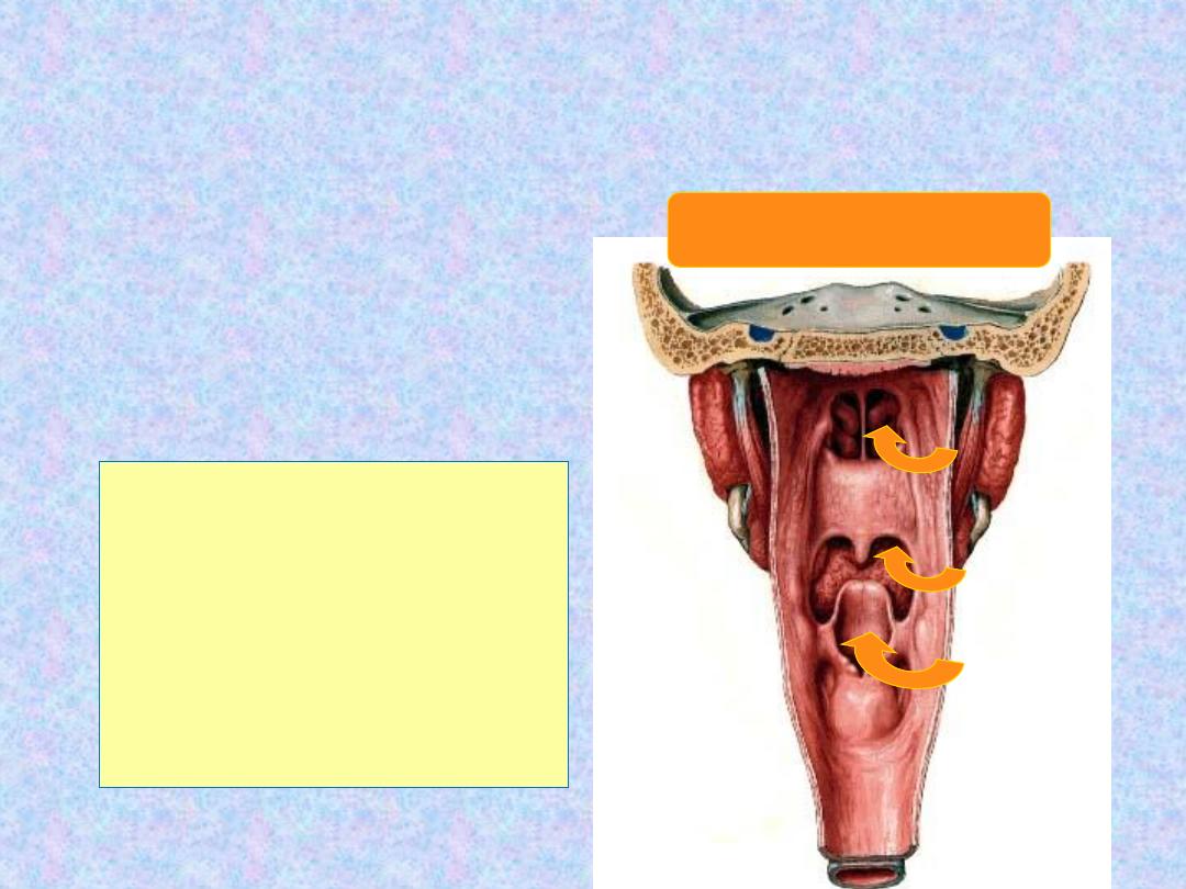
Seen from behind
Seen from behind

1-
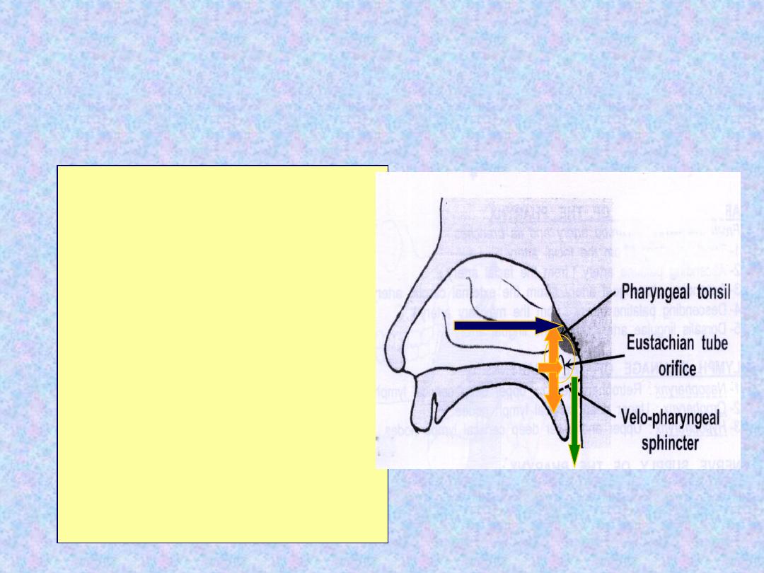
Nasopharynx:
This extends from the
base of the skull to the soft palate. At
the junction of the roof and posterior
wall lies a small mass of lymphoid tissue
called adenoids (nasopharyngeal tonsil).
On the lateral wall, there are the
openings of the Eustachian tubes.
Behind which are hollows called the
fossa of Rosenmuller
, which is the site
of nasopharyngeal malignancy.
Nasopharynx
-
Behind the nasal cavity
-
Extends from skull
Base superiorly to the
hard palate inferiorly
-
Communicates
inferiorly with the
oropharynx through the
velo-pharyngeal
sphincter
-
The nasopharyngeal
tonsil lies in the roof
-
The pharyngeal opening
of ET lies in the lateral
wall
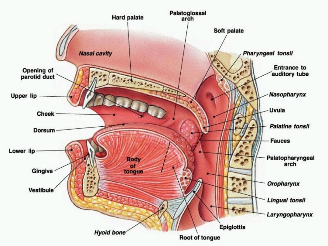
2. Oropharynx
: This extends from the
level of soft palate to the upper border of
epiglottis and opens anteriorly into the
oral cavity. The palatine tonsils are
situated in it's lateral wall.
3-
Hypopharyx:
This extends from the tip
of epiglottis to the upper end of
oesophagus and communicates anteriorly
with the larynx and below with the
oesophagus. It’s divided into 3 parts:

a. Pyriform fossae
: are two potential spaces on
each side of the pharynx forming a lateral food
channel during the act of swallowing.
b. Postcricoid area:
lies behind the cricoid
cartilage and encircled by the cricopharyngeus
muscle which forms the upper oesophageal
sphincter.
c. Posterior pharyngeal wall
: extends from the
hyoid bone to the oesophageal inlet.
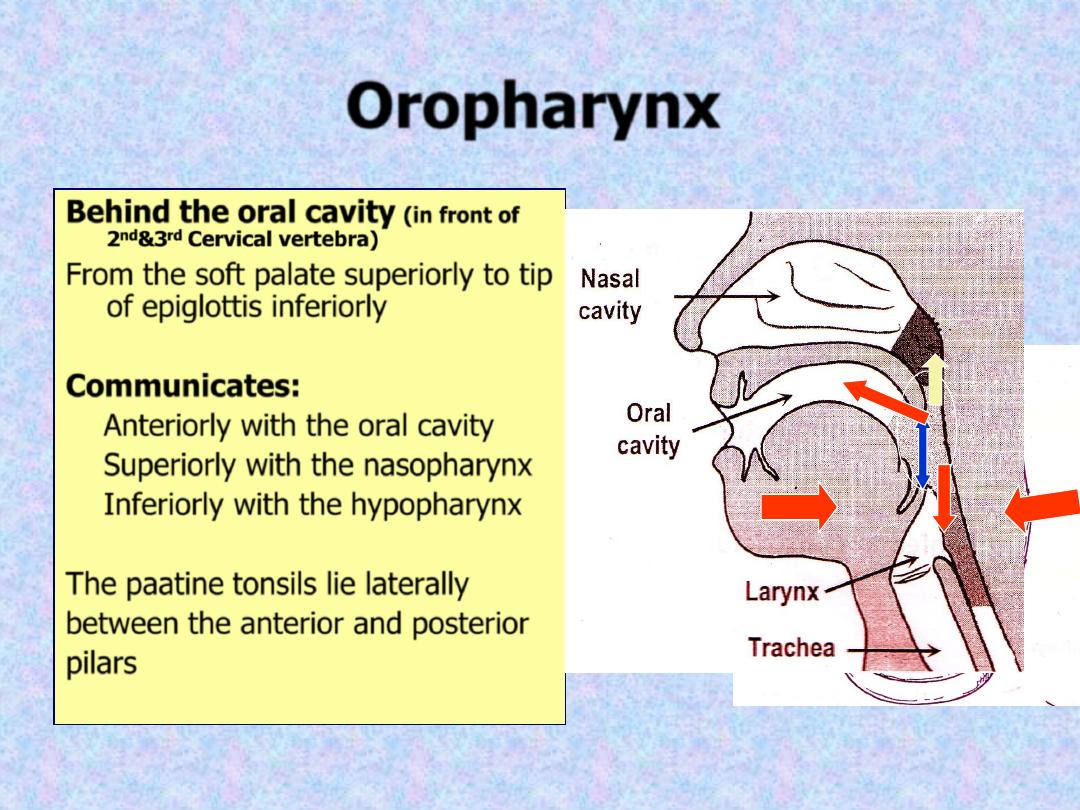
Oropharynx
Behind the oral cavity
(in front of
2
nd
&3
rd
Cervical vertebra)
From the soft palate superiorly to tip
of epiglottis inferiorly
Communicates:
Anteriorly with the oral cavity
Superiorly with the nasopharynx
Inferiorly with the hypopharynx
The paatine tonsils lie laterally
between the anterior and posterior
pilars
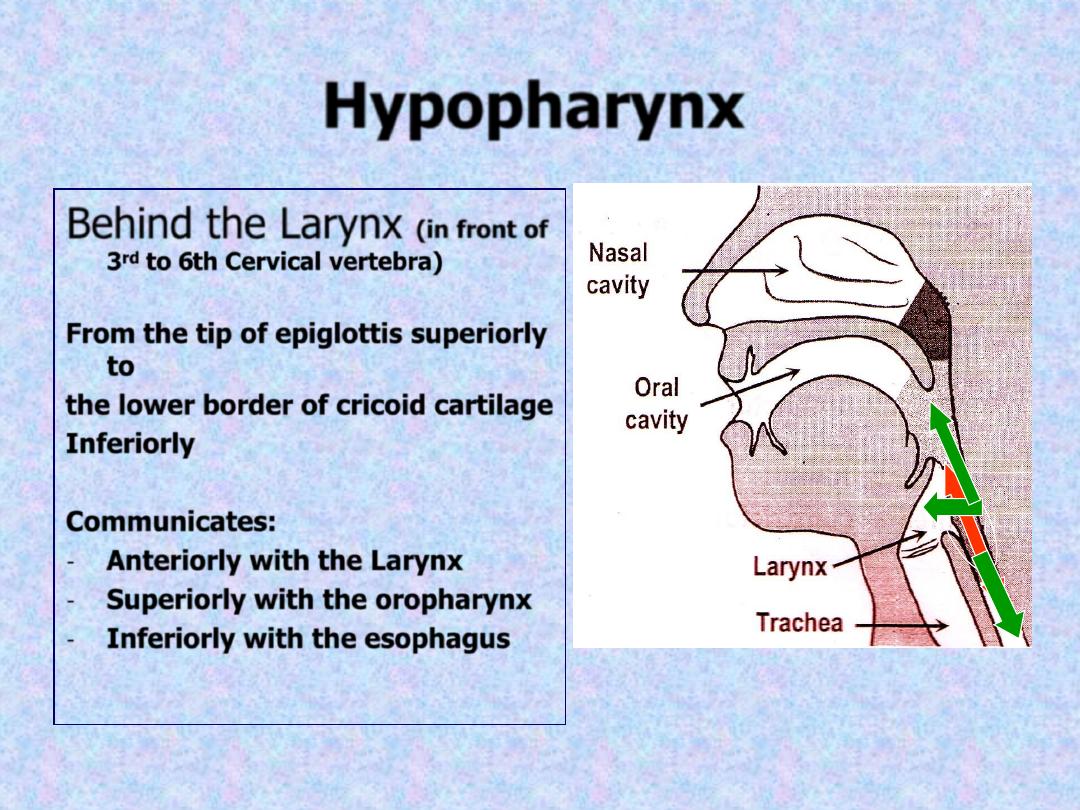
Hypopharynx
Behind the Larynx
(in front of
3
rd
to 6th Cervical vertebra)
From the tip of epiglottis superiorly
to
the lower border of cricoid cartilage
Inferiorly
Communicates:
-
Anteriorly with the Larynx
-
Superiorly with the oropharynx
-
Inferiorly with the esophagus
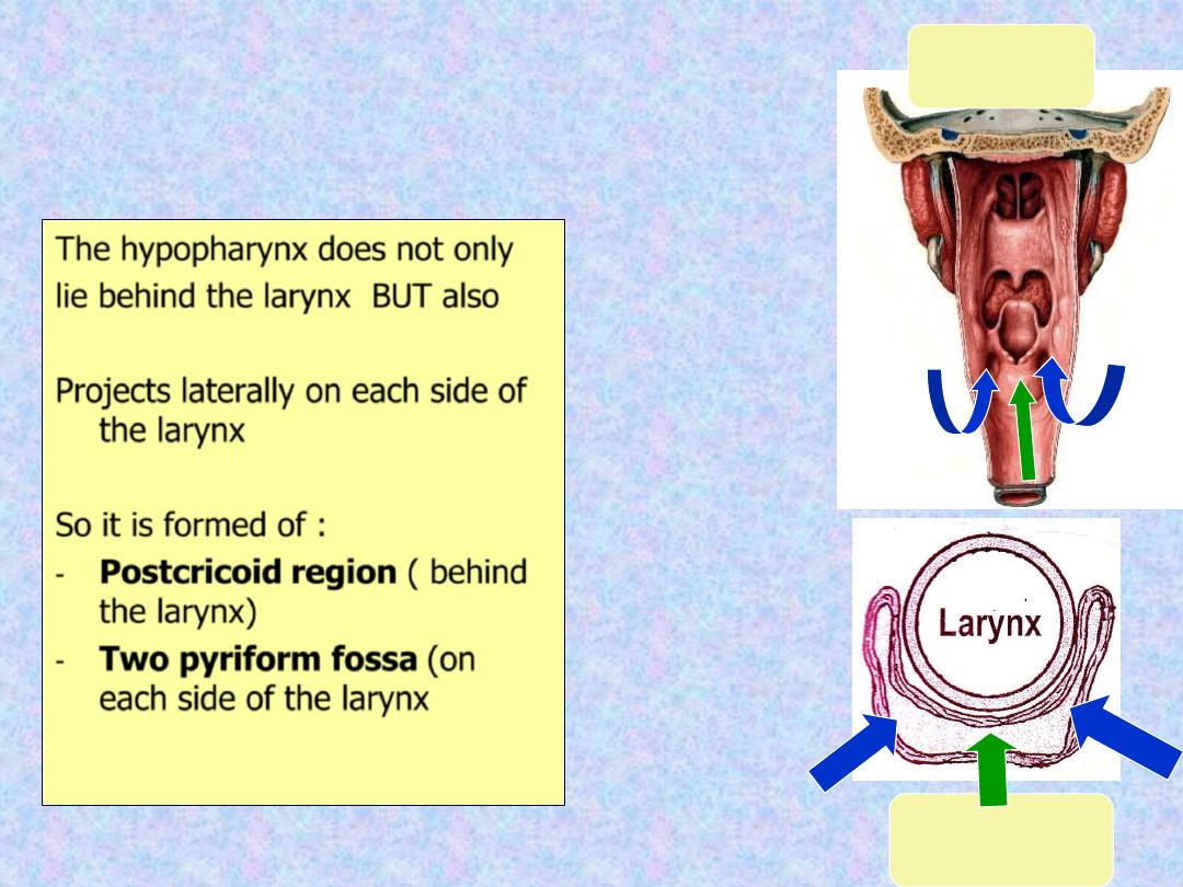
The hypopharynx does not only
lie behind the larynx BUT also
Projects laterally on each side of
the larynx
So it is formed of :
-
Postcricoid region ( behind
the larynx)
-
Two pyriform fossa (on
each side of the larynx
Seen from
behind
Cross section
The lining epithelium is stratified squamous
except in the nasopharynx, where
columnar epithelium is found.
This fascia is strengthened posteriorly by a
strong band called the median raphae.
This raphae is attached above to the base
of the skull and gives insertion to the
constrictor muscles.
I-
Circular (outer):
which consist of 3
constrictor muscles overlapping one
another from below upwards.
1. Superior constrictor
: it arises from the
medial pterygoid plate, pterygoid hamulus
and pterygomandibular ligament.
2. Middle constrictor
: it arises from the
hyoid bone and stylohyoid ligament.
3. Inferior constrictor
: has 2 parts:
a. Thyropharyngeus (oblique): arises from
the oblique line of thyroid cartilage and
the cricothyroid muscle.
b. Cricopharyngeus (transverse): arises from
the cricoid cartilage and passes
transversely backwards forming the
upper oesophageal sphincter.
All the constrictor muscles are inserted
posteriorly into the median pharyngeal
raphae.
Functions
The constrictor muscles propel the bolus of
food down into the esophagus
Cricopharygeus (lower fibers of the inferior
constrictor) act as a sphincter, preventing
the entry of air into the esophagus
between the acts of swallowing
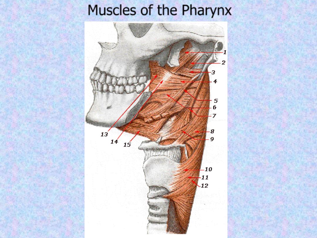
Muscles of the Pharynx
Killian dehiscence
: this is a potential gap
between the fibers of the thyropharyngeus
and
cricopharyngeus.
The
mucous
membrane may bulge between these two
muscles when there is incoordination of
the pharyngeal peristaltic waves (the
cricopharyngeus doesn’t relax at the
appropriate time during the 2nd stage of
deglutition) forming the pharyngeal
pouch.
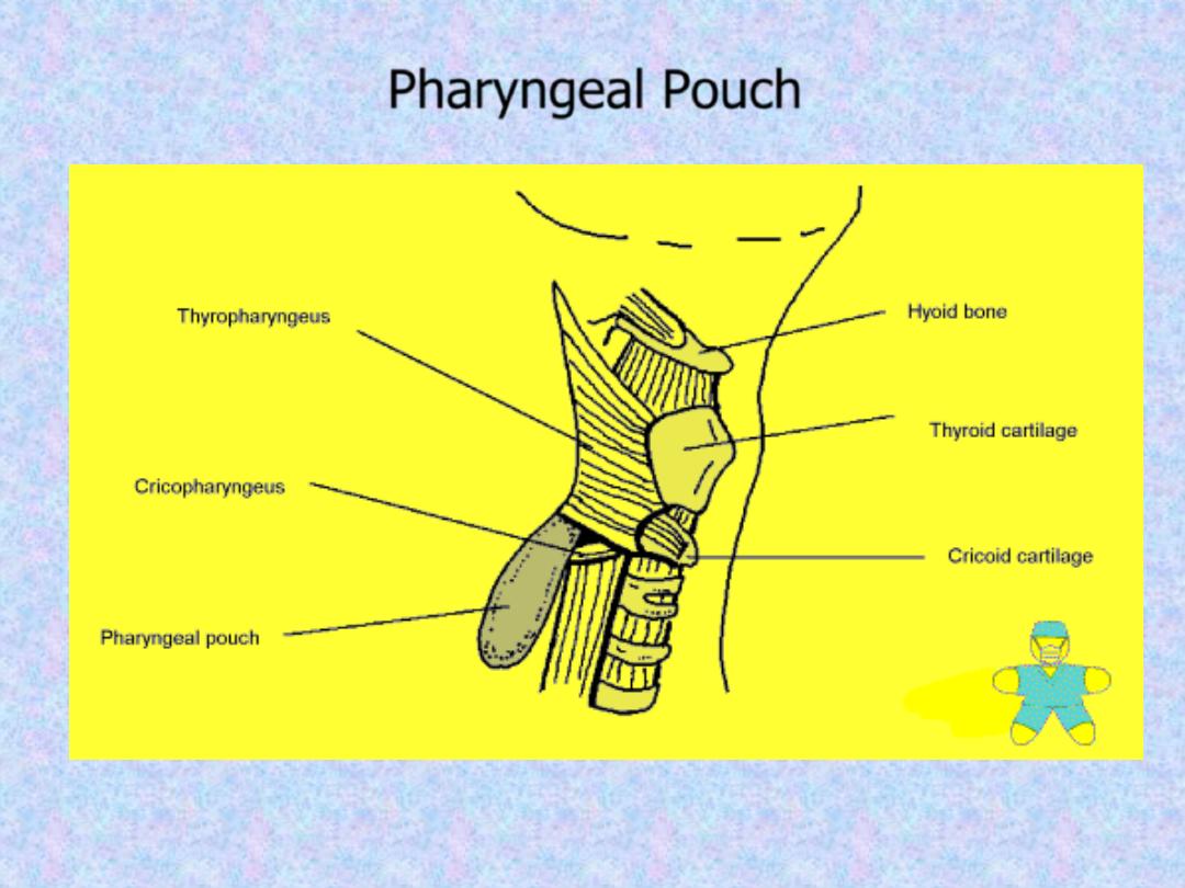
Pharyngeal Pouch
.
II-
Longitudinal (internal):
these
muscles elevate the larynx and shorten
the pharynx during deglutition:
1. Stylopharyngeus.
2. Salpingopharyngeus.
3. Palatopharyngeus
.

This fascia is loosely attached posteriorly
to the prevertebral fascia and laterally it's
connected to the styloid process and to
the carotid sheath.
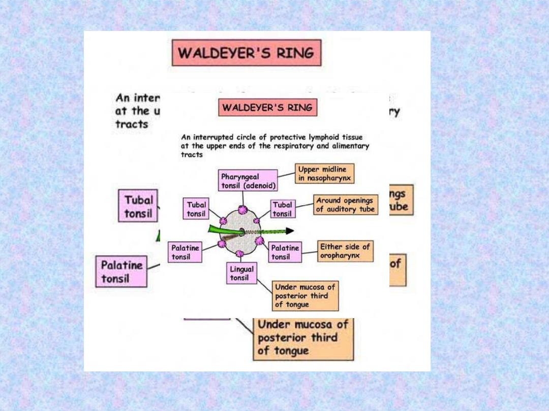
Subepithelial lymphoid tissue of the
pharynx (Waldeyer's ring)
Is a collection of sub-epithelial lymphoid
tissue around the entrance of the
respiratory and alimentary tracts.

1.
Nasopharyngeal
tonsil
(adenoid): Lies between the roof
and
posterior
wall
of
the
nasopharynx. The free surface
exhibits about 5 vertical fissures.
2. Tubal tonsils: lie behind the
openings of the Eustachian tubes.
3. Palatine tonsils: are two
masses of lymphoid tissue situated
on each side of the oropharynx.

4. Lingual tonsils: which is
embedded in the posterior 1/3 of the
tongue.
5. Lateral pharyngeal bands
behind the posterior tonsillar pillar.
6. Lymphoid nodules scattered on
the posterior pharyngeal wall.
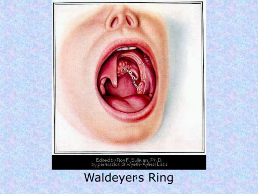
Waldeyer
,
s Ring

Hypertrophy of the lymphoid tissue
of Waldeyer's ring occurs in the
earlier years of childhood, probably
in response to upper respiratory
tract infection. Maximum bulk is
obtained at the age of about 6
years, thereafter, some regression in
size is to be expected, and in old
age it atrophies
.

The tympanic membrane separates the EAM
from the middle ear. It is thin, nearly oval
disk, forming an angle of about 55 with
the floor of the meatus. It has a pearly
grey colour with a triangular bright area;
the cone of light; extending from the
centre (umbo) downwards and forwards.
Waldeyer's ring is characterized by:
1. Sub-epithelial lymphoid tissue.
2. Lack a definite capsule.
3. They have efferent lymph vessels,
but no afferent vessels.
4. Function as one unit: when a
member of it is removed, the
others
parts
undergo
compensatory hypertrophy.

5. The exact function is unknown, but it's
thought that it has a protective function
by:
a. Formation of lymphocytes.
b. Secretion of antibodies, mainly lgA.
c. Localization of infection entering the body
by initial contact with incoming
organisms.

Are two masses of lymphoid tissue situated
on each side of the oropharynx. The
medial surface is exposed in the pharynx
and is pitted by a number of crypts. The
tonsil is related anteriorly and posteriorly
to
the
palatoglossus
and
palatopharyngeus muscles respectively.

Laterally the tonsil is enclosed by a dense
fibrous capsule separating the tonsil from
the superior constrictor muscle (tonsillar
bed). This capsule provide a convenient
plane of separation of the tonsil during
tonsillectomy.
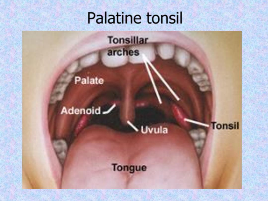
Palatine tonsil
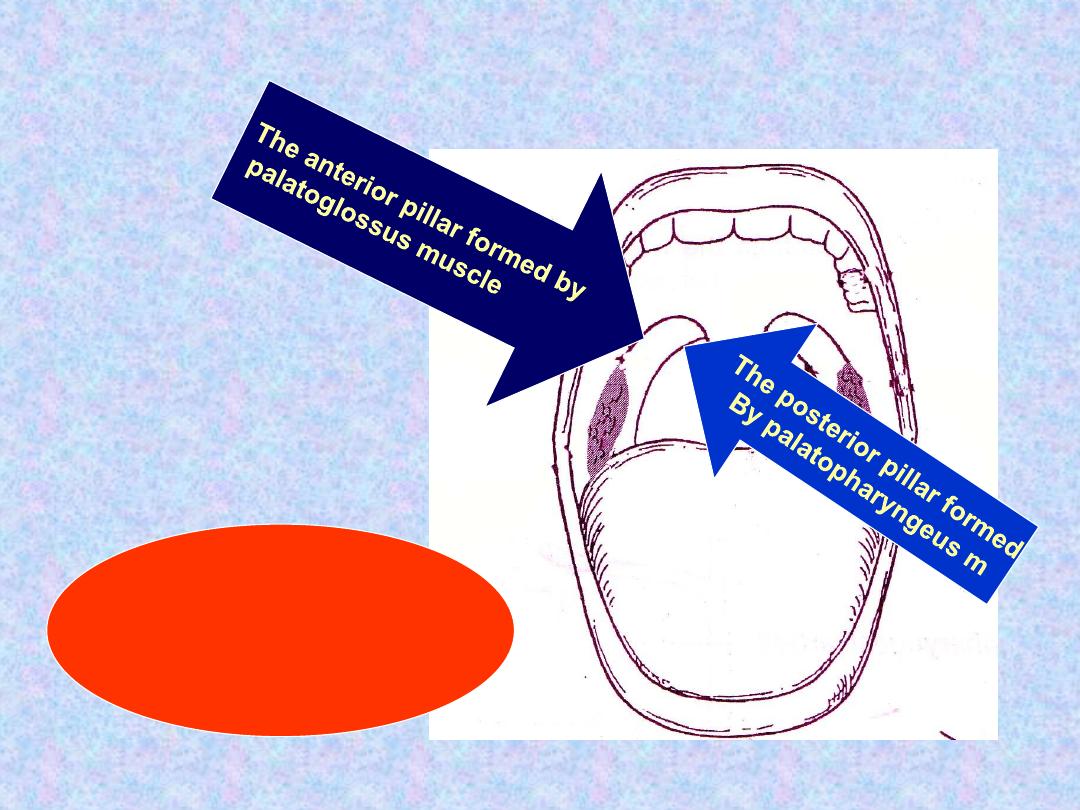
The tonsils lie between the
Two pillars

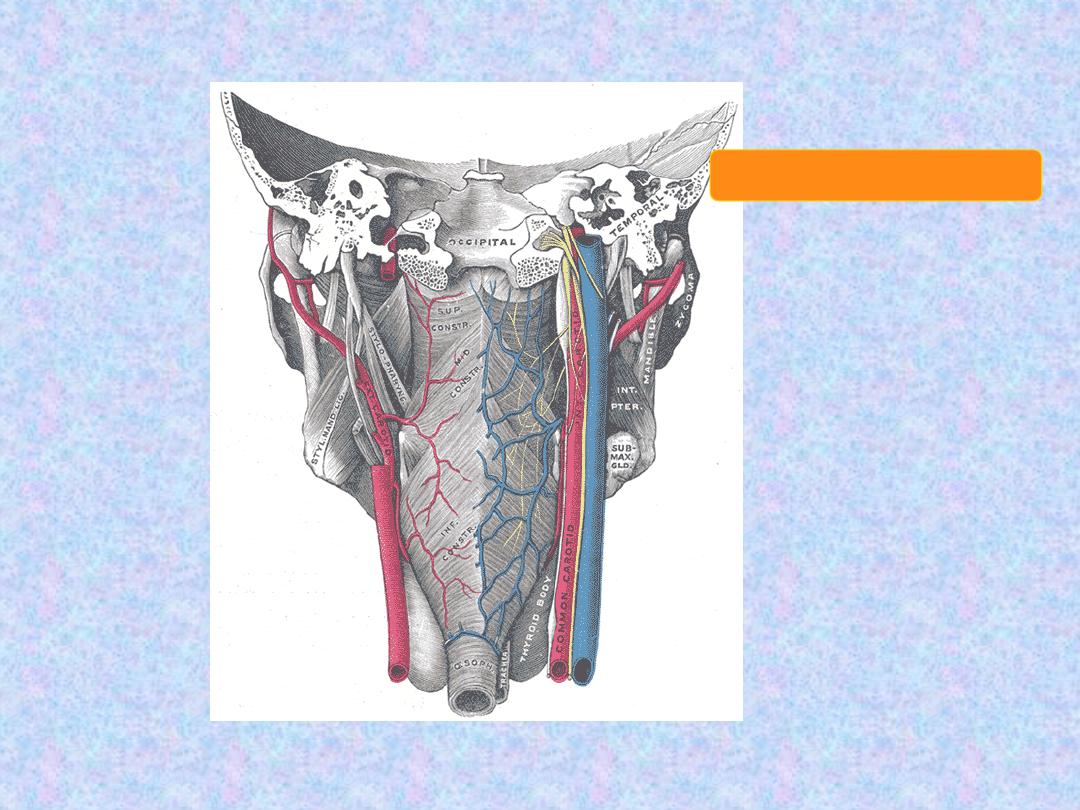
1. The main supply is the tonsillar branch of
the facial artery.
2. Lingual artery.
3. Ascending pharyngeal artery.
4. Greater palatine artery.
The venous drainage is to the
paratonsillar vein which drains to the
pharyngeal plexus. This plexus drains into
the internal jugular and anterior facial
veins.

Deep cervical chain of lymph nodes.

Sensory Nerve Supply
Nasopharynx: Maxillary nerve
Oropharyn x: Glossopharyngeal nerve
Laryngopharynx
: I
nternal laryngeal branch of the
vagus nerve
Motor Nerve Supply
All the muscles of pharynx, except the
stylopharyngeus, supplied by the
pharyngeal plexus. The plexus is formed
by the pharyngeal branches of the IX
and X nerves together with the
sympathetic fibers from the superior
cervical ganglion.
The stylopharyngeus is supplied by the
glossopharyngeal nerve

This space lies behind the pharynx and
extends from the base of the skull to the
superior mediastinum.
The anterior wall is formed by the posterior
pharyngeal wall and it's covering
buccopharyngeal fascia. The posterior wall
is formed by the cervical vertebrae and
their covering muscles and fascia.
Pharyngeal Wall
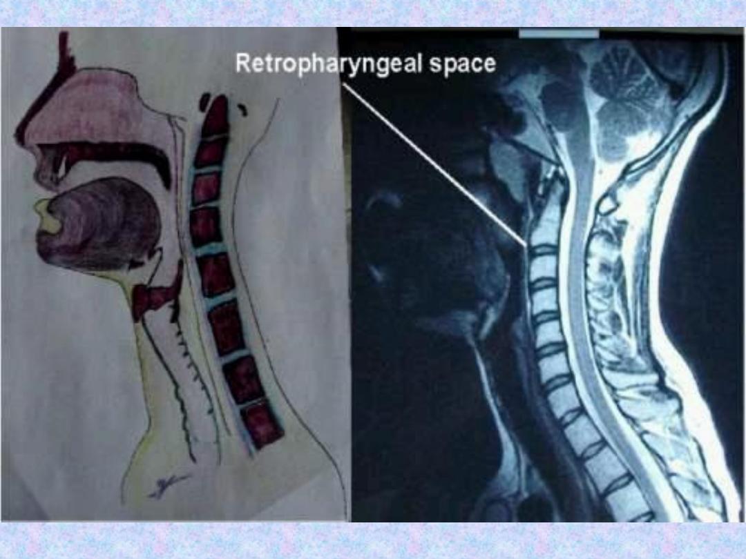

Contents:
retropharyngeal lymphnodes of
Rouviere. These are paired lymphnodes
separated from one another by a median
partition.
They
usually
disappear
spontaneously during the 3rd or 4th year
of life.
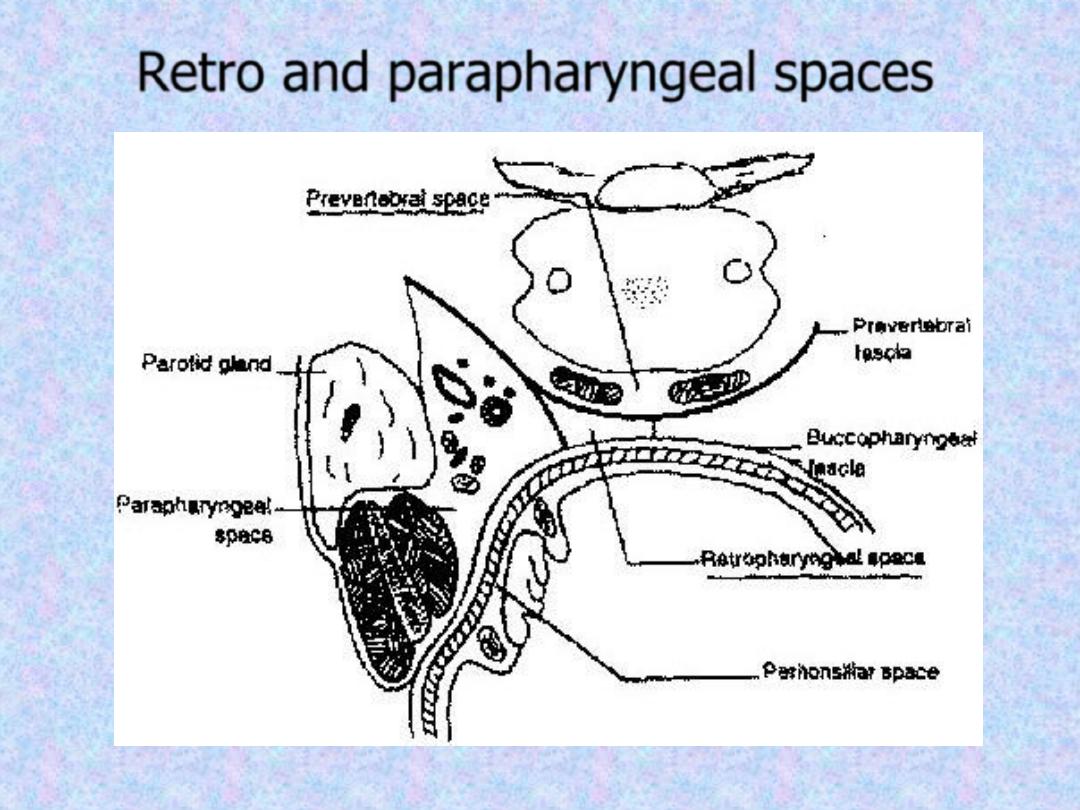
Retro and parapharyngeal spaces

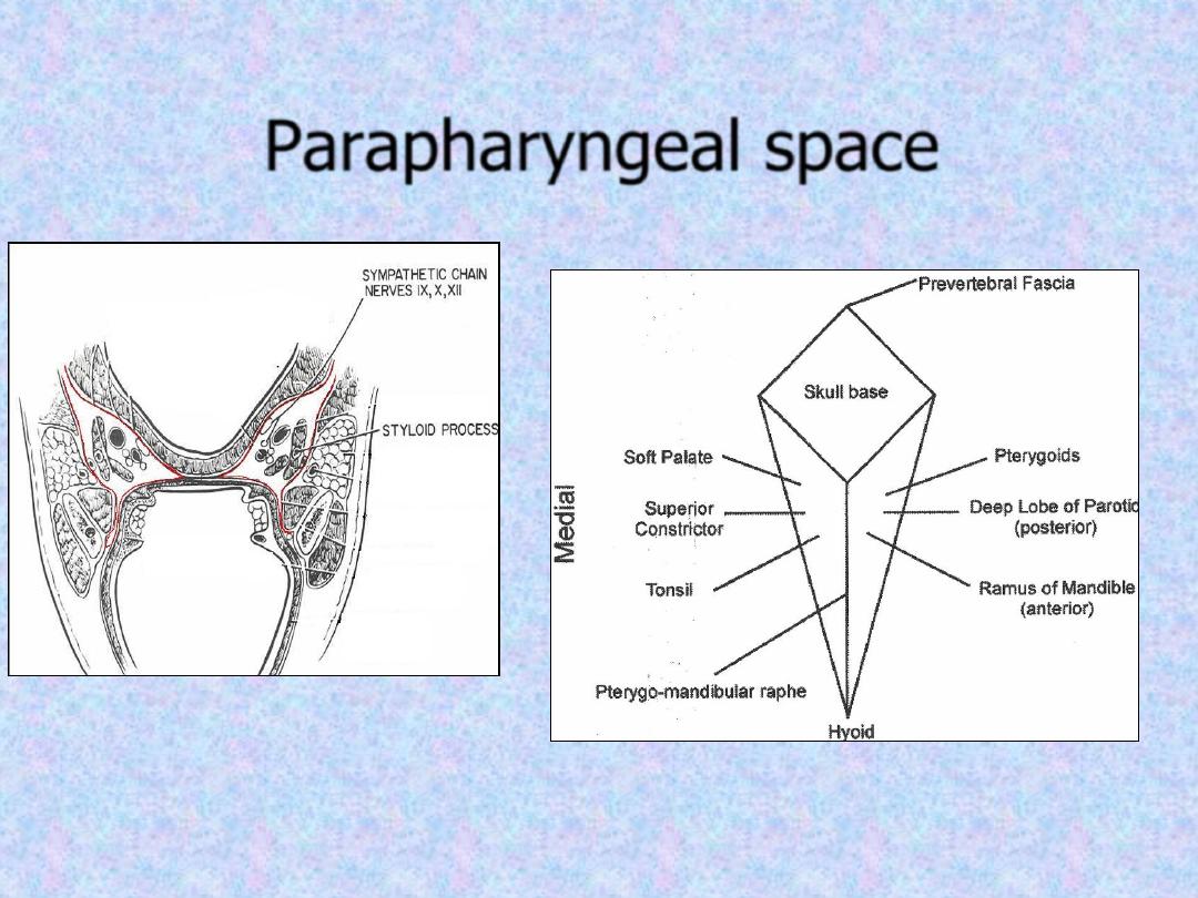
This potential space which resembles an
inverted pyramid lies lateral to the
pharynx and connects posteriorly with the
retropharyngeal space. It extends from
the base of the skull to the hyoid bone.
It's bounded medially by the superior
constrictor muscle. Laterally lies the
medial pterygoid muscle, the mandible
and the parotid gland. It's posterior wall is
the prevertebral muscles and fascia.

Contents
1. Deep cervical lymphnodes.
2. The last 4 cranial nerves and the cervical
sympathetic trunk.
3. Great vessels of the neck: carotid and
internal jugular vein.
Parapharyngeal space
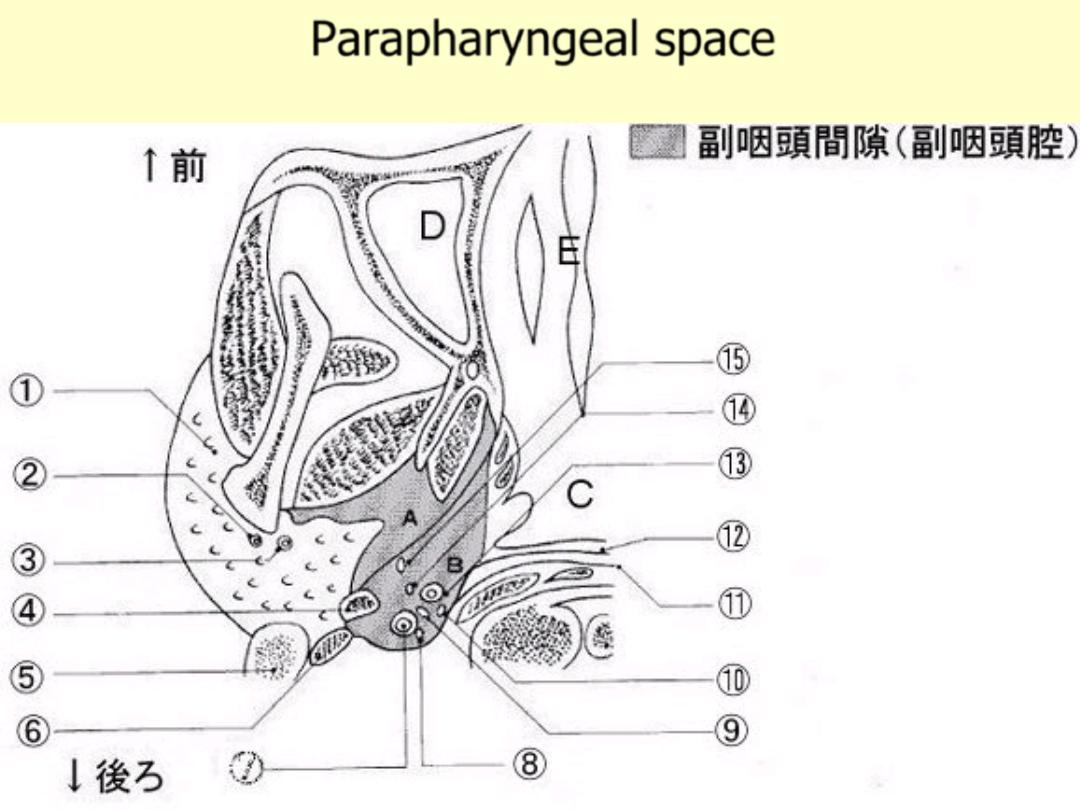
Parapharyngeal space
All are true about the pharynx EXCEPT:
a.
It is a fibromuscular tube 10-12 cm in length in
adults.
b.
It extends from the base of the skull to the level
of C6.
c. Pyriform fossae and postcricoid area are parts of
oropharynx.
d. It is divided anatomically into; nasopharynx,
oropharynx and hypopharynx.
Regarding anatomy of the pharynx:
A. Pharyngo- basilar fascia is the outermost layer.
B. Hypopharynx is lined by keratinized squamous
epithelium.
C. All constrictors muscles arise from posterior
pharyngeal raphe.
D. Oro-pharynx extends from the level of soft
palate to the tip of epiglottis.
E. Posterior tonsillar pillar is formed by palate-
glossus muscle.
All are contents of the
parapharyngeal space EXCEPT:
A/ Carotid artery.
B/ Internal jugular vein.
C/ Last 4 cranial nerves.
D/ Submandibular gland.
F/ Deep cervical LN.

Killian's dehiscence is present
between:
a. The skull base and superior constrictor
muscle.
b. Between the oblique and transverse
fibers of inferior constrictor
c. Between the superior and middle
constrictors.
d. Between the middle and superior
constrictors.
f. Between inferior constrictor and
oesophagus.