
Anatomy & physiology
of skin
Dr. Qassim S. Al Chalabi
F.A.B.H.S
(Dermatology and venereology)

Skin Structure
Skin is the single largest
organ in the human body.
It weighs an average of 4 kg and covers
an area of
2 m
2
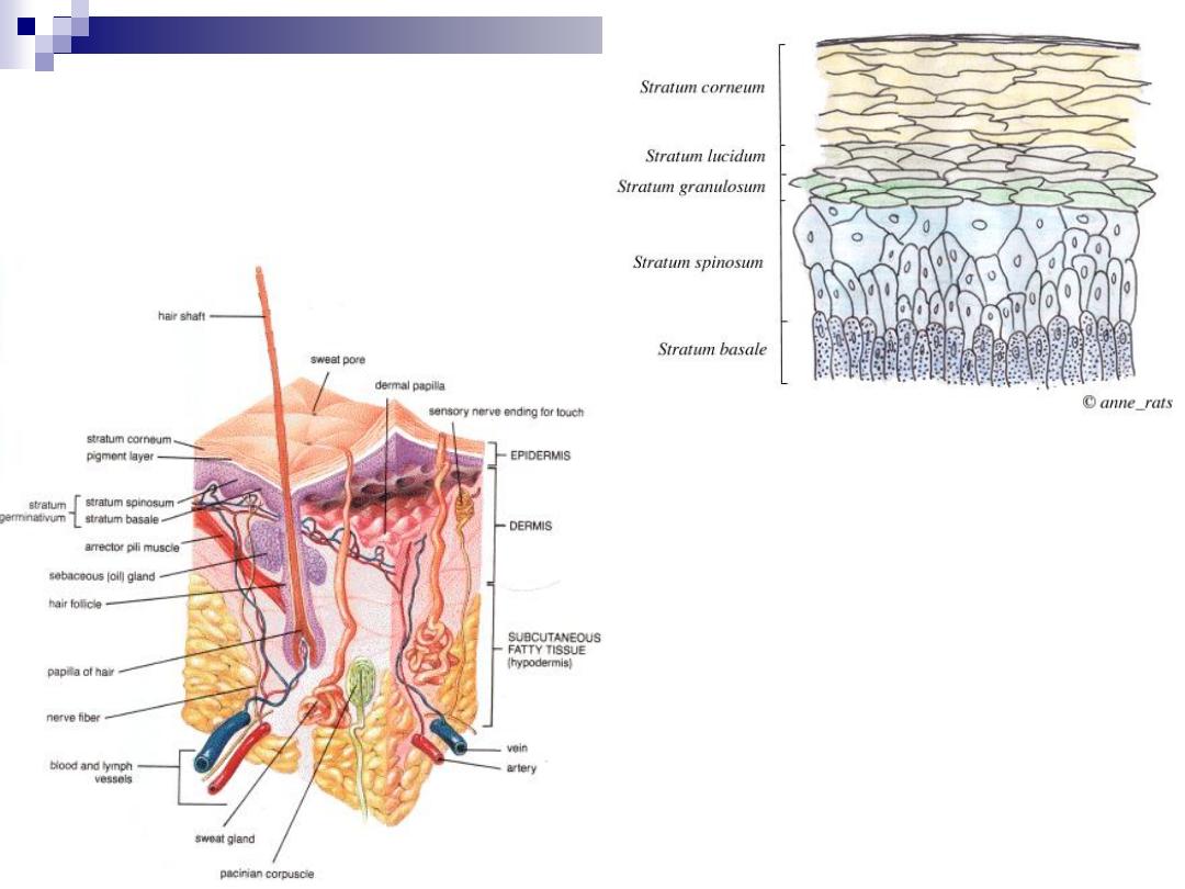
Three distinct layers
Epidermis: Composed of epithelial tissue
Dermis: Composed of a combination of connective
tissues
Hypodermis: usually contains abundant fat.
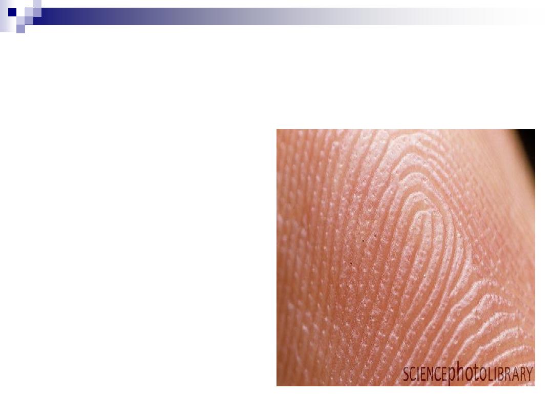
Epidermis
It’s outermost layer of skin
It’s thickness 0.1-1.0 mm
It’s a stratified squmaous
epithelium
It’s ectodermal in origin
It’s consist of:
Original cell (keratinocyte) 80
%
Immigrant cells:
Melanocyte 10%
Langerhan’s cell 2-4%
Merkle cell
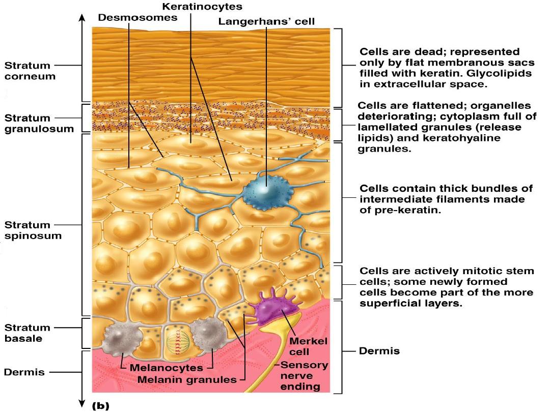

Stratum Basale
Basal cell layer: is a single row of cells where
mitosis takes place.
Not all basal cells have potential to divide, only (30%)
Stem cells give rise to transient amplifying cells
Once basal cell leaves basal layer in humans, normal
transit time to stratum corneum is at least 14 days, and
transit through stratum corneum to desquamation
requires 14 days, 28 days total.

Stratum Spinosum
Stratum Spinosum: Consists of 8-10 layers of
Keratinocytes.
Cells contain lamellar granules in which contain
glycoproteins and lipid precursors which are discharged
into intercellular space between granular and cornified
layer; forms (ceramide) or “mortar” which acts as
intercellular cement for corneocytes (“bricks”) thus
contributing to formation of cutaneous lipid barrier.
Composed of Keratinocytes attached to each other via
desmosomes.
Contains langerhans cells that aid in the immune
system response.

Stratum Granulosum
Stratum Granulosum: The middle layer
of 3-5 layers of cells that help form keratin.
Contains keratohyline granules that
produce a secretion These make up the
thick and tough peripheral protein coating
of the horny envelope.

Stratum Lucidum
Stratum Lucidum: Found only in the thick
skin of the palms and soles of the feet
Tends to be translucent.

Stratum Corneum
Stratum Corneum: The outermost layer of the
epidermis.
Contains up to 30 layers of the dead, scaly,
piled-up layers of flattened dead cells
(corneocytes)
– the bricks – separated by lipids
– the mortar – in the intercellular space.
Provides protection.
Continuously shed and replaced.

Cells of the epiderms
Merckle’s
cell
Langerhans
cell
Melanocyte
Few
Next common
Most common
Prevalence
-
2-4%
10%
Percentage
St.Basalis
St. Spinosum
St. basalis
Location in epidermis
Unknown
Bone marrow
Neural crest
Origin
Sensory
function
Immune
surveillance
Production of
melanin
Function
MM, lip,
digit
All skin
All skin
Type of skin
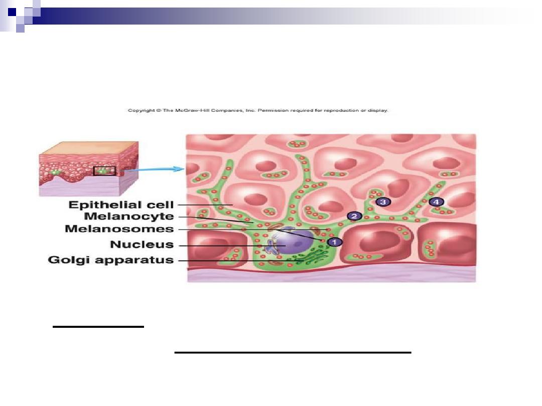
Melanocyte
Dendritic cell derived from neural crest
localized to basal layer of epidermis
Functions include: melanin production.
(epidermal melanin unit = approximately 36
keratinocytes in contact with one melanocyte), and
transfer pigment to keratinocytes
Melanin absorbs and scatters ultraviolet radiation (280-
400 nm), and thus protects from UV-induced DNA
mutations.
Special stains: silver nitrate, S100, HMB-45

Skin Color
Skin color is determined by 3 pigments:
Melanin: Provides brown, black and tan hues
Carotene: Provides yellowish hues
Hemoglobin: Found in red blood cells;
provides pink and flesh tones.
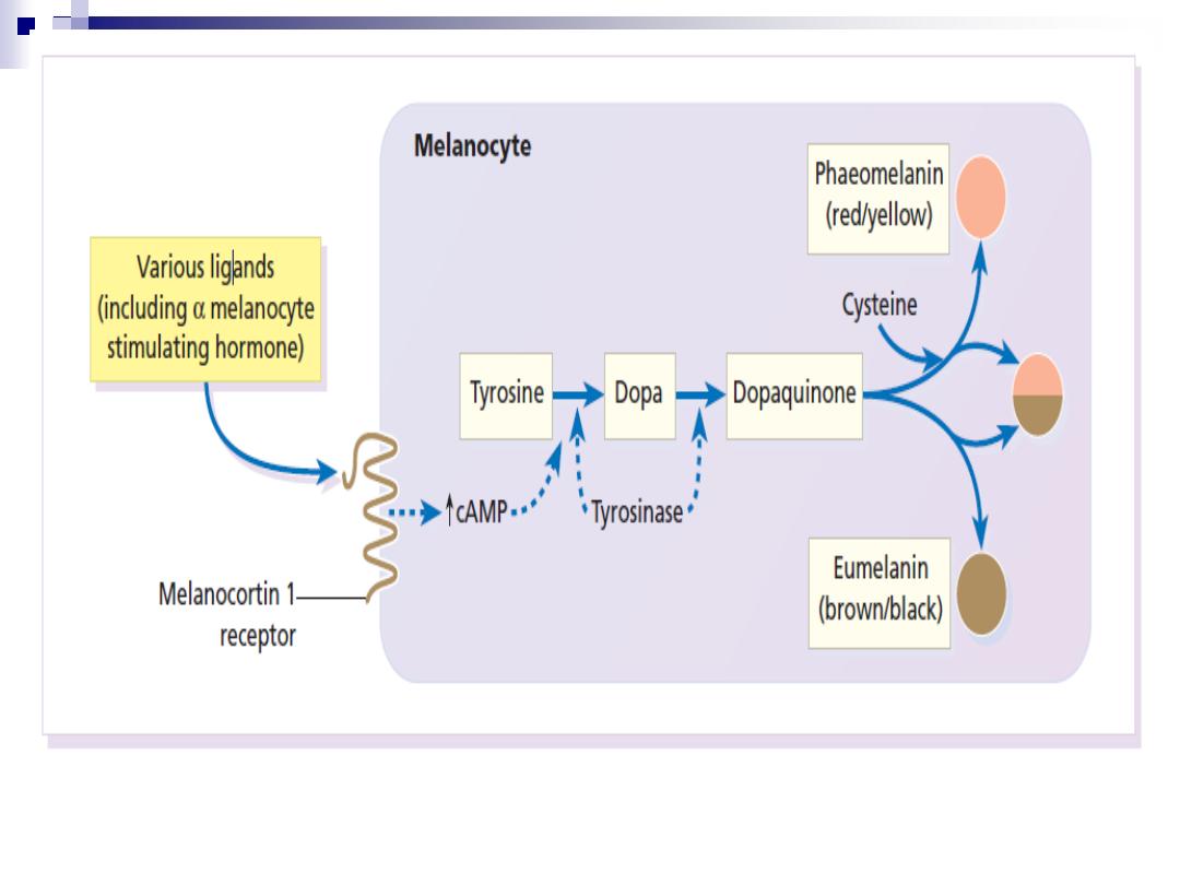
The pathway of melanin synthesis

Melanogensis control
Genetic (constitutional)
UV (facultative)
MSH
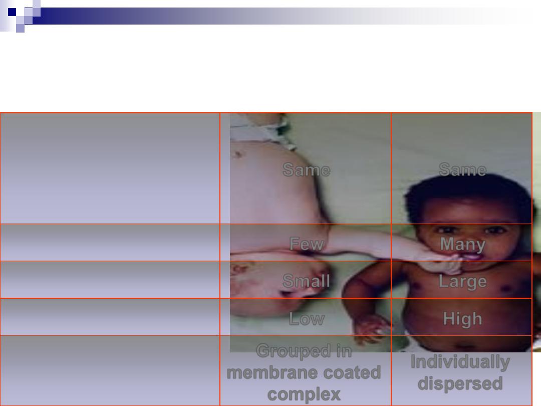
Difference between White &
dark individual
Same
Same
Melanocyte number
Many
Few
Melansome number
Large
Small
Melanosome size
High
Low
Degree of melanization
Individually
dispersed
Grouped in
membrane coated
complex
Distribution in
keratinocyte
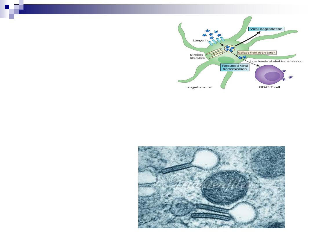
Langerhan’s cell
Bone marrow
–derived dendritic cell.
Location in epidermis: St. sipnosum
Langerhans cells constitute 2-4% of the total epidermal cell population.
Characteristic finding on electron microscopy
– Birbeck granules- tennis
racket shaped bodies in the cell.
Function: Antigen presenting cells, immune surveillance.
Functionally impaired by ultraviolet radiation and topical steroid.
Special stain: S100, CD1a
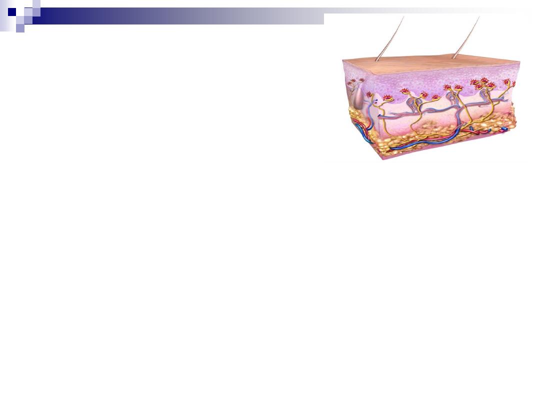
Merckel’s cell
Shape: Non dendritic cell
Site: MM, lip, digit & near hair
Location in epidermis: St. Basalis
Origin: Unknown
Function: Transducer of fine touch

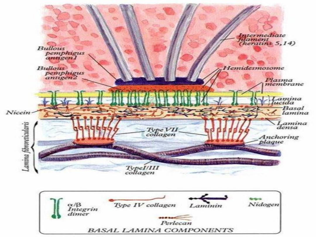
Basement Membrane Zone
Site of interaction between epidermis and
dermis
Three layers
1.) Lamina lucida, 2.) Lamina densa, 3.)
Sub-lamina densa
Hemidesmosomal adhesion complex: 1.)
hemidesmosomal plaque, 2.) anchoring
filaments, and 3.) anchoring fibrils

Dermis
Dermis: Composed primarily of connective
tissue, mainly collagen, elastic fibers and
ground substances.
Divides into 2 Categories:
Papillary Region: Superficial part that contains the
dermal papillae and comprises the wavy boundary
between the epidermis and dermis. Houses
capillaries, nerve endings, and Meissner corpuscles
(touch sensors).
Reticular Region: Deeper region made up of dense
irregular connective tissue. Contains many of the
glands, nail roots, and root follicles.

Hypodermis
Hypodermis: Known as the subcutaneous
tissue or superficial fascia.
The boundary of the dermis is indistinct, but it
is clear that there is more areolar and adipose
tissue than in the dermis.
Primarily serves as a storage site for large
blood vessels and fat.
Houses Lamellated or Pacinian corpuscles
(nerve endings sensitive to pressure).

Sebaceous
Appocrine
Eccrine
Hairy skin, free in
eyelid, lip, nipple,
genetalia
Axilla, areola,
Periumblical,
genetalia
All skin
Distribution
Multilobulated
Coiled
Coiled
Acinar part
Short & wide
Straight
Straight
Duct shape
Hair follicle
Hair follicle
Skin surface
Duct opening
Holocrine
Decapitation
Merocrine
Excretion
Oily
Milky & odorless
Isotonic
Secretion
Androgen
Adrenalin
Acetyl choline
Stimulant
Waterproof, lubricant
Anti-microbial.
Unknown ,Sex
stimulant
Thermoregulation
Function
Epidermal appendages
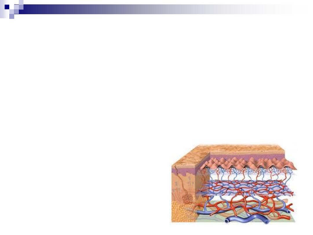
Blood supply of skin
Although the skin consumes little oxygen, its abundant blood supply
regulates body temperature.
Blood vessels lie in two main horizontal layers.
•
Superficial H.P.:
–
Lies at papillary dermis
–
Arterioles form capillary loop in dermal papillae
•
Deep H.P.:
–
Lies just above subcutis
–
Arterioles supply hair follicle
–
& sweat glands
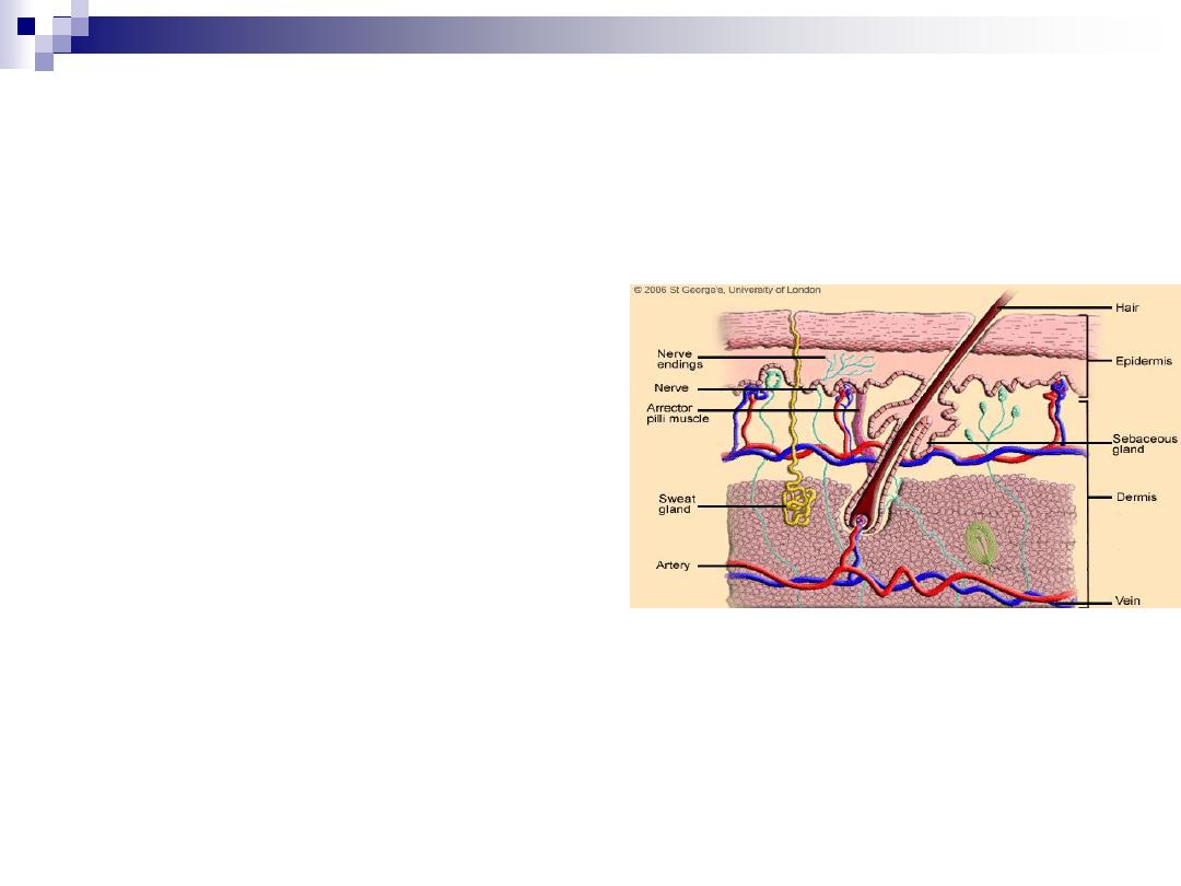
Nerve supply of skin
•
It’s supplied by 1.000.000 fibers
•
Enervation are:
–
Autonomic N.
–
Sensory N.
•
Sensory fibers are:
–
Myelinated fiber
–
Non-myelinated fiber (itching)
•
Nerve ending are:
–
Free (Nocecepters)
–
Specialized end organ (mechanoceptors)
•
Specialized end organ are: Meisseners & paccianian’s
corpuscles

6 Main Functions of the Skin
1. Protection: Physical and solar barrier.
1. Keratin protects tissues from heat, chemicals, injury,
& infection.
2. Melanin protects against solar radiation.
2. Vitamin D Synthesis: Vitamin D regulates
blood calcium levels
– production is stimulated
by ultraviolet radiation (UVB)
3. Extensive Sensory Functions: Detects heat,
cold, touch, texture, pleasure, vibration, and
tissue injury.

6 Main Functions of the Skin
4. Thermoregulation: Via sweat
production & blood flow; preserves
homeostasis.
5. Excretion & Absorption: Excretes &
absorbs lipid-soluble materials; sweat
allows secretion of small amounts of
waste materials including salt, carbon
dioxide, ammonia, and urea.
6. Reservoir: For 8-10% of adult blood flow

Most important
Good bye
And
Have a nice
day
