FERTILIZATION & IMPLANTATION
Dr.Sumeya

Obgectives
• This lecture explain spermatogenesis and
oogenesis then normal fertilization regarding
time and site .
• Also normal and abnormal implantation .
Gametogenesis
The creation of
highly specialized sex
cells
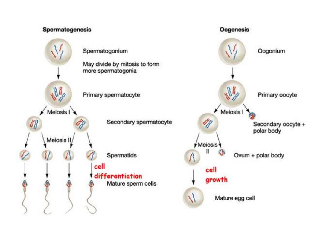
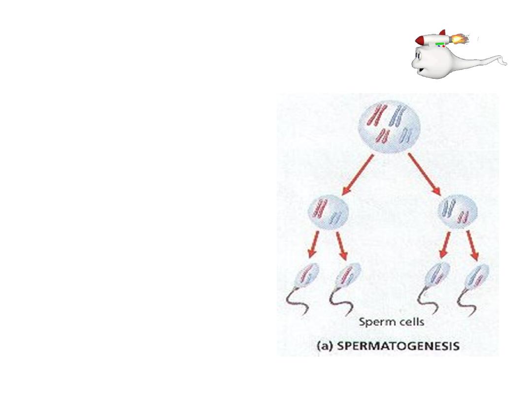
Spermatogenesis
The process of development of
spermatids
from
the
male
primordial germ cells and their
differentiation into spermatozoa.
Under
stimulation
anterior
pituitary
gonadotropic
hormones.
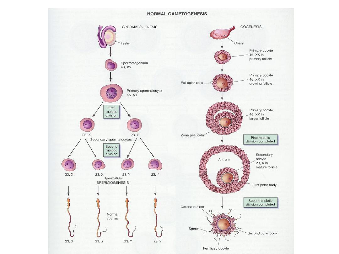
PROCESS :
1.
The
Primordial
germ
cells
develop into
spermatogonia
.

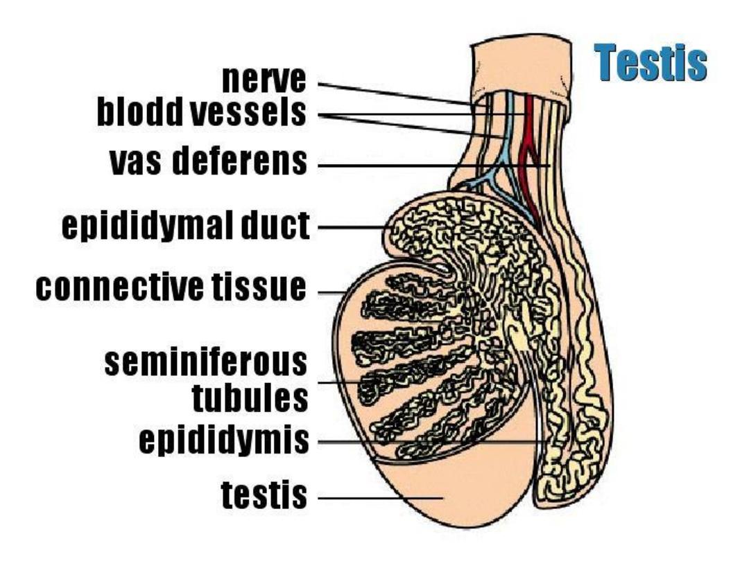
2.This occurs after
puberty,
and they remain in the wall of the
Seminiferous Tubule .
3.Spermatogonia form primary spermatocytes.
4.They remain in the prophase of
1
st
meiotic
division
for 16 days.
5. Each contains 22 pairs of autosomes and one pair of sex chromosome
XY.
6.Then
divides
into
two
secondary
spermatocytes.
(Meiotic
div.
completed).
7.Each secondary spermatocyte has equal cromosomes
.(22+X)
or
(22+Y).
8.Each of these divides again(2
nd
meiotic div), thus forming 4 Spermatids.
9.Each containing equal cytoplasm and
, HAPLOID
chromosomes….
TWO with 23X & TWO with 23Y
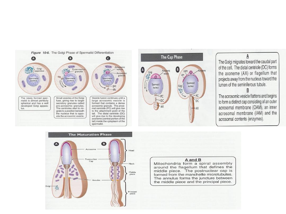
Spermiogenesis
Spermatids undergo morphological changes to form spermatozoa
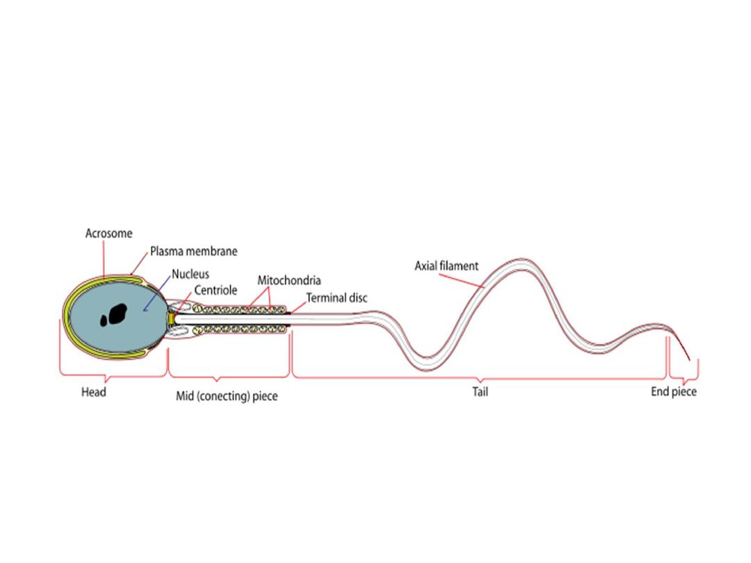

Structure Of The Sperm
The Head
• has two important features.
1. The acrosome
(derived from Golgi apparatus) contains
hydrolytic
enzymes
which are released when the sperm reaches an ovum. These
enzymes digest the outer membrane of the egg (proteins and complex
sugars) , allowing penetration of the sperm.
2. The nucleus
(haploid) contains a single set of
chromosomes
derived
from the male. This will include either an 'X' or 'Y' chromosome,
because of the way the XY separate during meiosis.

The Middle Piece
•
behind the head, contains numerous
mitochondria
. These respire
sugars in the semen to generate ATP in order to provide the
energy
for
movement of the tail.
The Tail
•
(Flagellum) contains microfilaments running the length of the tail
(arranged in the usual 9 + 2 )Rhythmic contraction of the filaments
causes the tail to wave and move against the fluid environment,
providing forward
motion
.
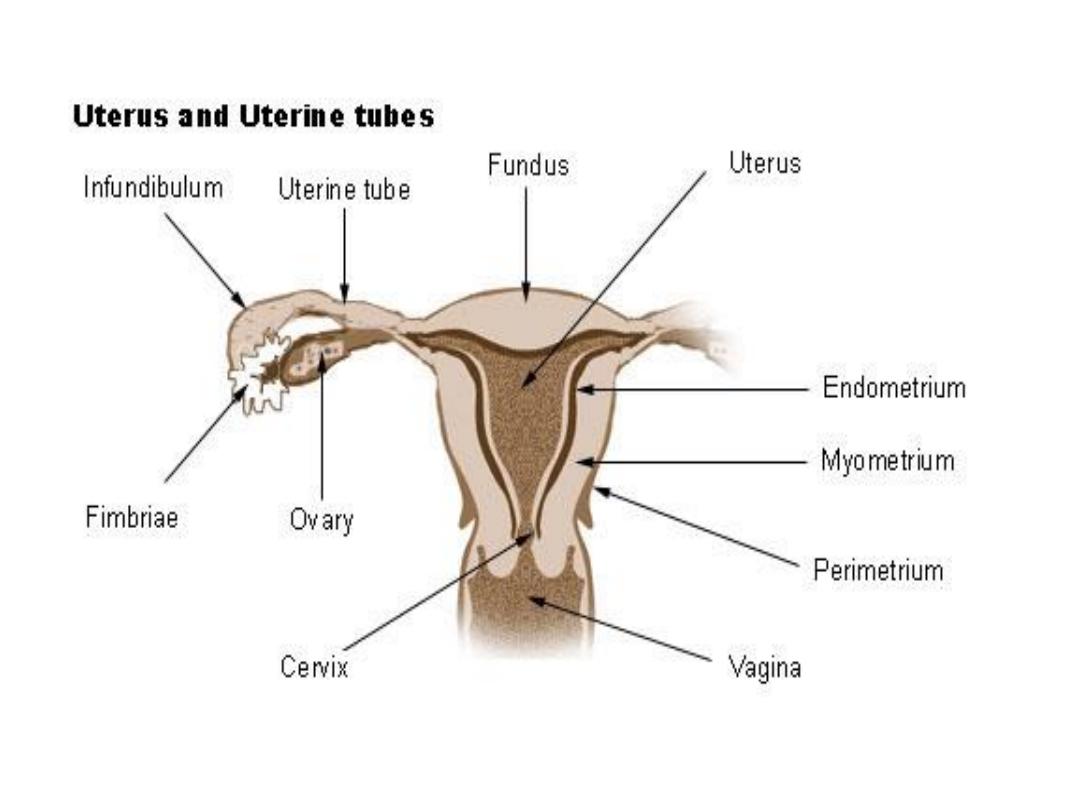
Female Genital System
The Ovary: female sex gland, produce ova.
The Uterus: in which the fetus develop.
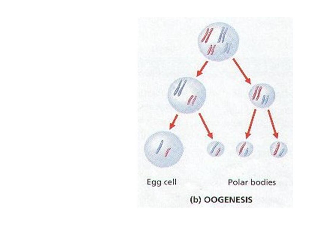
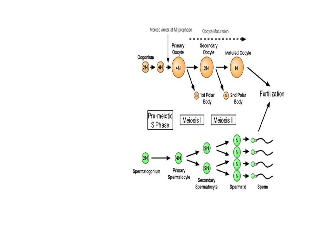
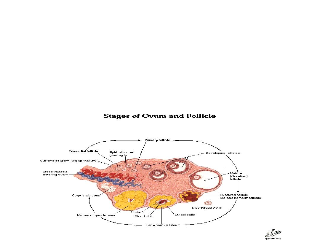
Oogenesis
Formation
of
female
gametes,
it
means
differentiation
of
female
primordial
germ
cell
(oogonium)
into
mature
ovum.
Oogonia(2N)
increased
in
the ovary through mitotic
division.
Oogonia
(2N)
grow
to
primary
oocytes
(2N)
intrauterine in primordial
follicle.
primary oocytes (2N) enter
meioses Ι but arrest until
preovulation complete it to
produce secondary oocytes
(N) & first polar body (N).
Then, complete the second
meioses to form mature ova
(N) and second polar body at
fertilization.

• The total number of primary oocytes at birth is estimated to vary
from 600,000 to 800,000.
•
During childhood, most oocytes become atretic; only approximately
40,000 are present by the beginning of puberty, and fewer than 500
will be ovulated. Some oocytes that reach maturity late in life have
been dormant in the diplotene stage of the first meiotic division for
40 years or more before ovulation. Whether the diplotene stage is
the most suitable phase to protect the oocyte against environmental
influences is unknown.
• The fact that the risk of having children with chromosomal
abnormalities increases with maternal age indicates that primary
oocytes are vulnerable to damage as they age.
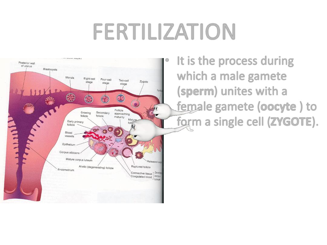
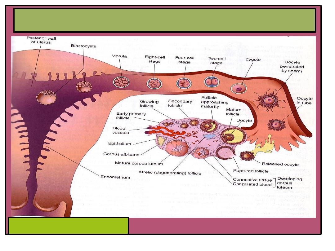
FERTILIZATION
• It is the process during
which a male gamete
(sperm) unites with a
female gamete (oocyte ) to
form a single cell (
ZYGOTE).
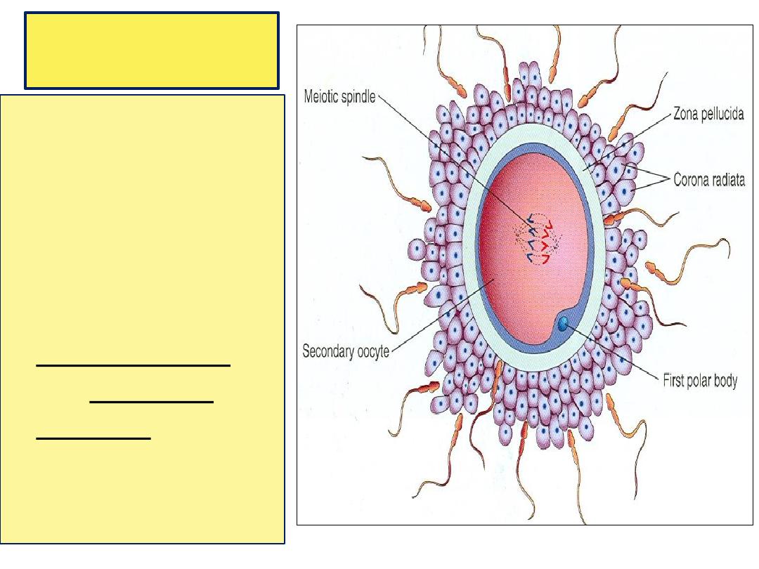
Fertilization
• Begins with a
contact
between
the sperm & the
ovum.
• Ends up with
intermingling of
the maternal and
paternal
chromosomes.
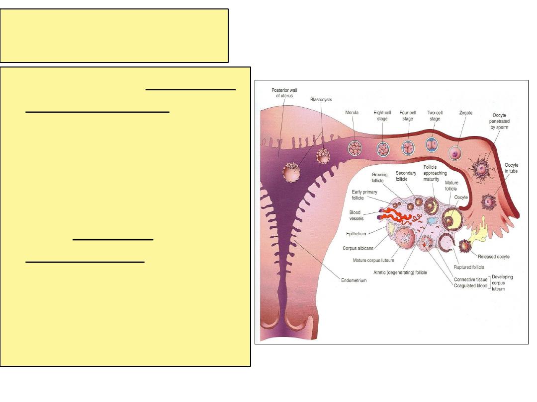
Site
• Usually in the ampulla of
the uterine tube.
• Ampulla is the longest
and widest part.
• Fertilization may occur in
other parts of tubes.
• Does not occur in the
uterine cavity.
• Chemical signals
from
oocyte attract the
sperms.
Spermatozoa
• Spermatozoa are not able to
fertilize the oocyte immediately
upon arrival in the female
genital tract but must undergo
• (1) capacitation and (2) the
acrosome reaction to acquire
this capability.
• Capacitation
is a period of
conditioning in the female
reproductive tract that in the
human lasts approximately 7
hours.

• capacitation occurs in the uterine tube and involves
epithelial interactions between the sperm and the
mucosal surface of the tube. During this time, a
glycoprotein coat and seminal plasma proteins are
removed from the plasma membrane that overlies the
acrosomal region of the spermatozoa.
• Only capacitated sperm can pass through the corona
cells and undergo the acrosome reaction.
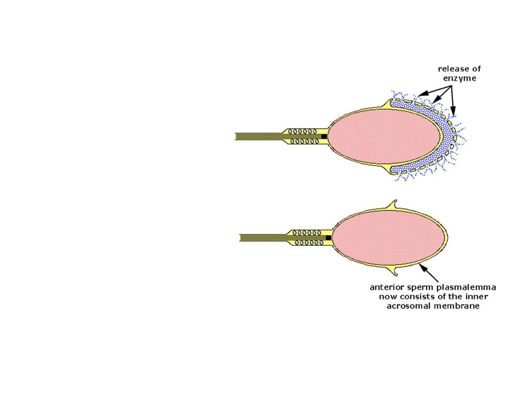
The acrosome
reaction,
occurs after binding to
the zona pellucida, is
induced by zona
proteins. This reaction
culminates in the release
of enzymes needed to
penetrate the zona
pellucida, including
acrosin- and trypsin-like
substances
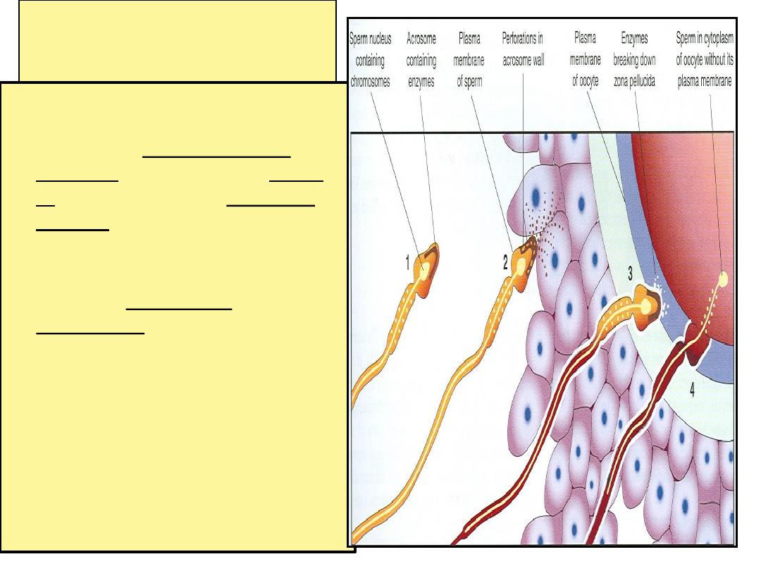
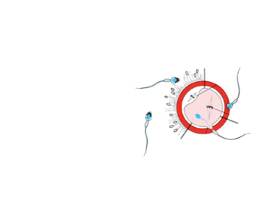
Phases of Fertilization
• 1- Passage of sperm through
corona radiata,
under the
effect of : hyaluronidase
enzyme from sperms, tubal
E. and movement of tail of
sperm.
• 2- Penetration of the zona
pellucida by head of sperms
through acrosine E. from
acrosome of one sperm.
• 3- Fusion of the plasma
membrane of the oocyte and
that of the sperm.
so
sperm’s plasma membrane
remains behind
.
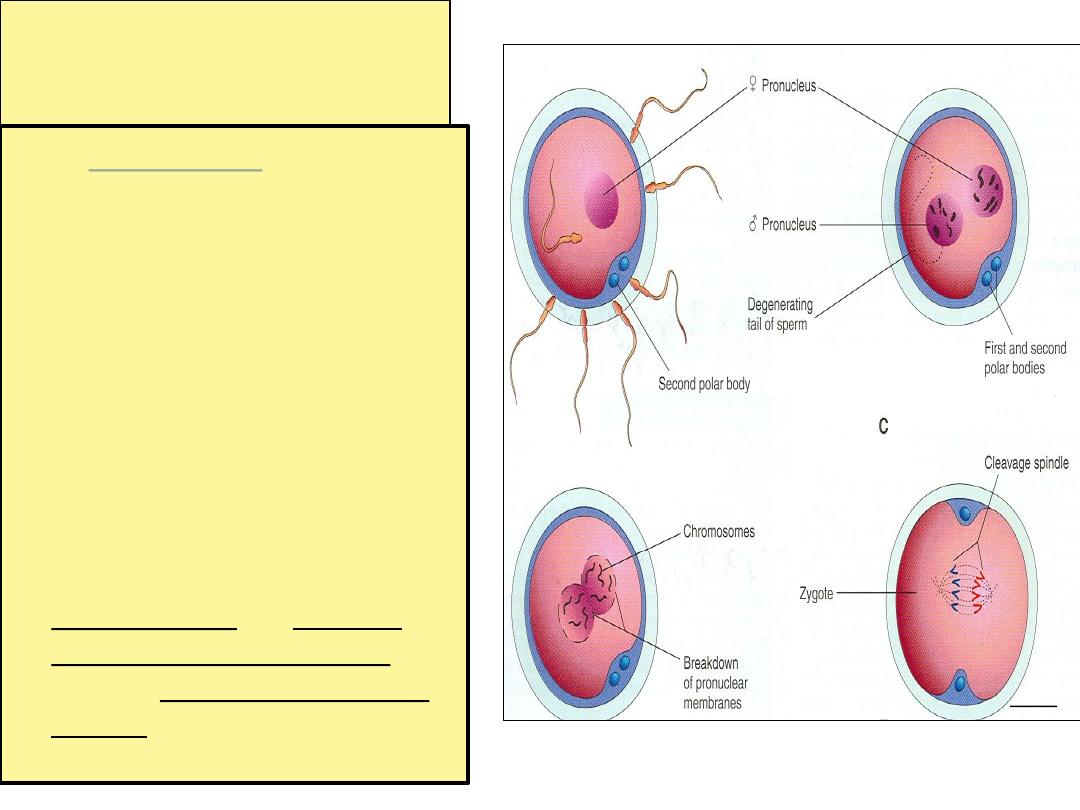
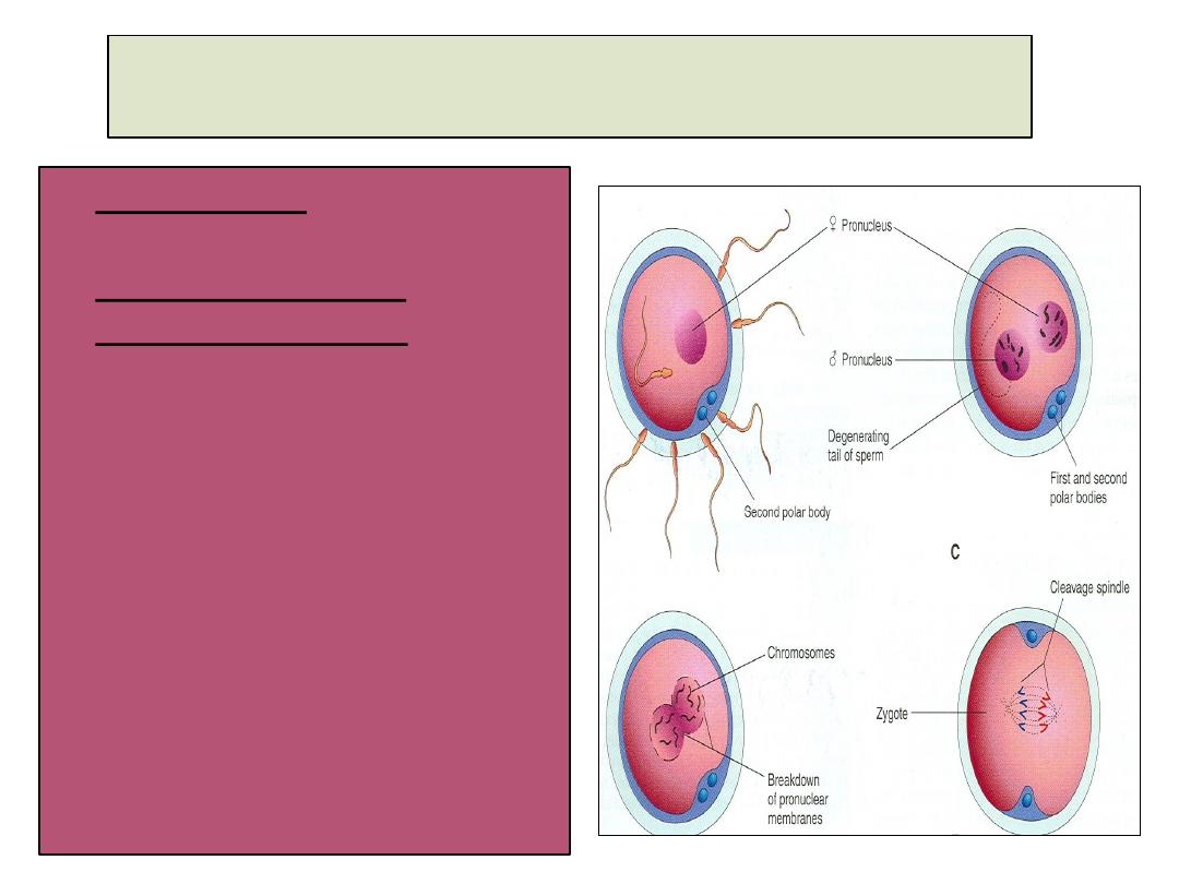
Phases of Fertilization
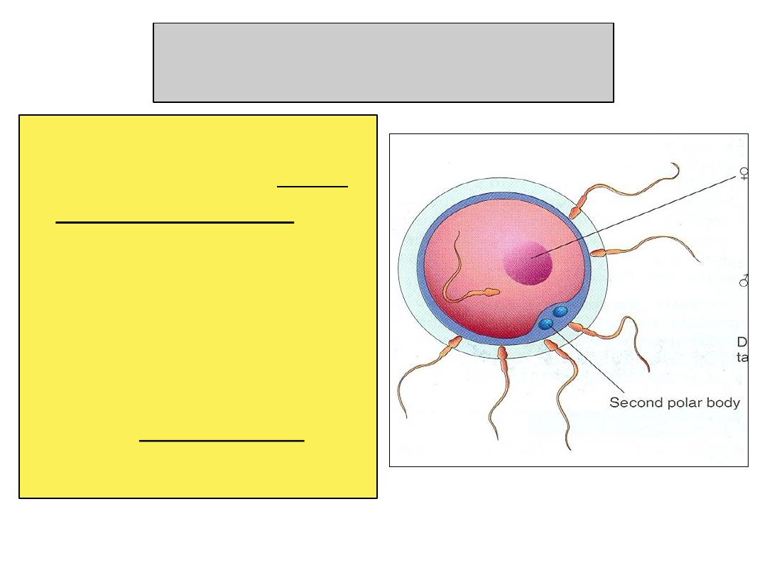
• 4- Completion of the
second meiotic division &
formation of the female
pronucleus.
• 5- Formation of the male
pronucleus :
It is a swollen nucleus of the
sperm.
Its tail is detached and
degenerated.
Zona reaction : it is a change in
properties of zona pellucida that
makes it impermeable to other
sperms.
zygote
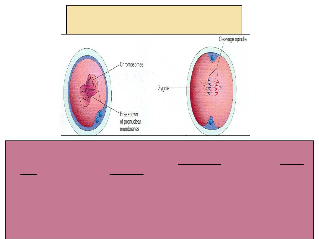
Chromosomes in zygote
• Zygote is genetically unique.
• Half of its
chromosomes
come from the father and the other
half comes from the mother.
• zygote
contains 46 chromosomes (diploid).
• New combination
is formed which is different from either of the
parents.
Sex of the Embryo
• Embryo's chromosomal
sex
is determined at the
time of fertilization.
• Sex is determined by the
type of sperm (X
or Y) that fertilizes the
oocyte.
• So, it is the father whose
gamete decides the sex.
Results of Fertilization
• Stimulates the
penetrated oocyte to
complete its 2
nd
meiotic division.
• Restores the normal
diploid number of
chromosomes in the
zygote (46).
• Determines the
chromosomal sex of the
embryo.
• Initiates cleavage (cell
division) of the zygote.
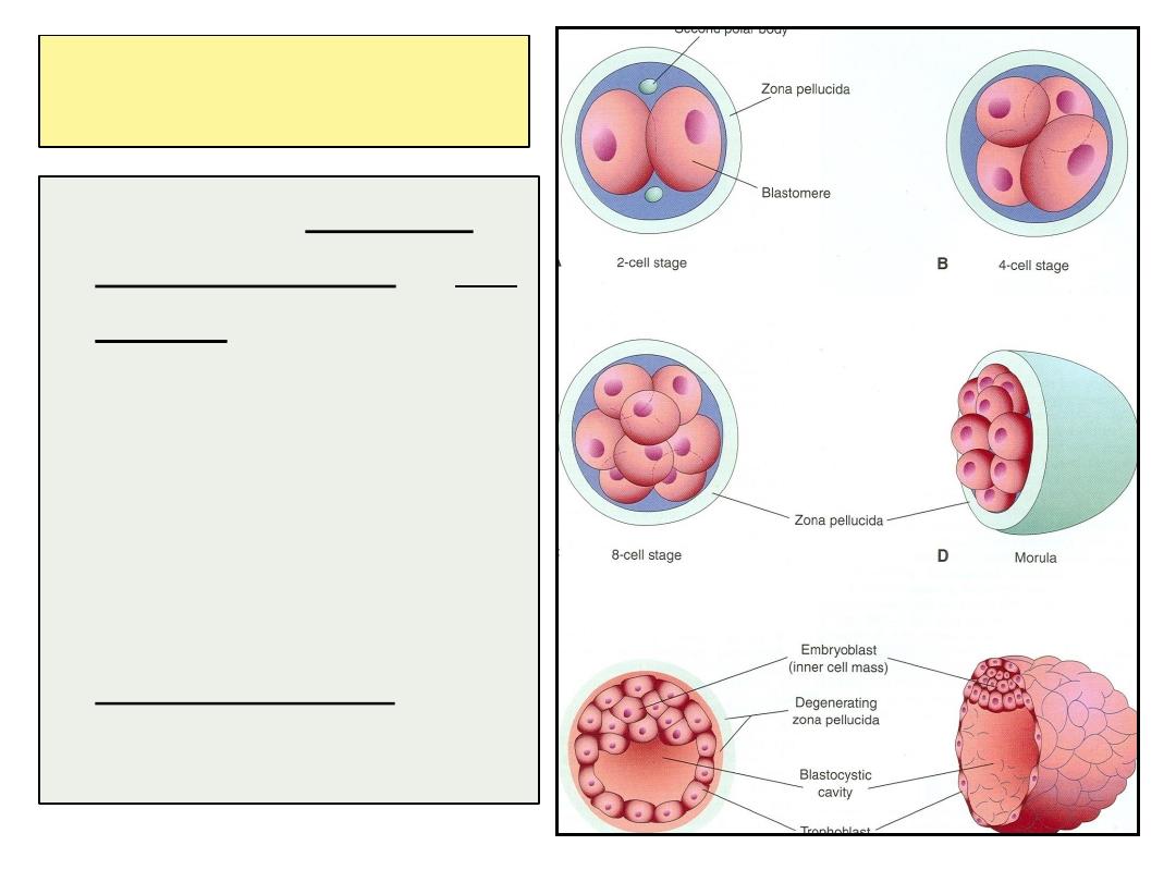
Cleavage of Zygote
• Consists of repeated
mitotic divisions of the
zygote.
• Rapid increase in the
number of the cells.
• These smaller
embryonic cells are
called
Blastomeres.
• Normally occurs in the
uterine tube.
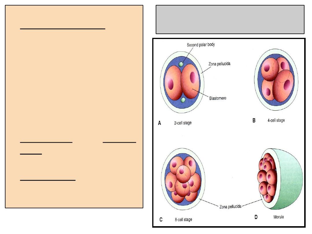
Cleavage of Zygote
• It begins about
30
hours
after fertilization.
• Zygote divides into 2,
then 4, then 8, then 16
cells.
• Zygote lies within the
thick
zona pellucida
during cleavage.
• Zygote migrates in the
uterine tube from its
lateral end to its medial
end.
• Zona pellucida
is
translucent under light
microscope.
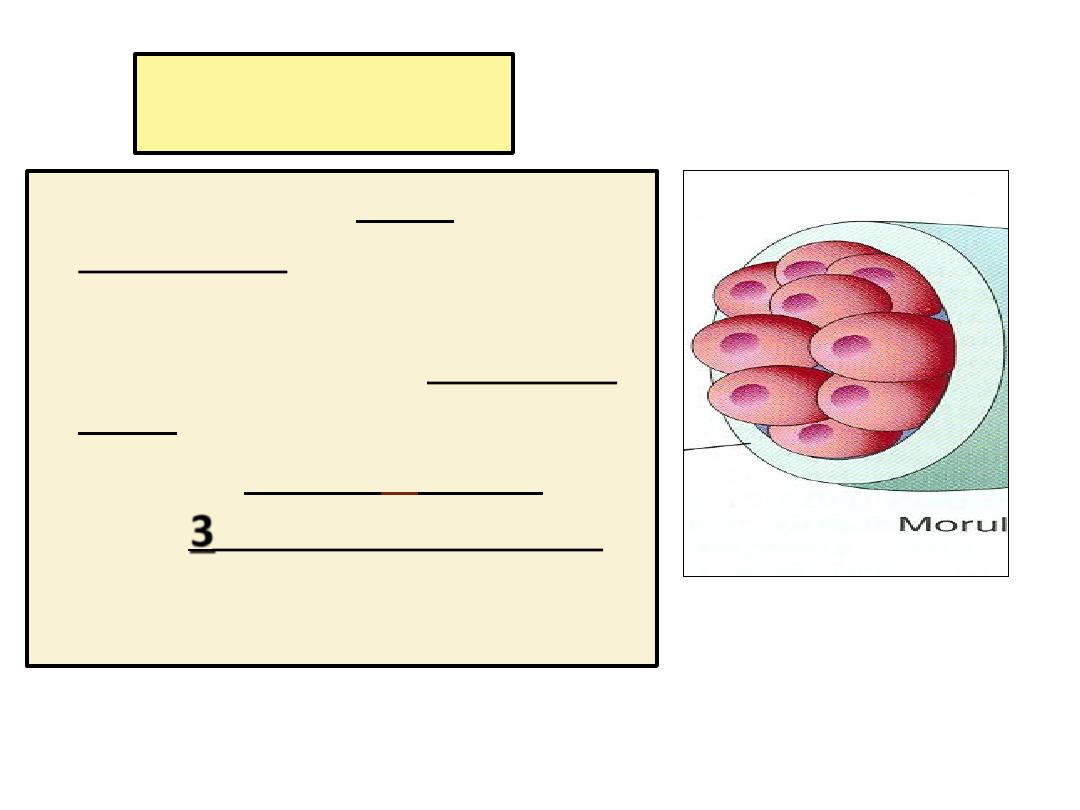
Morula
• When there are 16-32
blastomeres the developing
human is called
MORULA.
• The Morula reaches the uterine
cavity at this stage.
• Spherical Morula
is
formed
about
3
days after fertilization.
• Formation of blastocyst :
• The Morula reaches the uterine cavity by the 4
th
day
after fertilization, & remains free for one or two days.
Fluid passes from uterine cavity to the Morula.
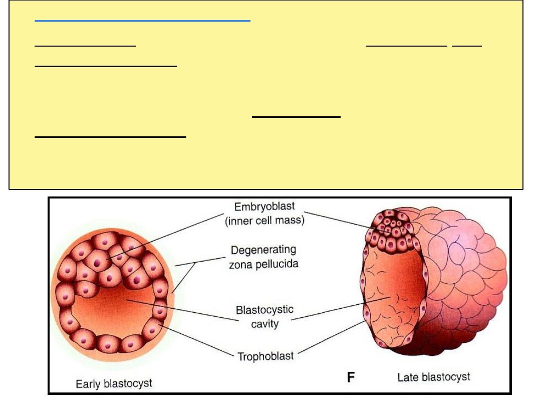
• Now the Morula is called Blastocyst, its cavity is called
blastocystic cavity, its cells divided into
Embryoblast
&
Trophoblast.
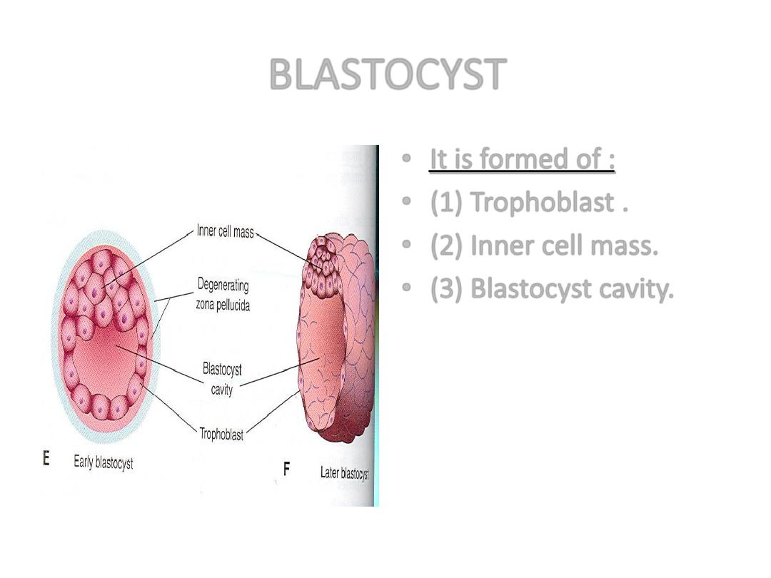
BLASTOCYST
• It is formed of :
• (1) Trophoblast .
• (2) Inner cell mass.
• (3) Blastocyst cavity.
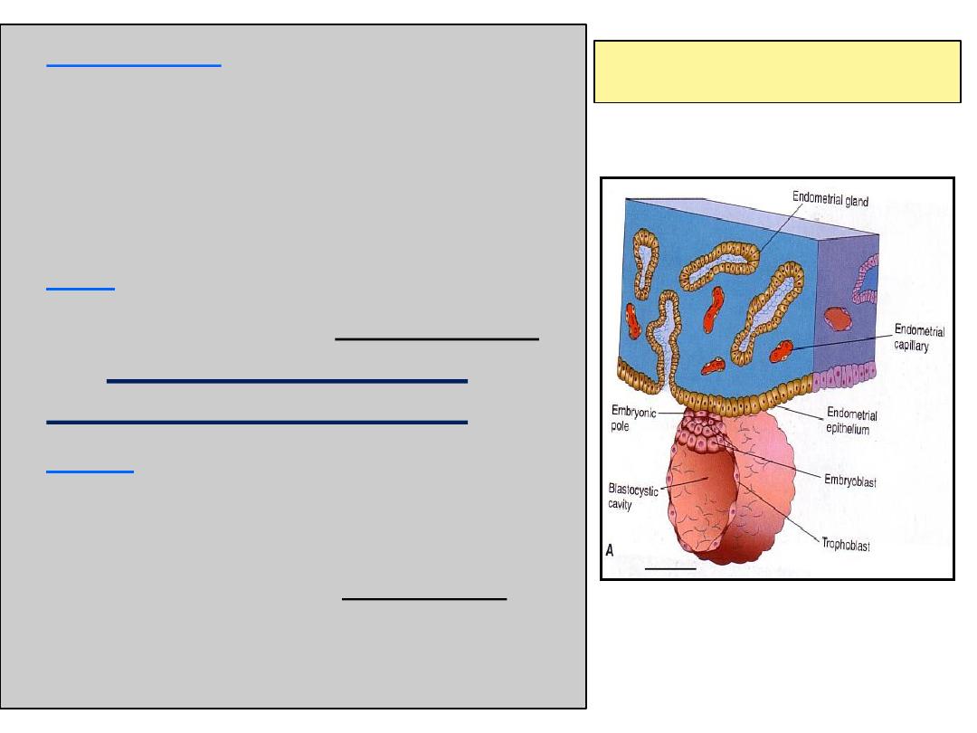
• Definition :
• It is the process by which the
Blastocyst penetrates the
superficial
(Compact) layer of the
endometrium of the uterus.
• Site:
• The normal site of implantation is
the
posterior wall of the
uterus near the fundus
.
• Time:
• It
begins
about the 6
th
day after
fertilization.
• It is
completed
by the 11th or 12th day
.
IMPLANTATION
6
th
day
By the 5
th
day the
Zona pellucida
degenerates.
Blastocyst begins
implantation
by the 6
th
day.
Trophoblast cells
penetrate the epithelium of the endometrium.
Penetration results from proteolytic enzymes (eg.COX-2)
produced by the
trophoblast
.
6
th
day
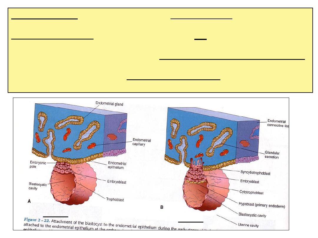
7
th
day
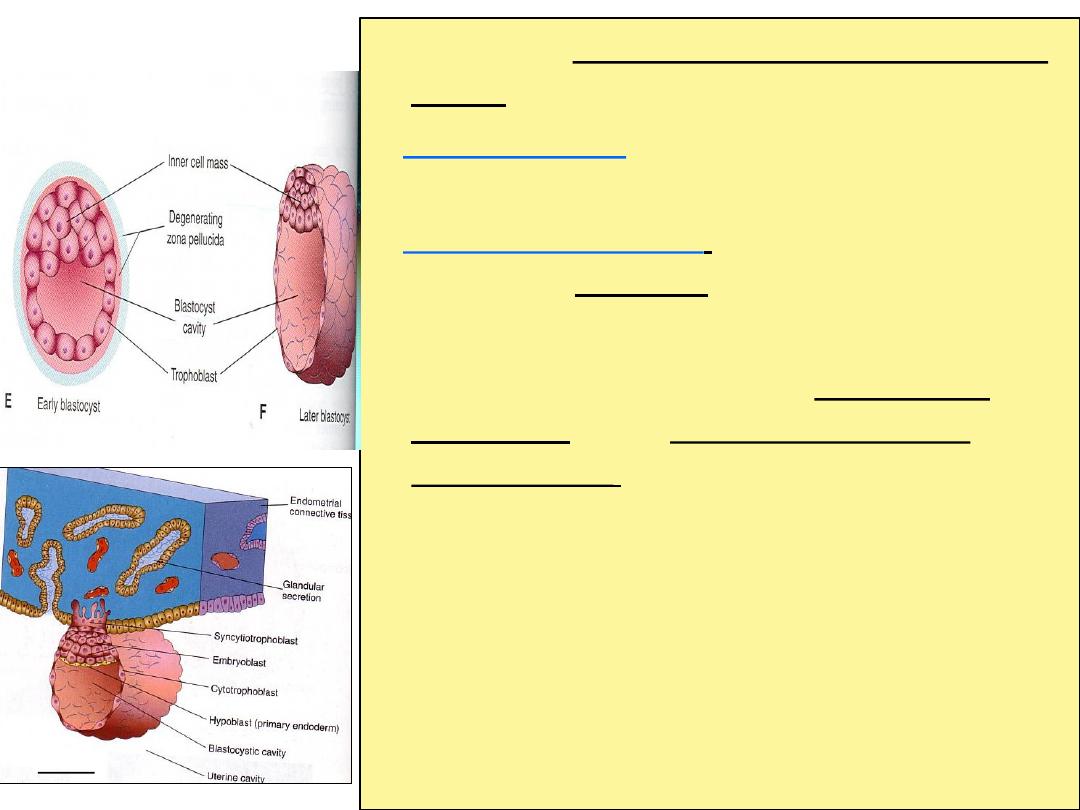
• By 7
th
day,
Trophoblast differentiated into 2
layers:
Cytotrophblast,
inner layer, mononucleated
mitotically active cells.
Syncytiotrophoblast
(outer multinucleated
mass, with indistinct cell boundary.
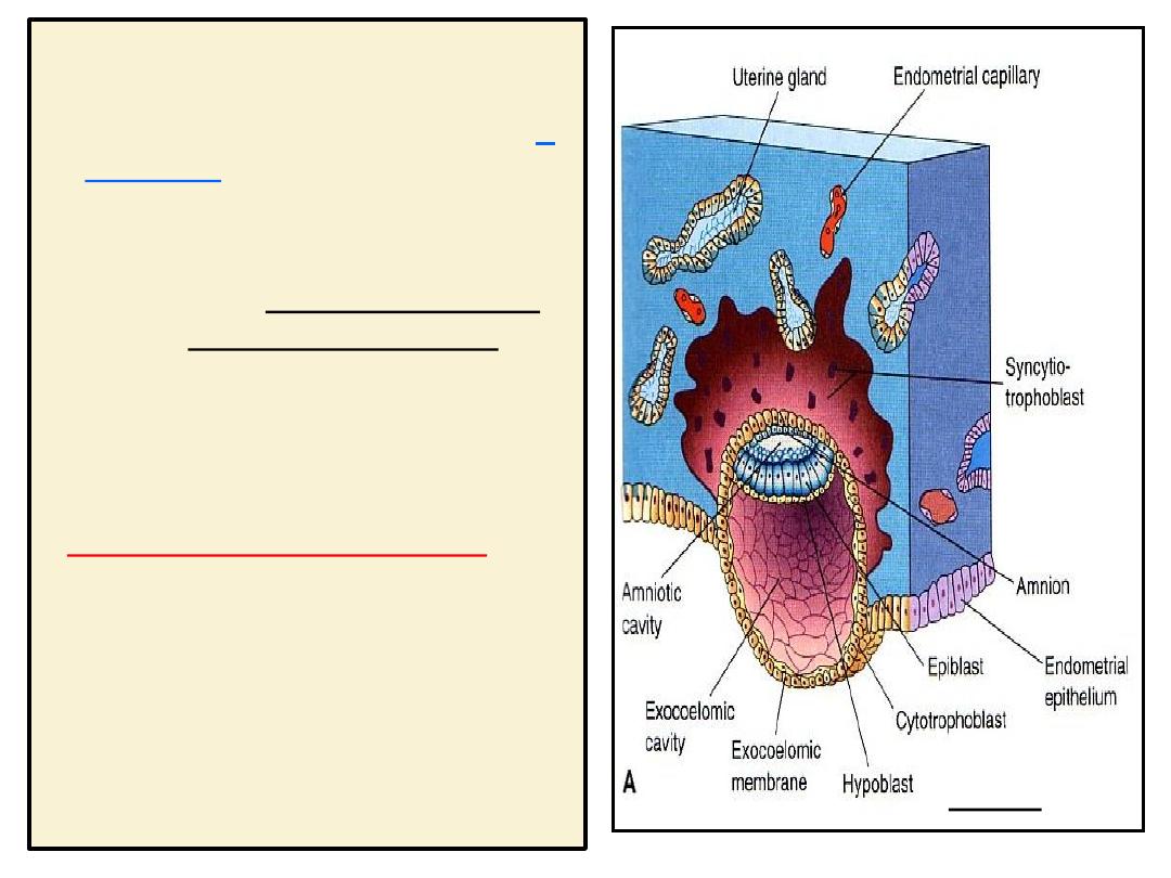
• By 8
th
day
the blastocyst is superficially
embedded in the compact layer of the
endometrium (by the end of 1
st
week,the
blastocyst is superficially implanted in
endometrium).
7
th
day
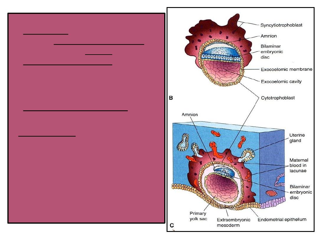
• Blood-filled Lacunae
appear in
the Syncytiotrophoblast
which communicate forming
a
network by the 10th
day or 11
th
day.
• Syncytiotrophoblast
erodes
the endothelial lining
of the maternal capillaries
which known as sinusoids.
Now blood of maternal
capillaries reaches the lacunae
so
Uteroplacental circulation
is established by
11
th
or 12
th
day.
8
th
day
Endometrial cells undergo
apoptosis (programmed cell
death) to facilitates invasion of
endometrium by the
Syncytiotrophoblast.
Syncytiotrophoblast engulf
these degenerated cells for
nutrition of the embryo.
Implantation
can be detected by:
1- Ultrasonography.
2- hCG
(human chorionic
gonadotrophin which is
secreted by the
Syncytiotrophoblast) about
the
end of 2
nd
week
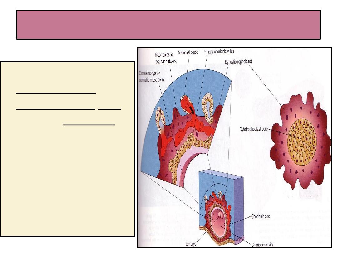
Formation of The Primary Chorionic villi
• By the 13
th
day
Proliferation of
Cytotrophblast cells
produce extension
inside the
Syncytiotropho-blast
to form
primary the
chorionic villi.

THANK YOU