Second Week of
Development: Bilaminar Germ DiscDr.Sumeya
DAY 8
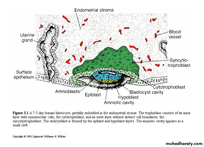
At the eighth day of development, the blastocyst ispartially embedded in the endometrial stroma. In
the area over the embryoblast, the trophoblast has
differentiated into two layers: (1) an inner layer of
mononucleated cells, the cytotrophoblast, and
(2) an outer multinucleated zone without distinct
cell boundaries, the syncytiotrophoblast.
•
Mitotic figures are found in the cytotrophoblast but not in the syncytiotrophoblast. Thus, cells in the cytotrophoblast divide and migrate into the syncytiotrophoblast, where they fuse and lose their individual cell membranes.
The syncytiotrophoblast is responsible for hormone production. hCG maintains the corpus luteum in the ovary, allowing it to continue to produce P+E.
Produces P+ E to
maintain the pregnancy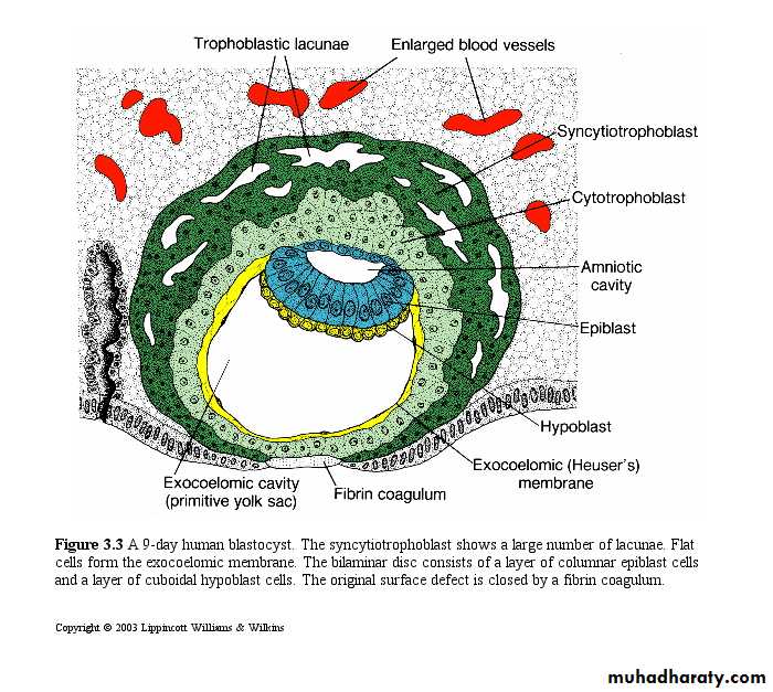
Ovary
UterushCG
• The cells of the embryoblast will also differentiate into 2 layers:
• The epiblast- a layer of high, columnar cells adjacent to the amniotic cavity.• The hypoblast- A layer of small cuboidal cells adjacent to the blastocyst cavity.
• Together these layers form a flat disc.
•
. At the same time, a small cavity appears within the epiblast. This cavity enlarges to become the amniotic cavity.
Epiblast cells adjacent to the cytotrophoblast are called amnioblasts; together with the rest of the epiblast, they line the amniotic cavity .
The endometrial stroma adjacent to the implantation site is edematous and highly vascular. The large, tortuous glands secrete abundant glycogen and mucus.
DAY 9
The blastocyst is more deeply embedded in the endometrium, and the penetration defect in the surface epithelium is closed by a fibrin coagulum .The trophoblast shows considerable progress in development, particularly at the embryonic pole, where vacuoles appear in the syncytium. When these vacuoles fuse, they form large lacunae, and this phase of trophoblast development is thus known as the lacunar stage.
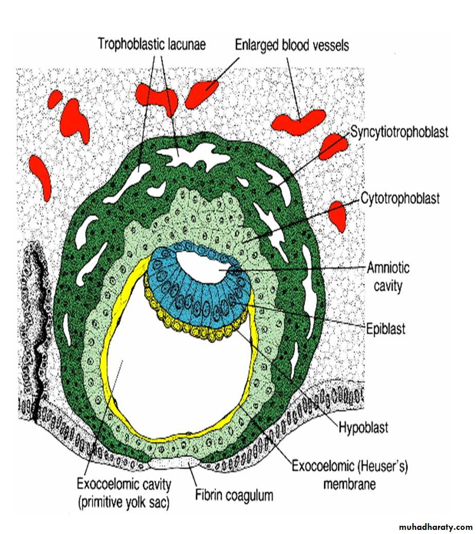
At the abembryonic pole, meanwhile, fl attened cells probably originating from the hypoblast form a thin membrane, the exocoelomic (Heuser’s) membrane that lines the inner surface of the cytotrophoblast .
This membrane, together with the hypoblast, forms the lining of the exocoelomic cavity, or primitive yolk sac.
DAY 11 AND 12
The blastocyst is more deeply embedded in the
endometrium, and the penetration defect in the
surface epithelium is closed by a fi brin coagulum. The trophoblast is characterized by
lacunar spaces in the syncytium that form an intercommunicating network.
cells of the syncytiotrophoblast penetrate deeper into the stroma and erode the endothelial lining of the maternal capillaries.
These capillaries, which are congested and dilated, are known as sinusoids.
The syncytial lacunae become continuous with the sinusoids, and maternal blood enters the lacunar system.
As the trophoblast continues to erode more and more sinusoids, maternal blood begins to fl ow through the trophoblastic system, establishing the uteroplacental circulation.
New cells appear between the yolk sac and the cytotrophoblast
They form a layer of loose connective tissue: extraembryonic mesoderm.Cavities or spaces appear in the extra-embryonic mesoderm
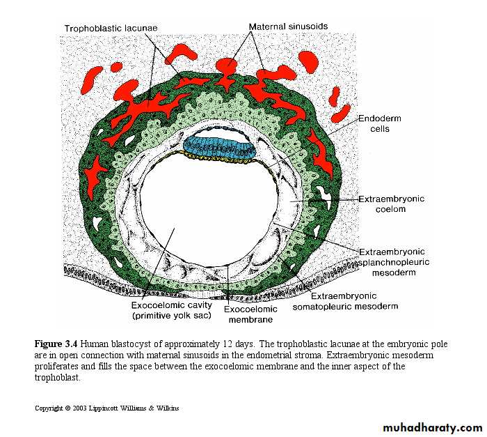
• The cavities form a new space- the chorionic cavity
• The cavity divides the extraembryonic mesoderm into the
• Extraembryonic somatic mesoderm- lining trophoblast and amnion
• Extraembryonic splanchnic mesoderm- lines the yolk sac
Growth of the bilaminar disc is relatively slow compared with that of the trophoblast; consequently, the disc remains very small .
Cells of the endometrium, meanwhile, become polyhedral and loaded with glycogen and lipids; intercellular spaces are filled with extravasate, and the tissue is edematous.
These changes, known as the decidua reaction, at fi rst are confi ned to the area immediately surrounding the implantation site but soon occur throughout the endometrium.
DAY 13

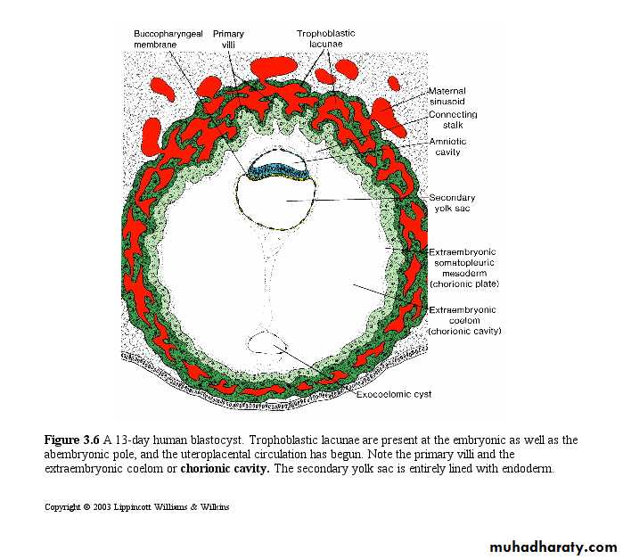
By the 13th day of development, the surface
defect in the endometrium has usually healed.however, bleeding occurs at the implantation site as a result of increased blood flow into the lacunar spaces. Because this bleeding occurs near the 28th day of the menstrual cycle , it may be confused with normal menstrual bleeding and, therefore , may cause inaccuracy in determining the expected delivery date.
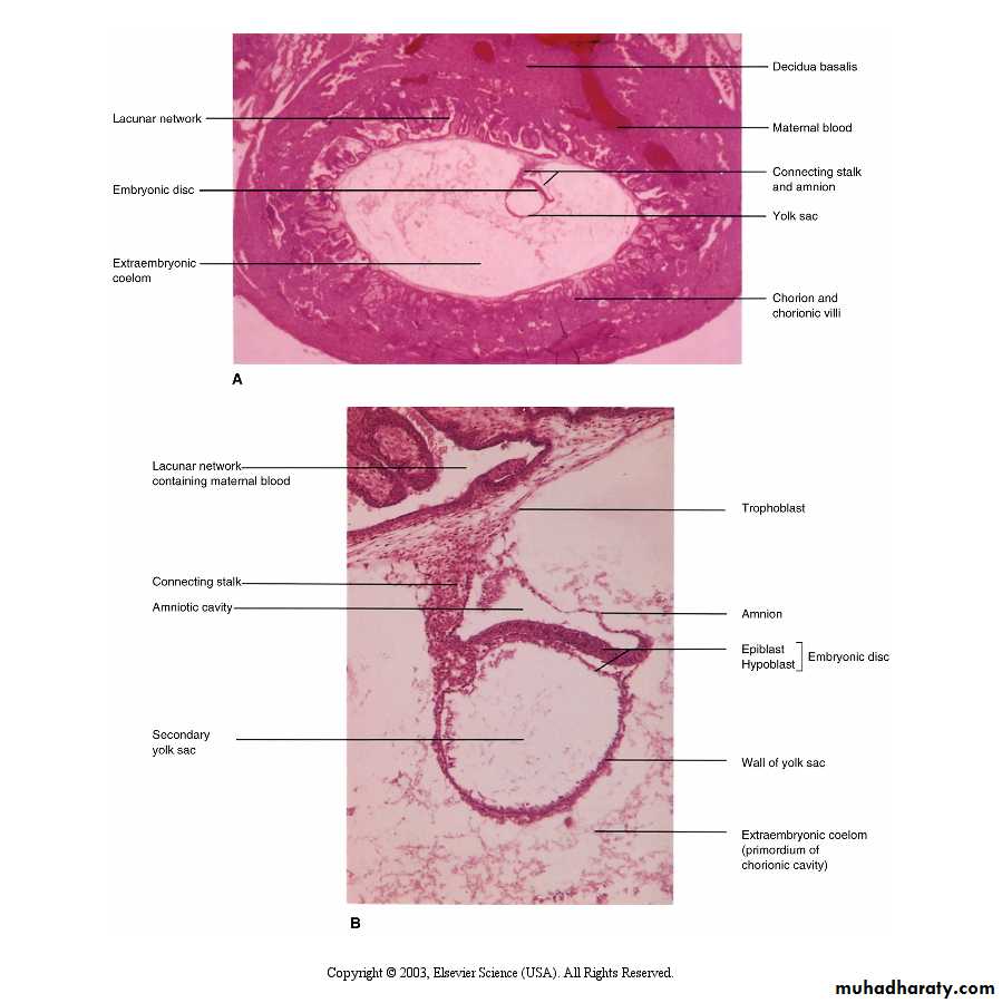
The trophoblast is characterized by villous structures. Cells of the cytotrophoblast proliferate locally and penetrate into the syncytiotrophoblast ,forming cellular columns surrounded by syncytium .
Cellular column with the syncytial covering are known as primary villi .
In the meantime , the hypoblast produces additional cells that migrate along the inside of the exocoelomic membrane. These cells proliferate and gradually form a new cavity within the exocoelomic cavity (secondary yolk sac or definitive yolk sac )
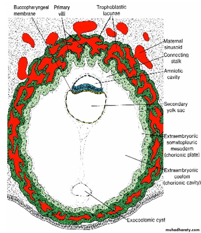
During its formation ,large portions of the exocoelomic cavity are pinched off. These portions are represented by exocoelomic cysts , which are often found in the extraembryonic coelom or chorionic cavity.
Meanwhile , the extraembryonic coelom expands and form a large cavity ,the chorionic cavity .The extraembryonic mesoderm lining the inside of the cytotrophoblast is then known as the chorionic plate .
The only place where extraembyonic mesoderm traverse the chorionic cavity is in the connecting stalk . With the development of blood vessels, the stalk becomes umbilical cord.
At the end of the second week:
Trophoblast has had a period of growth- greater than the embryoblast.The 2 layer bilaminar disc is formed and will give rise to other tissues and structures.
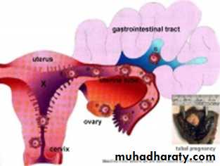
Abnormal Implantation
Ectopic Pregnancy
• occurs if implantation is in uterine tube or outside the uterus (external surface of uterus, ovary, gastrointestinal tract, mesentry, peritoneal wall)
Ectopic Pregnancy
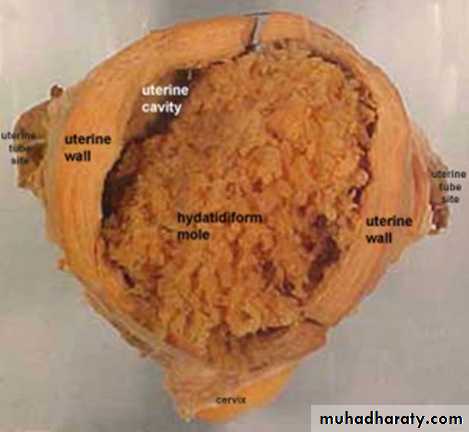
Hydatidiform Mole
the conceptus trophoblast layers proliferates and not the embryoblast,A haploid sperm fertilizing an egg without a female pronucleus
The tumour has a "grape-like" placental appearance without enclosed embryo formation.
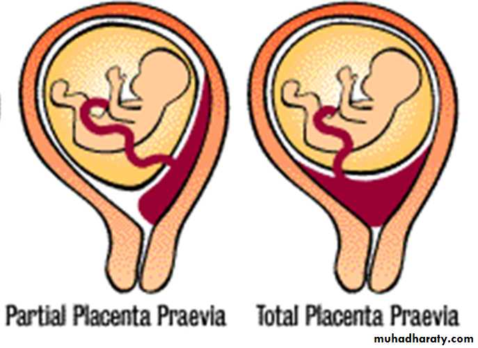
Abnormal Placental Sites
Uterine:Implantation in the lower segment leads to placenta praevia