X – Ray beam
shapeand
position
Central ray: - in the beam X – ray photons that traveling in very center of the cone of radiation, called [central ray] and this is commonly used to fix and locate the position of X – ray beam.
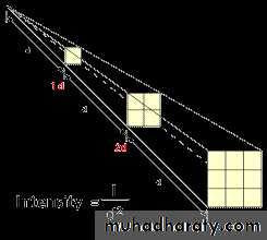
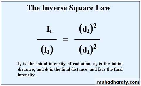
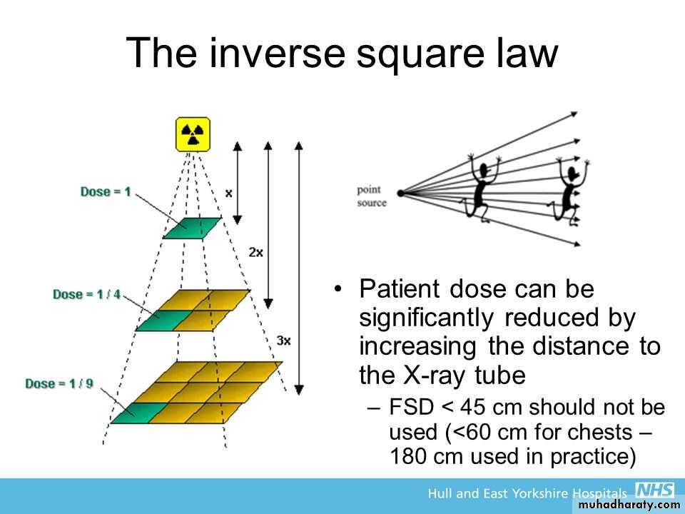
Inverse square law:- the low stated that (the intensity of radiation inversely proportional with the square of distance measured from the source of radiation to the point of measuring )
I : intensity
D: Distance(D2) 2
I ×(D1)1
Energy conversion to x – ray:-
X – ray photons result from conversion of kinetic energy of cathode electrons into X – ray photon energy, and this basically accomplished by 2 ways:-First:-
Electrons can be brought to stop on the surface of the anode (target) through collision with tungsten atoms of the target giving up all of their kinetic energy to X – ray photons such photons will be of high energy (short wave length photons).
Some of cathode electrons giving up only part of their energy and the resultant X – ray photons have low energy (long wave length photons) and most of X – ray photons are created by this manner.
Second:-
When cathode electrons able to dislodge one or more orbital electrons of tungsten atom. For example cathode electron must posses more than 69,000 electron volts to dislodge K orbital electron of tungsten atom and this is only occurred when X – ray machine set at 70 Kilovolts or more. Then after the dislodgment of K electron from its shell an electron from L shell falls into the empty place of K shell.Rectification:-
The main supply to the X – ray machine of 240 volts has 2 functions:-A – Generate the high potential difference (kV) to accelerate the electrons across the x – ray tube via the step up transformer.
B – Provide the low – voltage current to heat the filament via the step – down transformer.
However, the incoming 240 volts is an alternating current with typical wave form as shown:-
Voltage
+
-
Time
Half of cycle is (+) and other half is (-) but for production of X – ray only the positive half of the cycle can be used to ensure that the electrons from the cathode filament are always drawn towards the anode target.Thus, the stepped – up high voltage applied across the X – ray tube needs to be rectified to eliminate the negative half of the cycle.
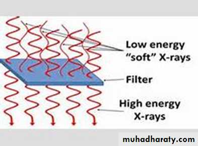
* Filtration:- X – ray used in dentistry must be able to penetrate dental hard tissues (teeth and bone). The longer wave length x-ray (soft X– ray) are not useful in diagnostic radiology thus removal of these long wave length photons from the beam by passing the beam through a filter made from Aluminum, this filter either built into the X-ray machine by manufacturer or added as an extra filter.
The effect of filtration on x – ray beam is absorption of most of long wave length photons (soft X – ray) so the resulting x – ray beam will consist of mainly X – ray photons of short wave length with high energy photons and high penetrating power that’s why they named (hard X – ray beam).
* Collimation:-
Is a process refers to control of size and shape of X – ray beam. In diagnostic radiography its essential to get the diameter of circular X – ray beam at patients skin surface is not great than 2.75 inches, while forrectangular X – ray beam the dimensions at the skin should be approximately 1½ × 2 inches.
Collimation can achieved by one of 2 methods:-
1.Using diaphragms (round or rectangular shape).
2.Using metal cylinders, cones and rectangular tubes.


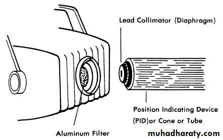
Diaphragm:-
Consists of a metal plate or disk made from lead. A hole is present in the center of the disk allow the beam to pass through it only.The shape of X – ray beam determine by the shape of the hole of diaphragm such diaphragm is placed over the opening in the head of X – ray machine.
X – Ray spectrum:-
X– Ray beam consist of many photons of different wave length because:
A - Electrons don’t give up all their kinetic energy in identical fashion.
B – Potential voltage across the X– ray tube changes constantly as the AC voltage varies to DC.
The minimum photon wavelength means power. That’s why X – ray beam is poly- different wave length.
Note:- X– ray photon wave length can calculated from this formula:-
12.35
kV=
Minimum photon wavelength in A˚
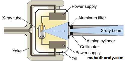
Tube head showing a recessed x-ray tube, components of the power supply, and oil that conducts heat away from the x-ray tube
Definition of terms used in X – ray interaction:-
Scattering: - change in direction of photon with or without a loss of energy.Absorption: - deposition of energy i.e. removal of energy from the beam.
Attenuation: - reduction in the intensity of X– ray beam caused by absorption and scattering attenuation = absorption + scattering.
Ionization: - removal of an electron from neutral atom.
Absorption of x – ray (interaction with matter):-
X–Ray are absorbed by any form of matter (solid, liquid, and gas) when photons reach an atom, 4things can happen:-1.X – Ray photons can pass through the atom without change occurred to either.
2.It can be deviated from its direction by the atom so the x – ray photon after deviation becomes a photon of scattered radiation.
3. X – Ray photon interacts with inner – shell electron of the tissues atom where the X – ray photon disappears and deposits all i ts energy this process is pure absorption. Now the inner – shell electron is ejected with considerable energy (now called a photo – electron) into the tissue for further interaction with other electrons of other tissue atoms.So this high – energy ejected photo electron be haves like the original high energy X – ray photons interact and eject other electrons as it passes through the tissues, these ejected electrons that are responsible for the majority of ionization interactions within the tissue and the possible resulting damage attributable to the X– rays.
4.When the X – ray photon interacts with free or loosely bond outer shell electron of tissue atom.
So the electron is ejected (called Compton recoil electron) with some energy from the X – ray photon i.e. there is some absorption and this ejected electron undergoes further ionization interaction within the tissue.
While the remainder of X – ray photon energy is scattered from its original path as scattered X– ray photon.
Incoming x – ray photon
Photon completely scatter with no loss energy
Photon totally absorbedPhoton scatter with some loss of energy
Photon transmitted unchanged
Primary radiation:- radiation emerging from the x – ray machine in form of collimated useful X – ray beam
Secondary radiation:- radiation result from interaction of primary beam with matter
Half – value layer:- it’s a method of monitoring the penetration quality of the X – ray beam. Determination of half – value layer is done by placing thin filtering material such as aluminum filter in front of the beam so we continue increase the thickness of filtering material until we have a thickness that reduce the number of X– ray photons in the beam passing through it to (one half) this will representing a half – value layer for such beam of radiation.
High half value:- layer the high penetrating ability of the beam. In oral diagnosis the acceptable value is approximately 2 mm of aluminum
X- ray measuring:-
1.Traditional unitsRoentgen:- is the amount of x-radiation or gamma radiation which will produce in one cc of air ions carrying one electrostatic unit of either sign .
rad: it is a roentgens absorbed dose
rem: it is a roentgens equivalent man dose.
RBE: is a relative biological effectiveness dose.
Curie
2. International System of Units (SI system)
*Coulomb per Kilogram (C/Kg)1C/Kg =3876 R 1R=2.58*10^-4C/Kg
*Gray (Gy):The gray (Gy) is the SI unit used to measure the energy imparted to irradiated matter and is defined as the absorbed radiation dose of one joule (J) per kg
1Gy=100 rad 1rad=0.01 Gy
*Sievert (Sv) : the SI unit of dose equivalent (the biological effect of ionizing radiation), equal to an effective dose of a joule of energy per kilogram of recipient mass.
1Sv=100rem 1rem=0.01Sv
*Becquerel (Bq) : One becquerel is defined as the activity of a quantity of radioactive material in which one nucleus decays per second.
Radiation Units and Conversion Factors
ExposureConventional Unit
SI Unit
Conversions
Exposure
roentgen (R)
coulomb/kg of air (C/kg)
1 C/kg = 3876 R
1 R = 258 uC/kg
Doserad (R)
gray (Gy)
1 Gy = 100 rad
Dose equivalent
rem
sievert (Sv)
1 Sv = 100 rem
Activity
curie (Ci)
becquerel (Bq)
1 mCi = 37 mBq