


Dr. Jamal Al-Saidy
Assistant Professor and
Consultant Orthopaedic Surgeon
osteochondritis

Definition
Group of disorders in which there is demarction, fragmentation or
separation and necrosis of bone and cartilage
Mainly adolescent and young adult.
During phases of increase physical activity.
May initiated by trauma or repetitive stress .
Occur in epiphysis or apophysis
There is no inflammation
Pathology
: Features of AVN
Pathgenesis…
still not completely understood
.
Impact injuries cause bleeding or oedema in the
subarticular bone.
Capillary compression or thrombosis and ischemia.
separation a necrotic oseteochondral fragment.

Types
:-
• Crushing
• Splitting
(osteochondritis dissecans )
• Pulling
( tractional apophysitis )

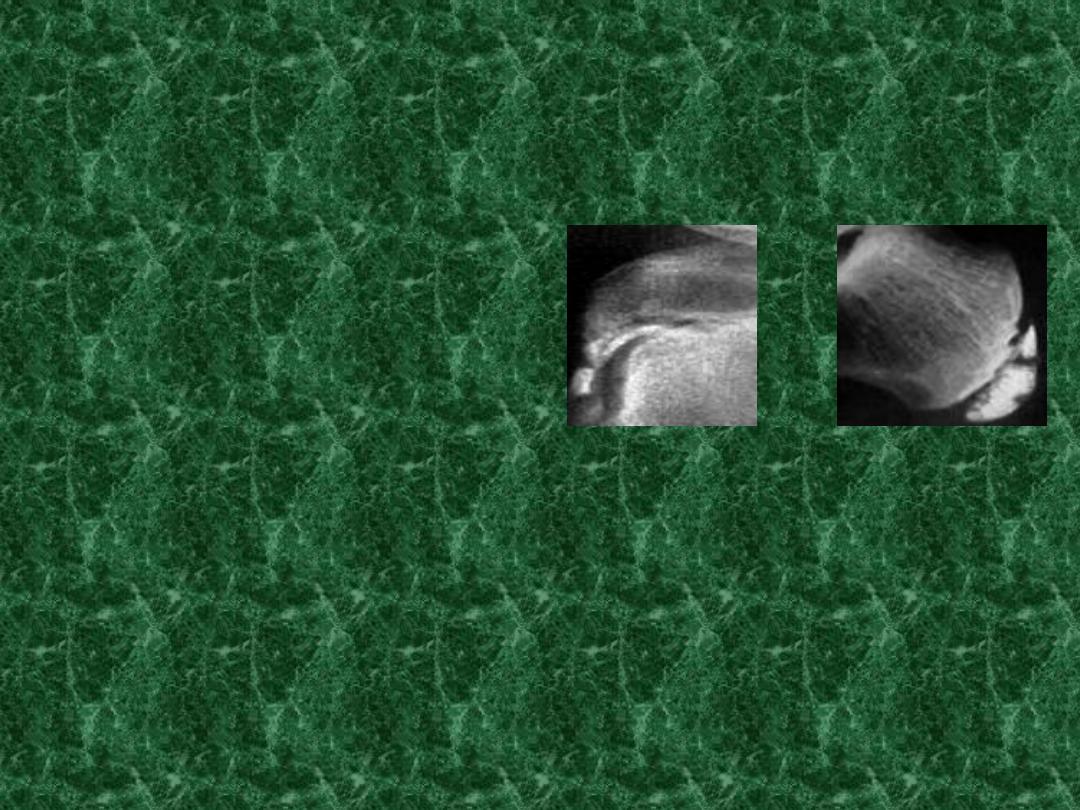
Crushing Osteochondritis
AVN of the ossification centre
in a long bone epiphysis or one of the
cuboidal bones of the wrist or foot
.
usually in late adolescent.
The path….
same as those in other form of osetonecrosis
bone death.
fragmentation.
distortion of the necrotic segment.
reactive new bone formation around the ischemic trabeculae.

Clinical F:
pain
limitation of movement.
Tenderness
x-ray
:
increase density.
Later stages distortion and collapse
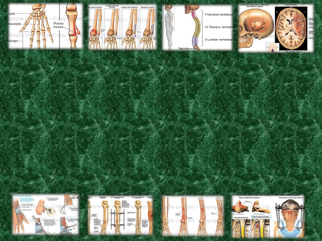
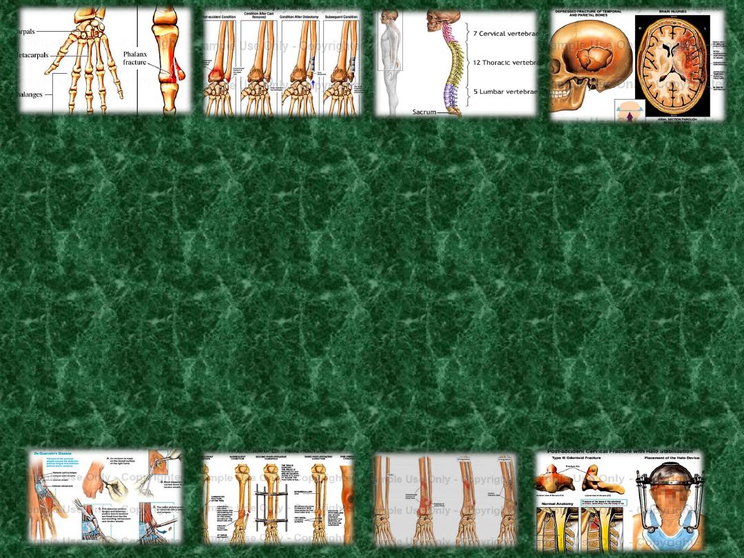
Examples
of a crushing Osteochondritis
Freiberg's d. of metatarsal.→ → → → → → →
köhler's d. of navicular. → → → → → →
Kienböck's d.of lunate. → → → →
Panner's d.of capitulum .
Scheuermann`s d.of vertebra
Perthes d. of femoral head.
S
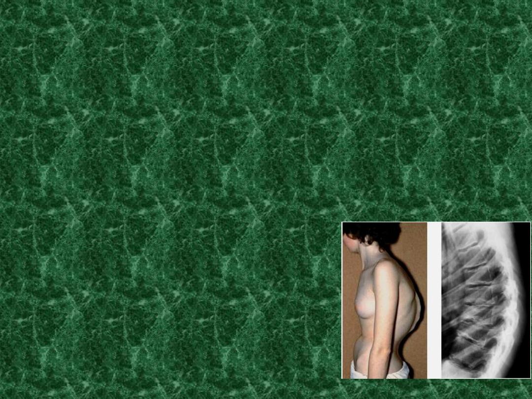
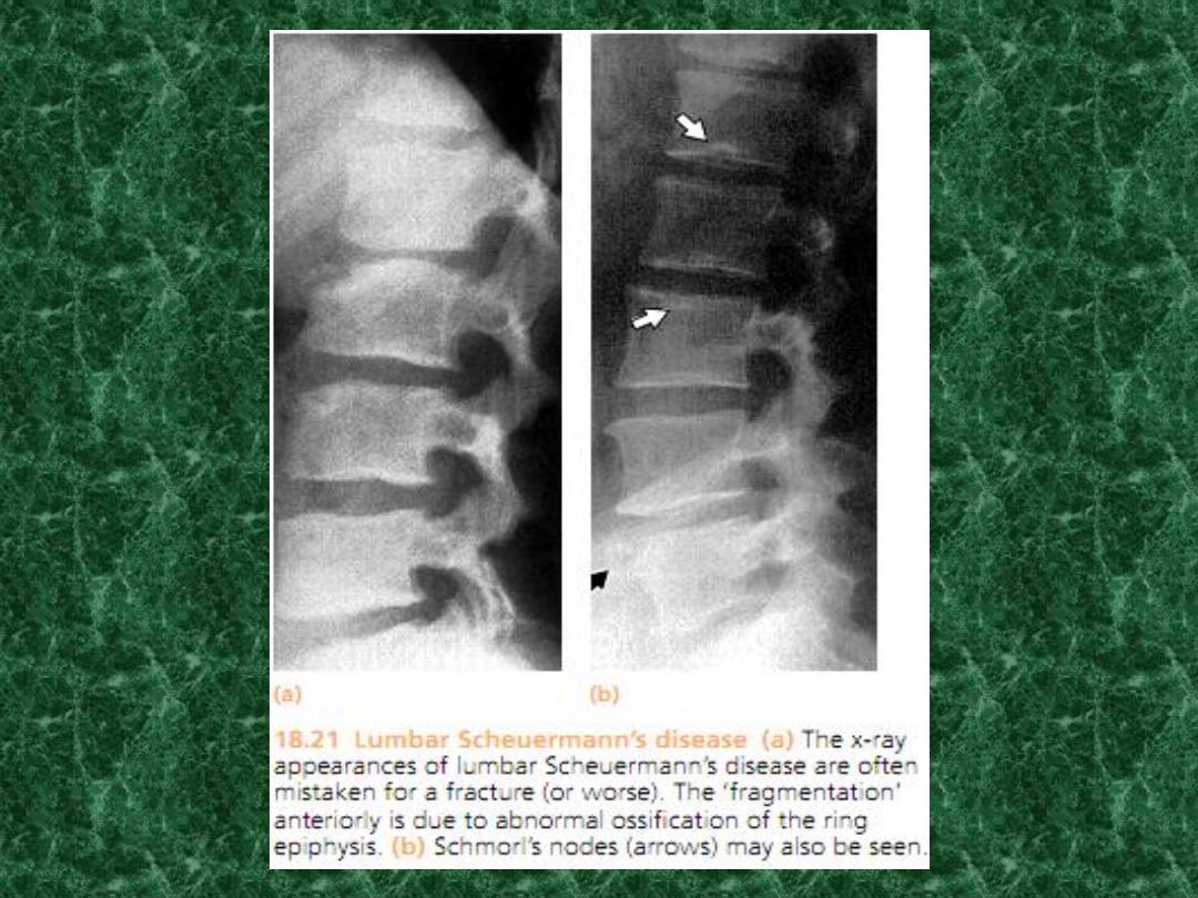
cheuermann's disease
• vertebral Osteochondritis,without clear evidence of bone
death.
• compression and fragmentation of the vertebral epiphyseal
plate lead to distorted growth of the vertebral body.
• occurs during adolescent and may cause back pain and
dorsal kyphosis(adolescent K.).
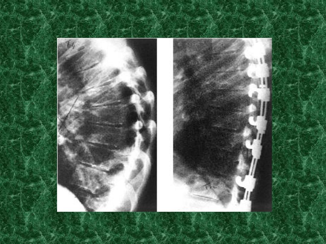
X-rays
• show irregularity of the vertebral
end plate.
• wedge-shape vertebrae(kyphosis).
S
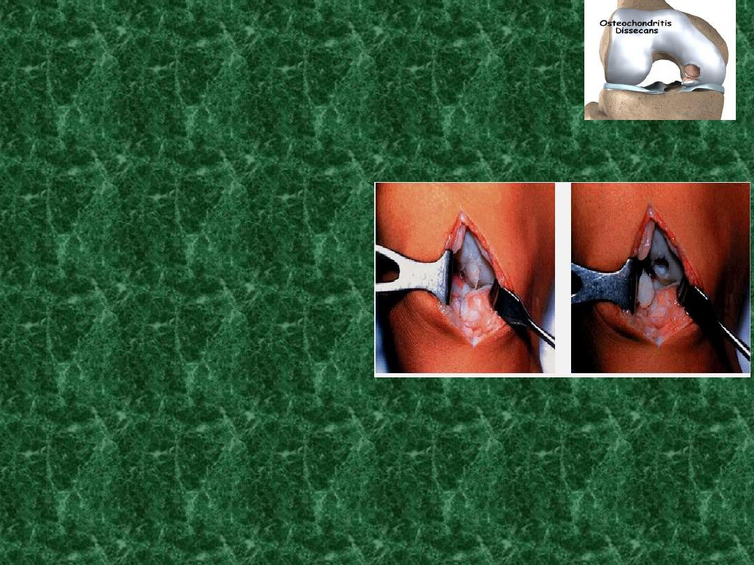
plitting-Osteochondritis
(Osteochondritis dissecans)
A small segment of articular cartilage and the
subjacent bone may separate(dissect) as an
avascular fragment.
In young adults,
usually man
Repeated minor trauma resulting in oseteochondral
fracture of a convex surface and the fragment loses its
blood supply.
Intermittent pain and joint effusion.
Detachment, cause loose body and may locking .
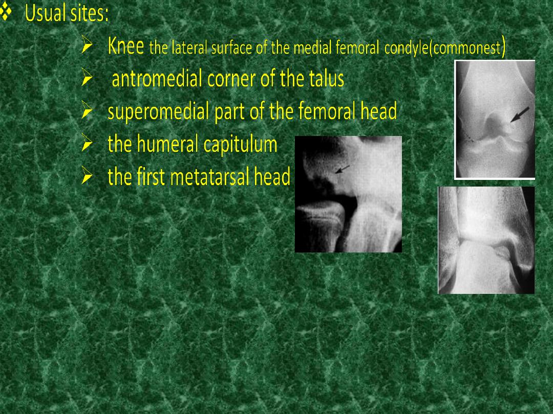
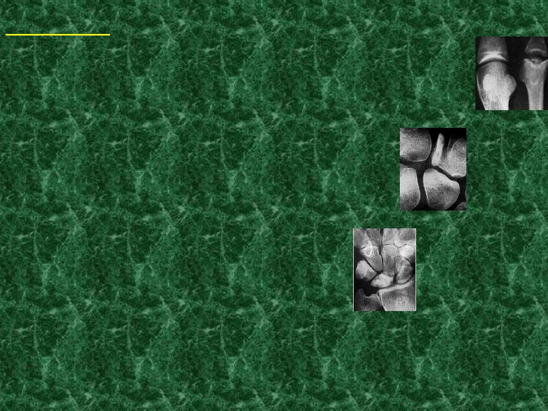
Imaging
x-ray :- dissecting fragment is defined by radiolucent line
of demarcation , when separated the crater may be
obvious.
early changes by MRI.
Radionuclide scanning show increase activity .
Treatment :-
in early stage , load reduction and restriction activity
In children complete healing may occur (2 years).
partially detached fragments may pinned .
completely detached and causes symptoms, either
fixed or removed.
arthroscopy.
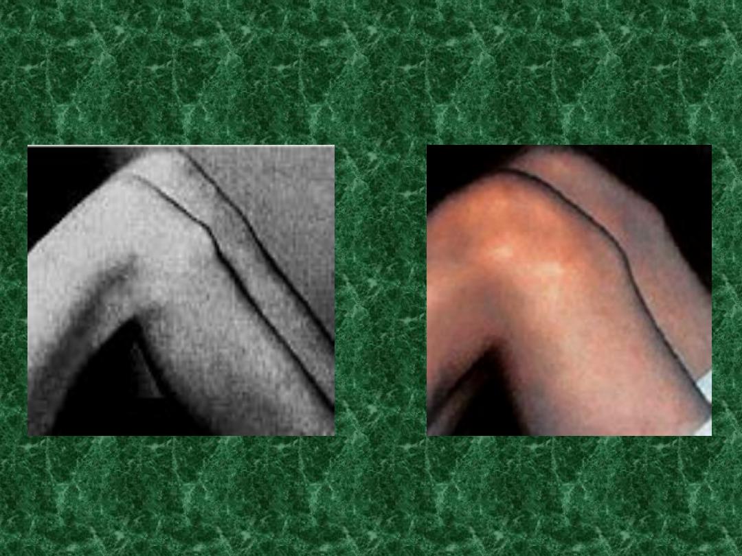
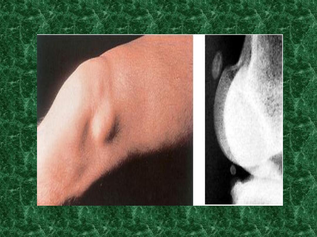
Pulling-Osteochondritis
(Traction apophysitis):-
due to stress on the physeal junction with unusual
traction forces from powerful tendons on unfused
apophysis.
Localized pain
increase density on x-ray
Sites:-
The tibial tuberosity ( Osgood's – schlatter's disease )
Calcaneal apophysis (Sever's disease).
the bone changes may be reaction to repetitive
local trauma rather than true necrosis.
Rx:rest…analgesia….Immobilization………………………
…………………..

YOU