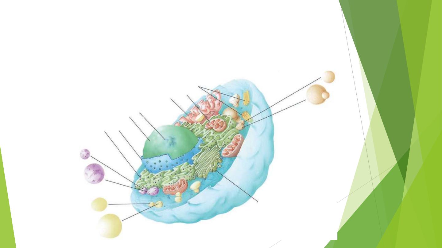
Cytoplasmic organelles
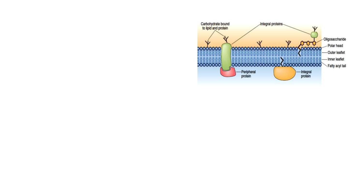
Plasma membrane
phospholipids
,
cholesterol
,
proteins
, and
oligosaccharide
chains
covalently linked to
phospholipid
and
protein
molecules.
7.5 nm thick
consists of two layers, known as the
lipid bilayer
that contain
associated
integral
and
peripheral
proteins.
The inner layer of the plasma membrane faces the cytoplasm, and
the outer layer faces the extracellular environment.
Structure
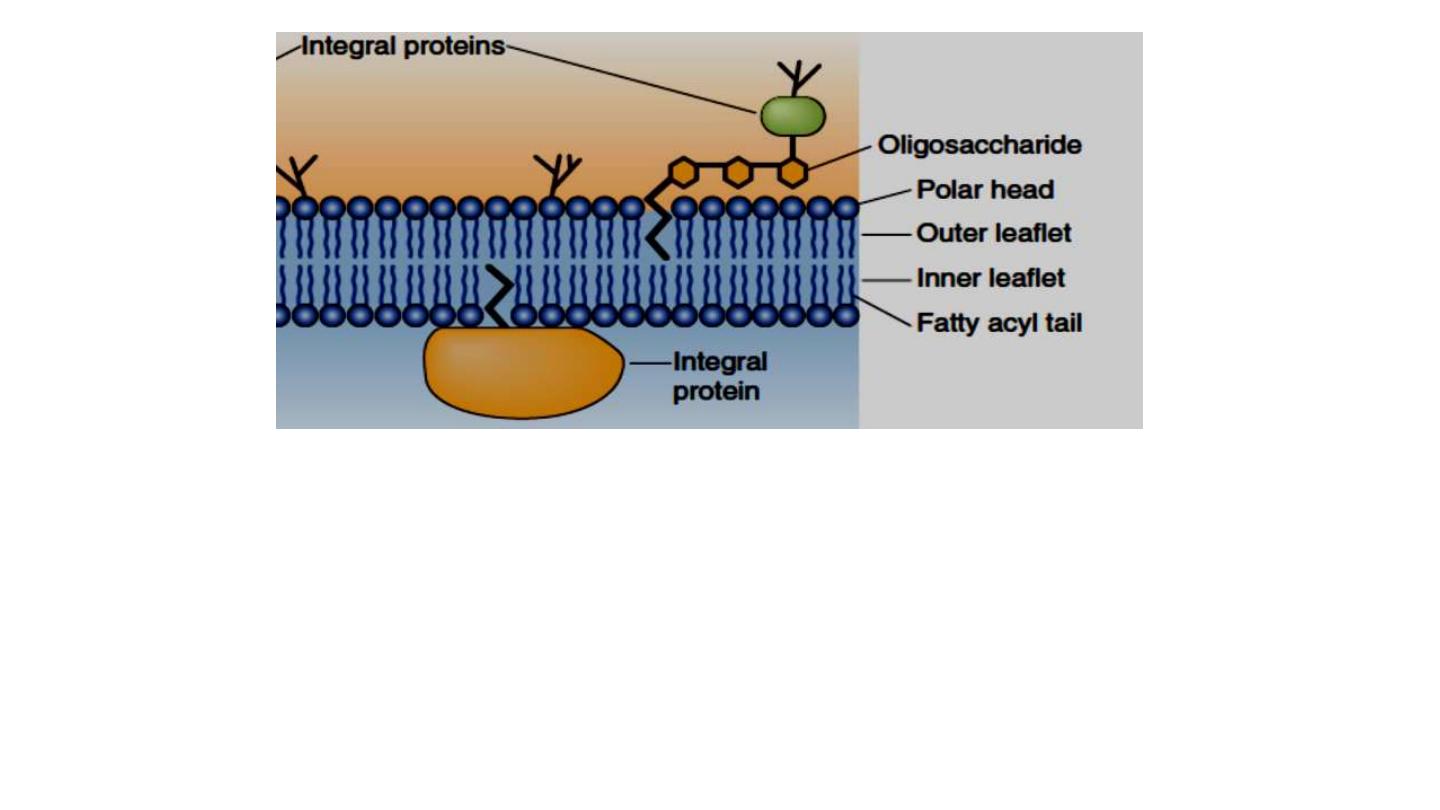
• Phospholipid molecules spontaneously orient to form a bilayer
in which the
hydrophobic tails
are pointed
inwards
. The
hydrophilic, ionic head
groups are in the
exterior
and are thus in
contact with the surrounding aqueous environment.

Structure
:
Cytoplasm represents
everything enclosed by the plasma membrane
,
with the exclusion of the nucleus.
• It consists of a viscous fluid medium that includes
salts, sugars,
lipids, vitamins, nucleotides, amino acids, RNA, and proteins
which
contain the
protein filaments, actin microfilaments, microtubules,
and intermediate filaments.
• These filaments function in animal and plant cells to provide
structural stability and contribute to cell movement.
Cytoplasm

Function:
1. Energy production through metabolic reactions,
2. Biosynthetic processes, and photosynthesis in plants.
3. Storage place of energy within the cell.
Cytosol
: is the fluid of the cytoplasm, refers only to the
protein-rich fluid environment, excluding the organelles.
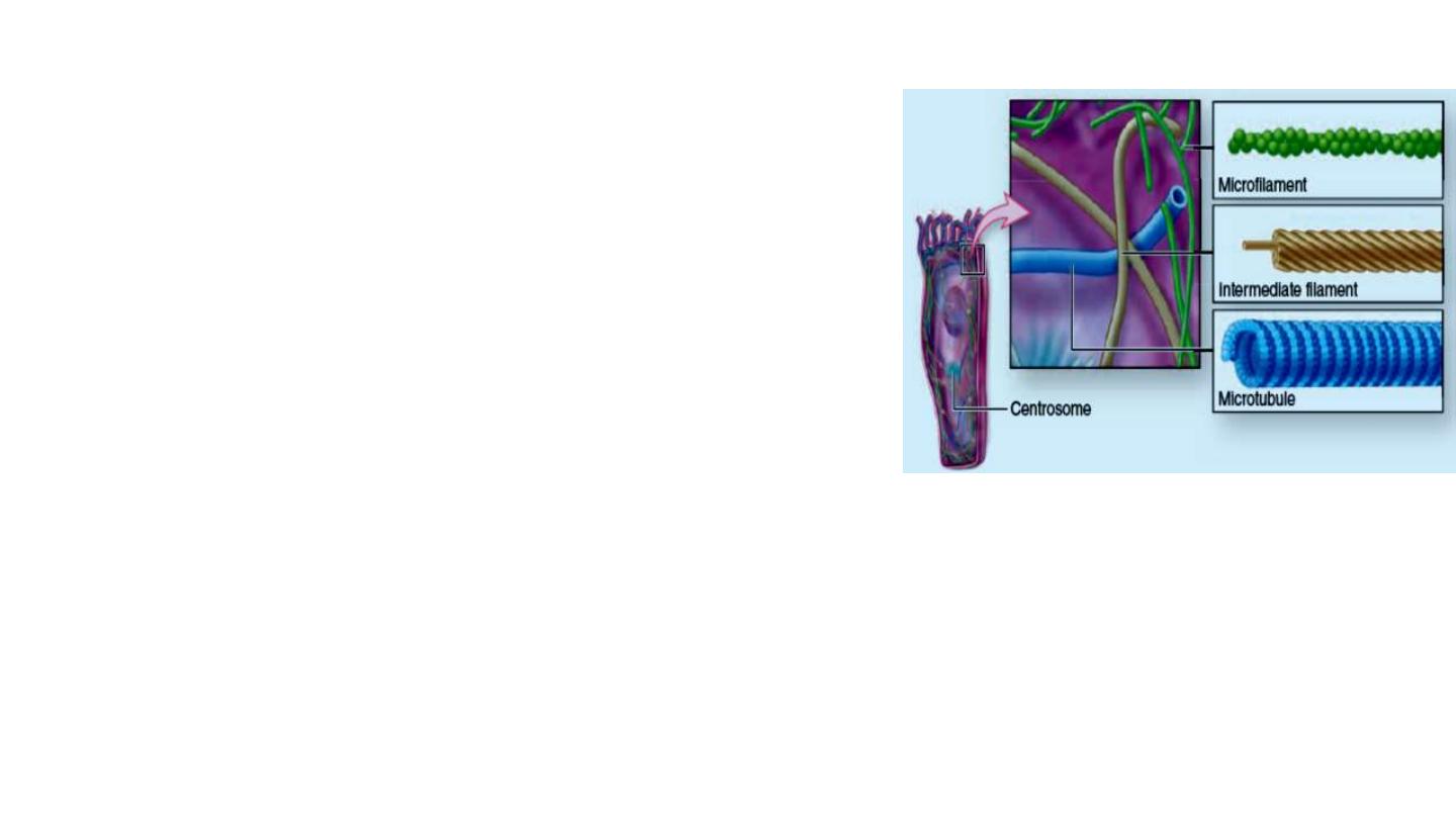
Structure:
Complex array of protein fibers found in three forms:
(1)
Microtubules
: hollow structure, with an outer
diameter of 25 nm and a wall 5 nm thick,
give the
rigidity
to help maintain cell shape.
Cytoskeleton
(2)
Microfilaments
: composed of actin, allow cellular
motility
and most contractile
activity in cells
(3)
Intermediate filaments
: intermediate in size between the other two and with a
diameter averaging 10 nm. The intermediate filaments are much more
stable
than
microtubules and actin filaments, composed of different protein subunits in different cell
types.

Functions of
Cytoskeleton
:
1. Structural
:
• Provides structural support to cell
• Stabilizes junctions between cells
2.
Movement
:
• Assists with cytosol streaming and cell motility;
• helps moving organelles and materials throughout cell;
• helps moving chromosomes during cell division.
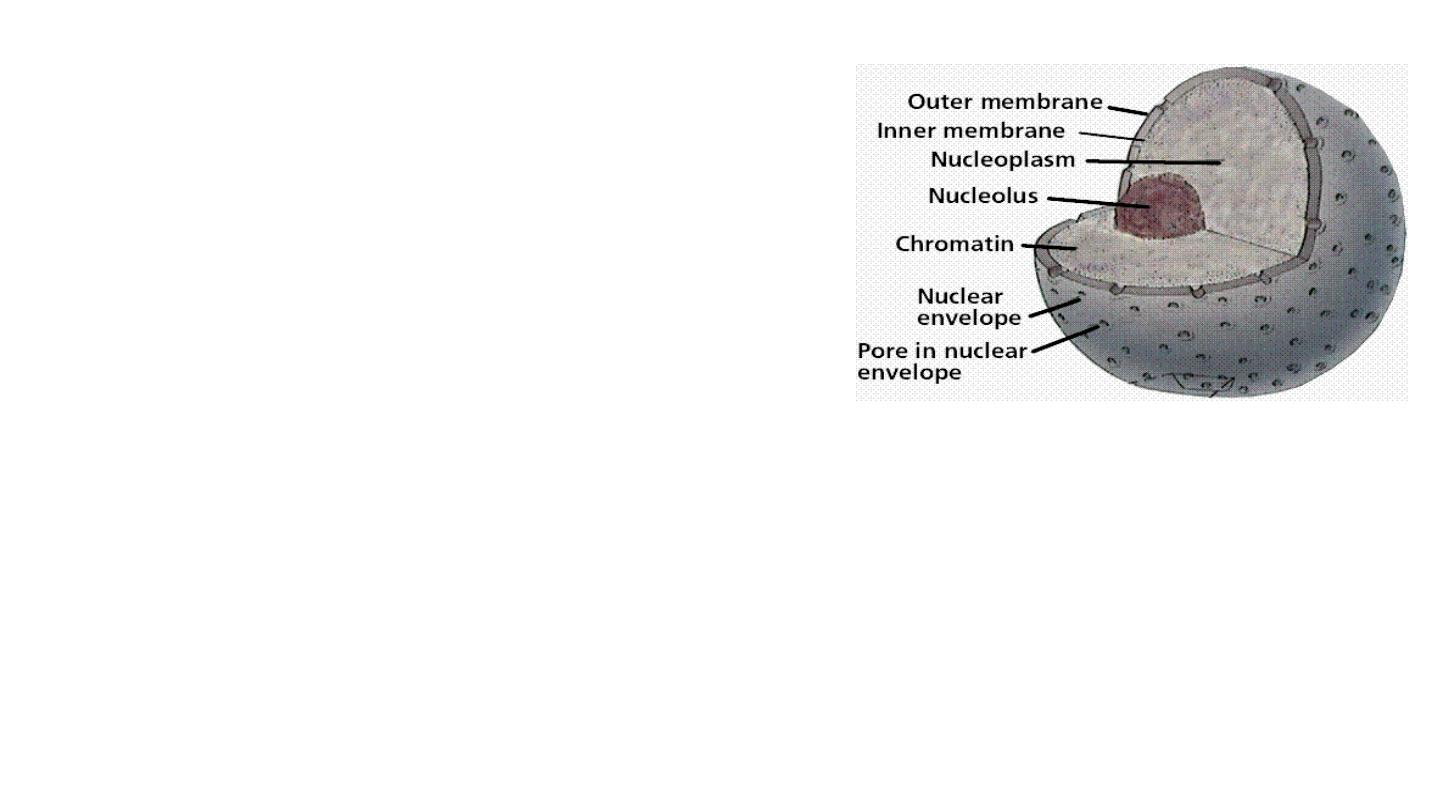
Structure: The nucleus, the largest organelle of
the
cell,
includes
the
nuclear
envelope,
nucleolus, nucleoplasm, and chromatin and
contains the genetic material encoded in the
(DNA) of chromosomes.
Nucleus
1. The nuclear envelope: surrounds the nuclear material and consists of
two parallel membranes separated by a narrow
perinuclear space
. These
membranes fuse at intervals, forming openings called
nuclear pores
in
the nuclear envelope.
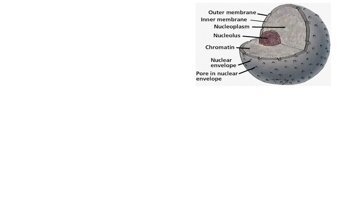
2. Nucleolus:
spherical
, highly
basophilic
, actively making
proteins
. The intense basophilia of nucleoli is due the
presence of
heterochromatin
and the presence of
densely
concentrated ribosomal RNA (rRNA)
that is transcribed,
processed, and complexed into ribosomal subunits in
nucleoli.

3. Nucleoplasm:
is
the
protoplasm
within
the
nuclear
envelope. It consists of a matrix and various types of
particles.
4. Chromatin: consists of
double-stranded DNA
complexed
with
histones
and
acidic proteins
. It resides within the
nucleus
as
heterochromatin
and
euchromatin
.
The
euchromatin/heterochromatin ratio
is higher in malignant
cells than in normal cells
??
.
• Chromatin is responsible for RNA synthesis.
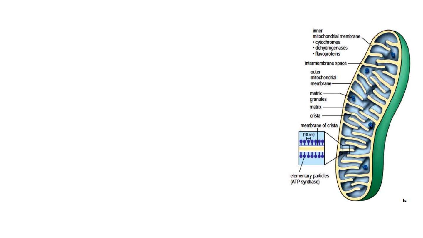
Mitochondria
Structure:
• rod-shaped organelles that are 0.2 um wide and
up to 7 um long.
• They occupy about
20%
of the cytoplasmic
volume.
• They possess an outer membrane, which
surrounds the organelle, and an inner
membrane, which folded to form
cristae
which
provide a large surface area for attachment of
enzymes involved in respiration.
• The
matrix space enclosed
by the inner
membrane is
rich in enzymes
and contains the
mitochondrial DNA
Function: mitochondria generate ATP.
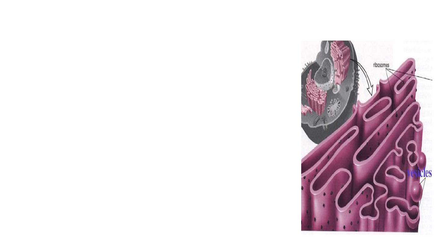
Structure:
• flattened sheets of membranes that extend throughout the
cytoplasm of eukaryotic cells and enclose a large
intracellular space called
lumen
.
• There is a continuum of the lumen between membranes of
the nuclear envelope.
• The rough endoplasmic reticulum (rough ER):
is close to
the nucleus, and is the site of
attachment of the ribosomes
.
• Ribosomes
are small and dense structures, 20 nm in
diameter, that are present in great numbers in the cell,
mostly attached to the surface of rough ER, but can float
free in the cytoplasm.
Endoplasmic Reticulum:

Function:
• Rough ER
responsible for
protein synthesis
.
• Smooth ER
is the primary site of
synthesis of lipids and sugars
• contains
degradative enzymes
, which
detoxify
many organic
molecules
• The rough ER
transitions into
a smooth endoplasmic reticulum (smooth
ER)
, which is generally more
tubular
and
lacks attached ribosomes.
• They are manufactured in the nucleolus of the nucleus on a DNA
template and are then transported to the cytoplasm.
Ribosomes are
the sites of protein synthesis.
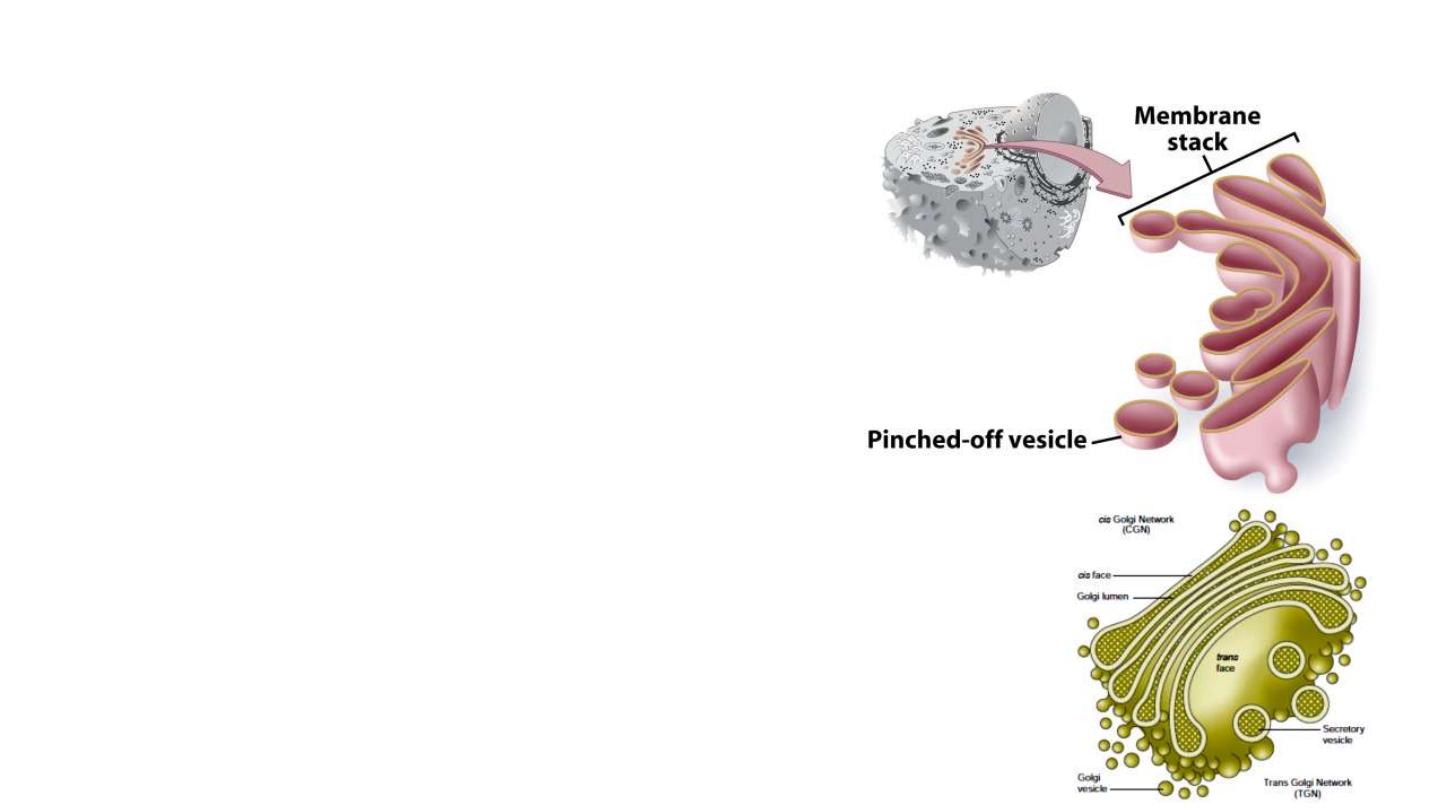
Golgi apparatus:
Structure.
• consists of several membrane-
bounded
cisternae
(
saccules
)
arranged in a
stack
• positioned and held in place by
microtubules.
• Cisternae are disk-shaped and
slightly curved, with flat centers
and dilated rims, but their size
and shape vary.
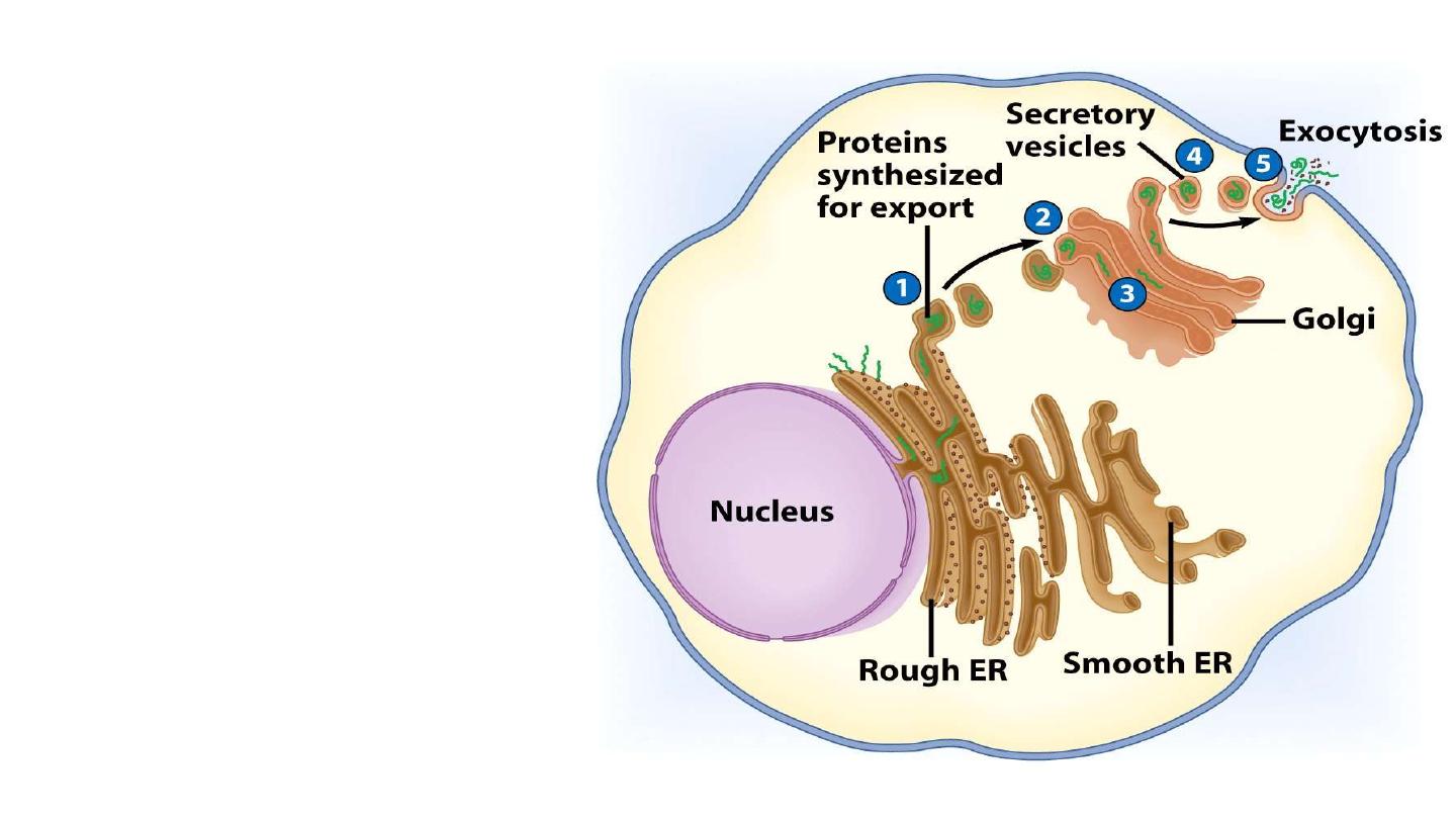
Function:
• modifying, sorting,
and packaging of
proteins for secretion
or
• delivery to other
organelles
or
• for
secretion outside
of the cell
.

Lysosomes:
These are vesicles of
hydrolytic enzymes
and are
single-
membrane bound
. They have an
acidic interior
and contain about
40
hydrolytic enzymes
involved in intracellular digestions.
Peroxisomes:
These are membrane-bound vesicles containing
oxidative enzymes
that
generate and destroy hydrogen peroxide
.
•
Peroxisomes participate in many different metabolic activities,
including the
oxidation of fatty acids
, the
breakdown of purines
, and the
biosynthesis of cholesterol
.


Thank you