
Date 15/6/2020
Subject :histology
Lecture 2
Glandular Epithelial Tissue
First year
Time(2-3)h
Dr. Sabreen Saleem AL –Sayigh(Ph.D.)

Glandular Epithelia
Glandular epithelia are formed by cells specialized to
produce secretion. The molecules to be secreted are
generally stored in the cells in small membrane-bound
vesicles called secretory granules
Glandular epithelial cells may synthesize, store, and secrete
proteins (eg, pancreas), lipids (eg, adrenal, sebaceous
glands), or complexes of carbohydrates and proteins (eg,
salivary glands). The mammary glands secrete all three
substances. Less common are the cells of glands that have
low synthesizing activity (eg, sweat glands) and that
secrete mostly substances transferred from the blood to the
lumen of the gland.
Types of Glandular Epithelia
The epithelia that form the glands of the body can be
classified according to various criteria. Unicellular glands
consist of isolated glandular cells, and multicellular glands
are composed of clusters of cells. An example of a
unicellular gland is the goblet cell of the lining of the
small intestine) or of the respiratory tract

Types of Glandular Epithelia
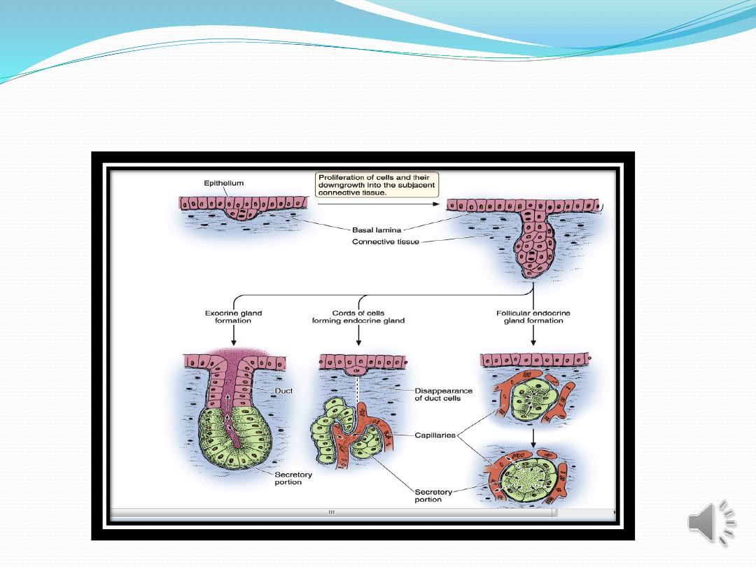
. Glands develop during fetal life from covering epithelia by
means of cell proliferation and invasion of the subjacent
connective tissue, followed by further differentiation ,Exocrine
glands retain their connection with the surface epithelium from
which they originated. This connection is transformed into
tubular ducts lined with epithelial cells through which the
glandular secretions pass to reach the surface. Endocrine glands
are glands whose connection with the surface is lost during
development. These glands are therefore ductless, and their
secretions are picked up and transported to their site of action by
the bloodstream rather than by a duct system (as show figure 3).
Types of Glandular Epithelia
Types of Glandular Epithelia
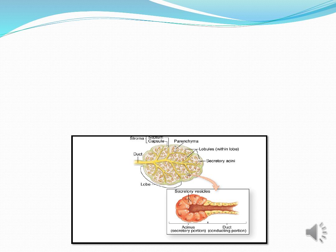
Multicellular glands, whether exocrine or endocrine, also
have connective tissue in a surrounding capsule and in
septa that divide the gland into lobules. These lobules
then subdivide, and in this way the connective tissue
separates and binds the glandular components together
as figure.

Types of Glandular Epithelia
Two types of endocrine glands can be recognized based on
the arrangement of their cells. The endocrine cells may
form anastomosing cords interspersed between dilated
blood capillaries (eg, adrenal gland, parathyroid, anterior
lobe of the pituitary or they may arrange themselves as
vesicles or follicles filled with non cellular material (eg, the
thyroid gland).
Exocrine glands have a secretory portion, which contains
the cells responsible for the secretory process, and ducts,
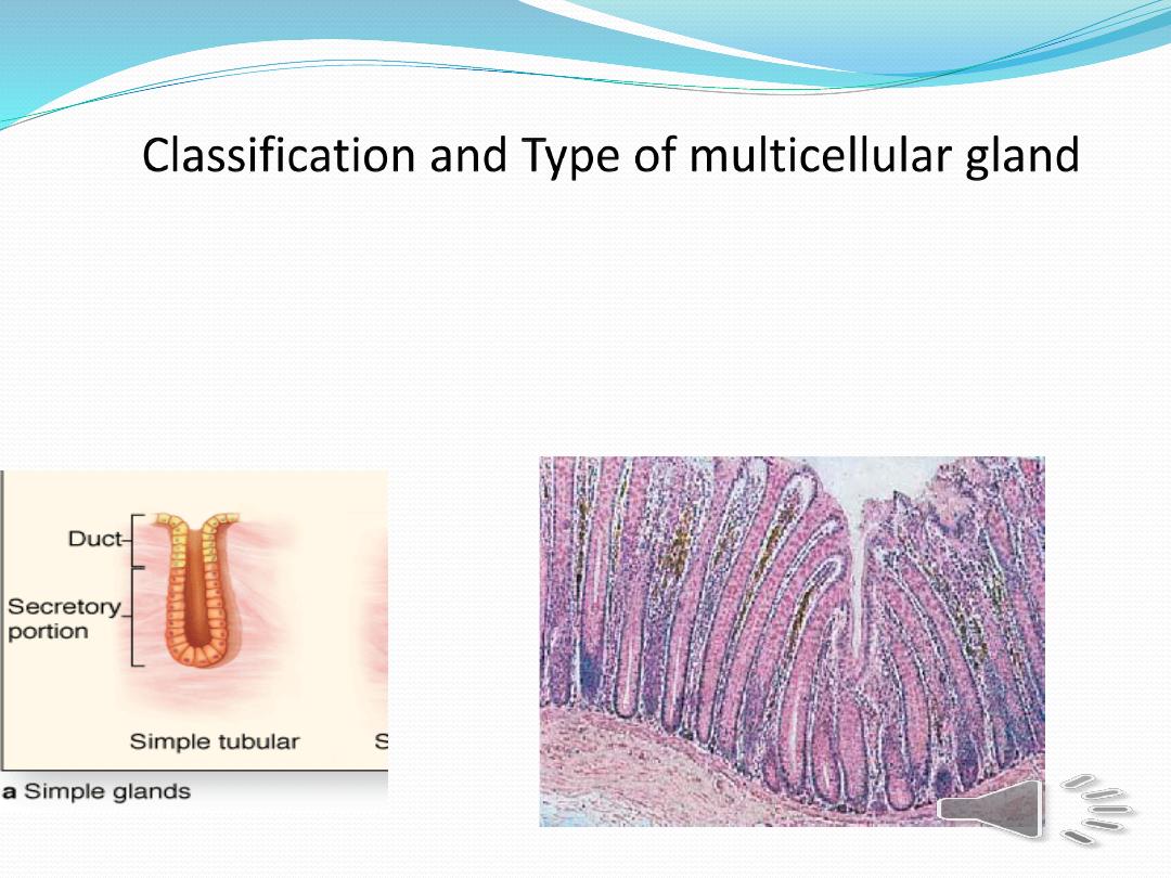
which transport the secretions Simple glands have only
one un branched duct, whereas compound glands have
ducts that branch repeatedly.
1
-
Simple tubular glands have only one un
branched duct that connects directly to surface(
e.g intestinal gland ).
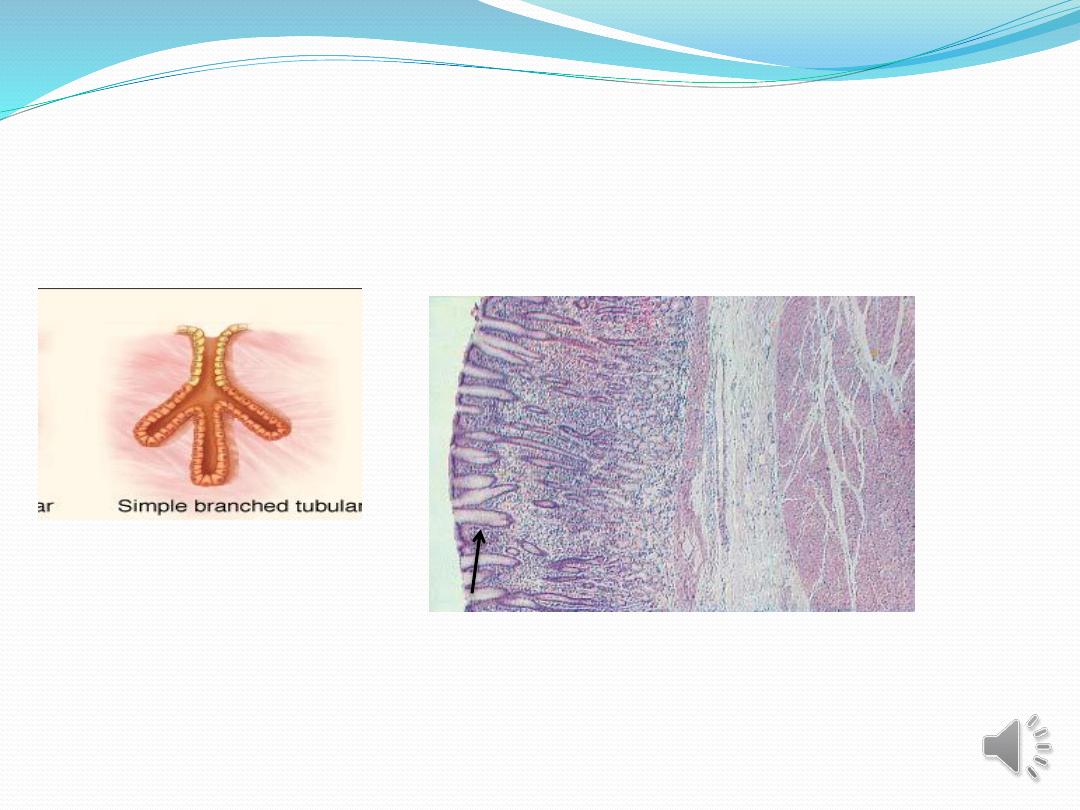
2-Simple branched tubular glands : tubular gland whose
secretory units branch ( e.g, fundic gland of stomach ).

3-Simple coiled tubular glands: Long un branched duct , the
secretory unit is long coiled tube (e.g., sweat gland).
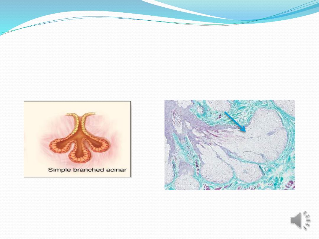
4-Simple branched acinar glands( alveolar). Secretory units are
branched and open into a single duct(e.g. sebaceous gland).
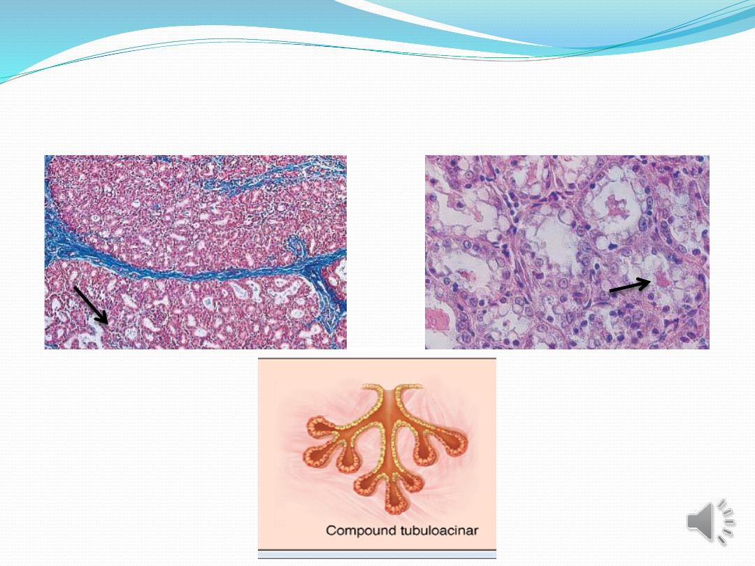
5- Compound tubulacinar(alveolar):Branching ducts with both
tubular and acinar secretory units (e.g., submaxillary salivary
gland).
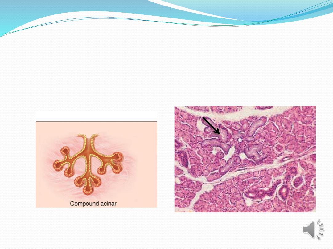
6- Compound acinar : Branching ducts with acinar secretory
unites(e.g,. Parotid salivary gland and mammary glands ).

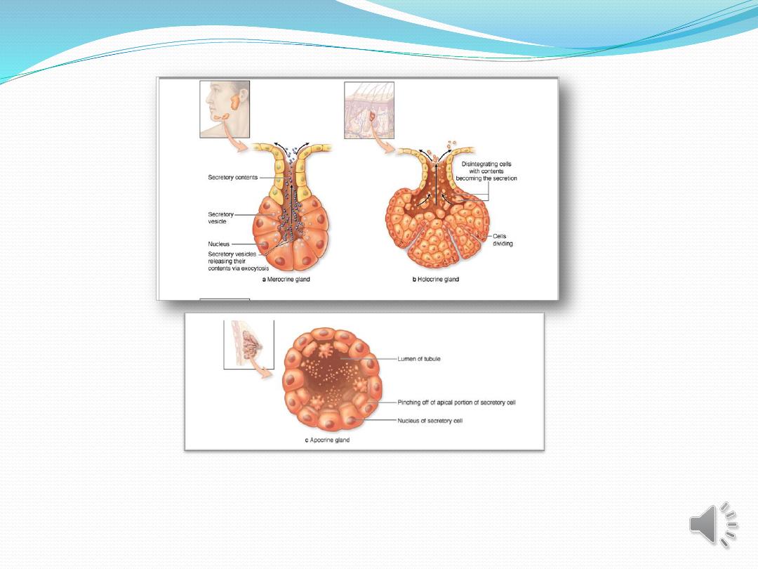
Exocrine glands are also classified functionally according to the
way the secretory products leave the cell :
1-
Merocrine secretion (sometimes called eccrine) involves
typical exocytosis of proteins or glycoproteins. This is the most
common mode of secretion. (Figure17)
2- Holocrine secretion involves the cell filling with secretory
product and then the whole cell being disrupted and shed. This
is best seen in the sebaceous glands of skin (Figure18).
3-, apocrine secretion, In an intermediate type ,the secretory
product is typically a large lipid droplet and is discharged
together with some of the apical cytoplasm and
plasmalemmaloss, is seen in mammary glands. (Figure19)
Figuer(19)

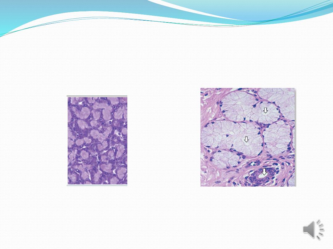
Exocrine glands with merocrine secretion can be
further categorized as either serous or mucous
according to the nature of the proteins or
glycoproteins secreted and the resulting staining
properties of the secretory cells. The acinar cells of the
pancreas and parotid salivary glands are examples of
the serous type which secrete digestive

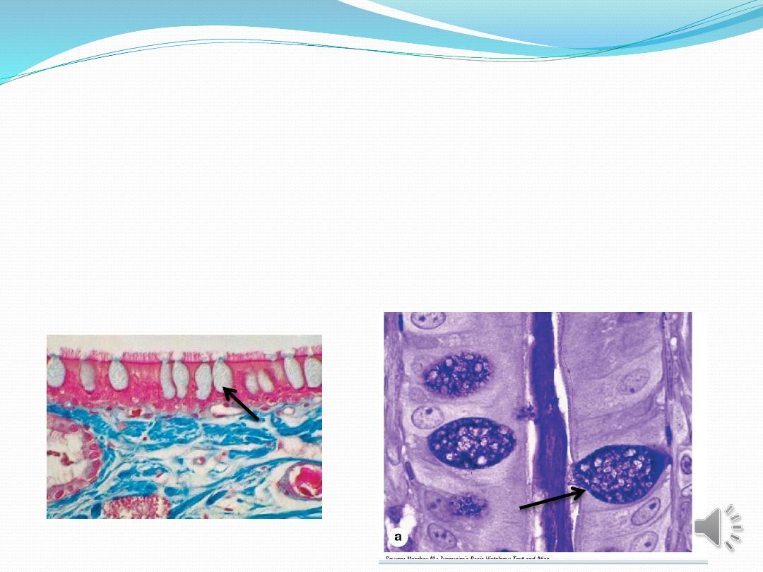
The basal ends of serous cells have well-developed
RER and Golgi complexes and the cells are filled
apically with secretory granules in different stages of
maturation as(figure20) ,Mucous cells, such as goblet
cells, while also rich in RER and Golgi complexes are
filled apically with secretory granules containing
strongly hydrophilic glycoproteins called mucins as
figure(21).
Figure(20)
figure(21)

References
Junqueira's ,L.C and Carreiro ,J.(2014):Basic Histology
,tText and Atlas3 th ed.McGraw-Hill Paulo,pp(70-90).
Junqueira's ,L.C and Carreiro ,J.(2017):Basic Histology
,tText and Atlas3 th ed.McGraw-Hill Paulo,pp(80-95