Skeletal connective tissue
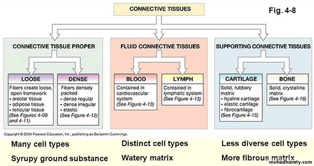
The skeletal system composed of specialized forms of supporting connective tissue.
Bone provides a rigid protective and supporting framework for most of the soft tissue of the body,cartilage provides semi-rigid support in limited sites such as the respiratory tree and external ear.
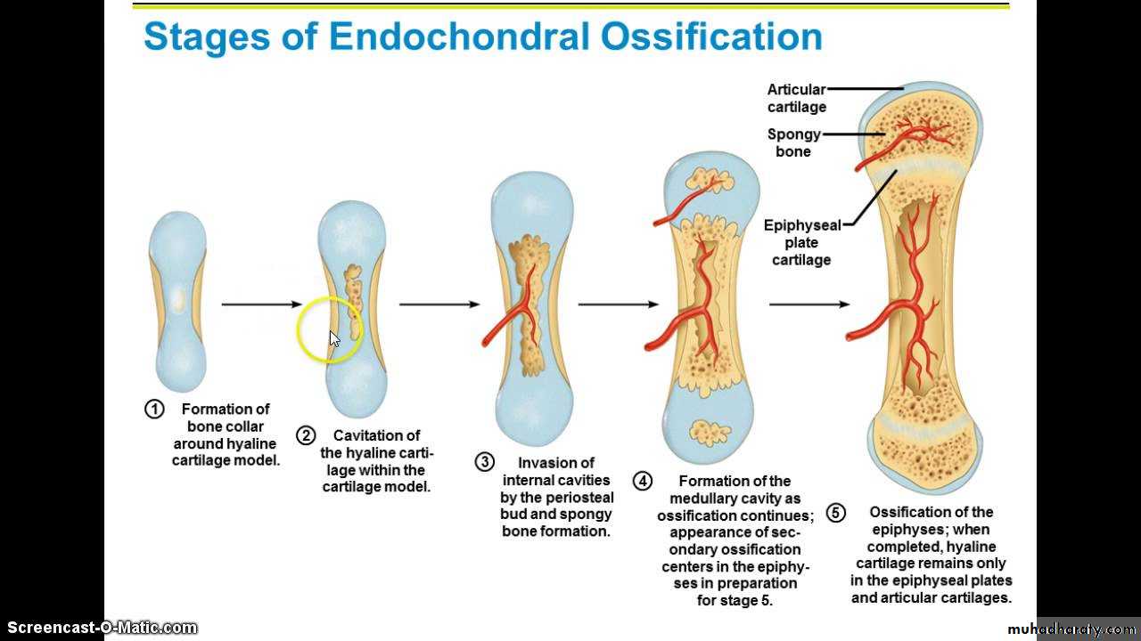
Cartilage formation is also precursor in the processes of bone formation by either the membranous or endochondral ossification processes.
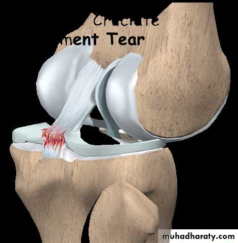
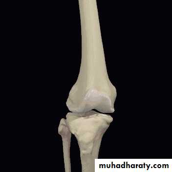
Joints are composite structure which joins the bones the skeleton , permit varying degrees of movement.
Ligaments are robust but flexible bands of colagenous tissue which contribute to the stability of joints.
Tendons provide strong pliable connections between muscles and their points of insertion into bones.

Cartilage
Cartilage extracellular matrix enriched with glycosaminoglycan’s and proteoglycans macromolecules that interact with collagen and elastic fibers.Variation in the composition of these matrix components produce 3 types of cartilage adapted to local biomechanical needs:
the firm consistency of the extracellular matrix in cartilage allows the tissue to bear mechanical stresses without permanent distortion.
Another function of cartilage is to support soft tissue.
cartilage is a shock –absorbing and sliding area for joints and facilitates bone movements Because it is smooth –surfaced and resilient.
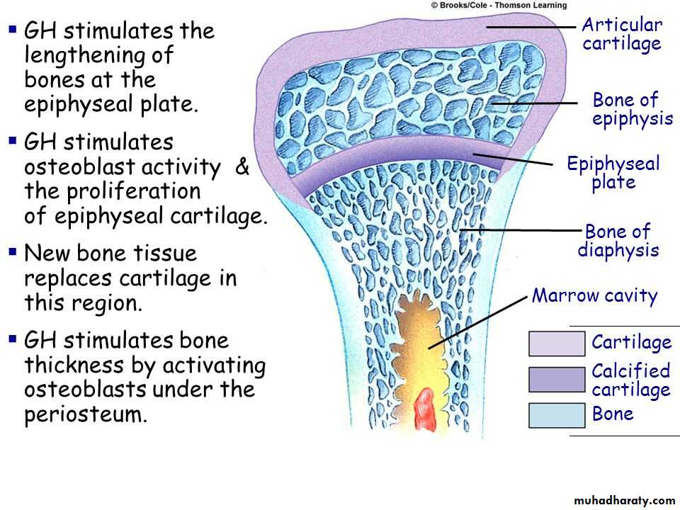
Cartilage is also essential for the development and growth of long bones both before and after birth.
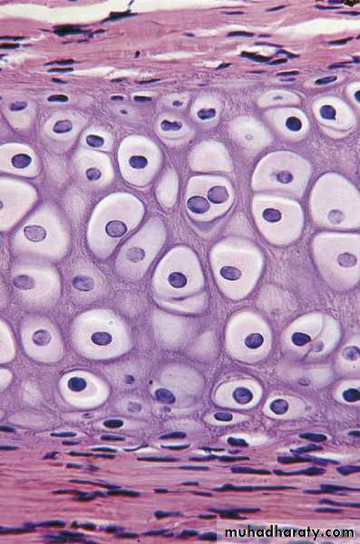
Cartilage consists of cells called chondrocytes and an extensive extracellular matrix composed of fibers and ground substance, chondrocytes synthesize and secrete the extracellular matrix and the cells themselves are located in matrix cavities called lacunae.
Collagen, hyaluronic acid, proteoglycans and small amounts of several glycoproteins are present in all types of cartilage matrix.
Cartilage has no lymphatic vessels or nerves.
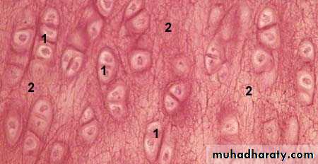
Hyaline cartilage
Hyaline cartilage is the most common and best studied of the 3 forms.
In the matrix, type II collagen is the principal collagen type.
Fresh Hyaline cartilage is bluish –white and translucent. In the embryo, it serves as a temporary skeleton until it’s gradually replaced by bone.
Hyaline cartilage
In adult mammals hyaline cartilage is located in the articular surfaces of the movable joints in the walls of larger respiratory passages (nose, larynx, trachea, bronchi), in the ventral ends of ribs where they articulate with the sternum and in the epiphyseal plate, where it is responsible for the longitudinal growth of bone.

Elastic cartilage:
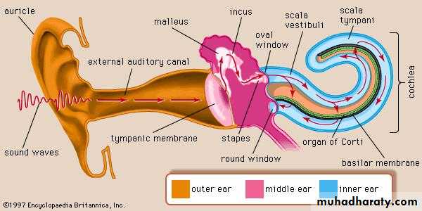
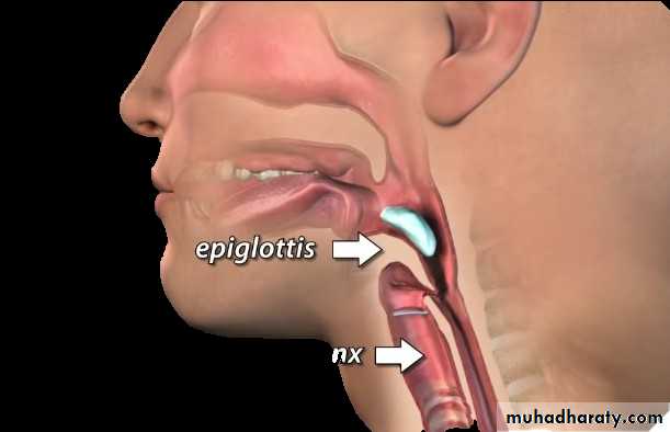
Elastic cartilage is identical to hyaline cartilage except that it contains an abundant fine elastic fibers in addition to collagen type II fibrils, frequently found to be gradually continuous with hyaline cartilageElastic cartilage is found in the auricle of the ear, the wall of the external auditory canals, the epiglottis, and the cuneiform cartilage in the larynx.
Fresh elastic cartilage has a yellowish color (presence of elastic fibers).
Elastic cartilage possesses a perichondrium.
Elastic cartilage :
Fibrocartilage:
Fibrocartilage is a tissue intermediate between dense connective tissue and hyaline cartilage. So it is always associated with dense connective tissue and the border areas between these two tissues are not clear out showing a gradual transition.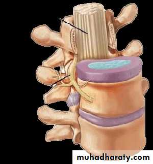
• Fibrocartilage, present in regions of the body subjected to pulling forces, is characterized by a matrix containing a dense network of coarse type I collagen fibers.
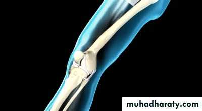
• Fibrocartilage is found in intervertebral disks, in attachments of certain ligaments to the cartilaginous surface of bones.
Fibrocartilage :
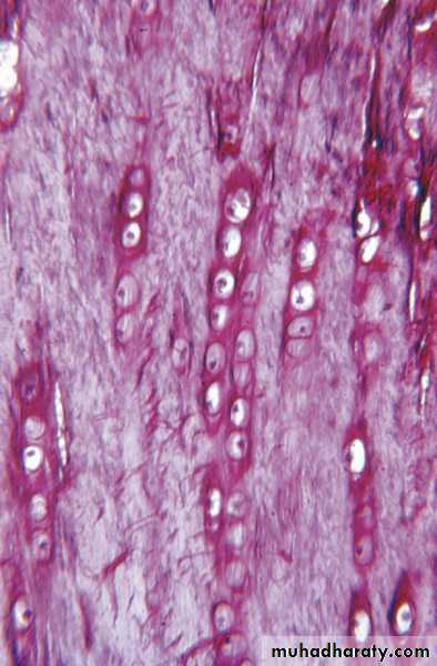
In Fibrocartilage the numerous collagen fibers either form irregular bundles between the groups of chondrocytes or are aligned in a parallel arrangement along the columns of chondrocytes.
the collagen bundles take up a direction parallel to those stresses. There is no identifiable perichondrium in Fibrocartilage
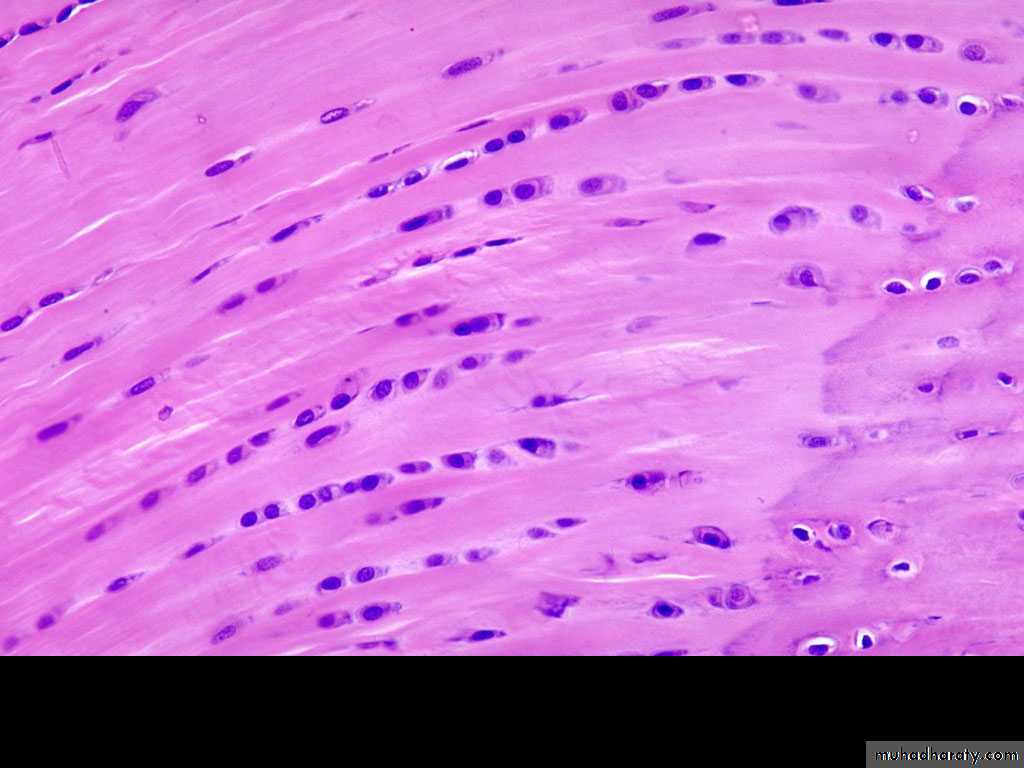
Fibrocartilage contains chondrocytes, either singly or in isogenous groups, usually arranged in long rows separated by coarse collagen type I, the Fibro cartilage matrix is acidophilic.