
CHEST ANATOMY THI-QAR UNIVERSITY
COLLEGE OF MEDICINE
LECTURE 4 2019/2020
Dr. Rafid AL-Temimi ; Clinical radiology ( CABM)
Page
1
Dr. Ahmed Abdulameer Daffar ; Thoracic & Vascular Surgeon ( FIBMS )
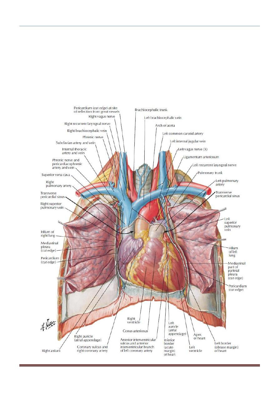
THE CHEST
PERICARDIUM
The pericardium is a fibroserous sac that encloses the heart and the roots of the
great vessels.
The pericardium lies within the middle mediastinum, posterior to the body of the
sternum and the 2nd to the 6
th
costal cartilages and anterior to the 5th to the 8th
thoracic vertebrae.
Fibrous Pericardium
The fibrous pericardium is the strong fibrous part of the sac.
It is firmly attached below to the central tendon of the diaphragm.
It fuses with the outer coats of the great blood vessels passing through it.
Serous Pericardium
The serous pericardium lines the fibrous pericardium and coats the heart.
It is divided into parietal and visceral layers.
The parietal layer lines the fibrous pericardium and is reflected around the roots
of the great vessels to become continuous with the visceral layer of serous
pericardium that closely covers the heart.
HEART:
The heart is a hollow muscular organ that is somewhat pyramid shaped and lies within the
pericardium in the mediastinum.
It is connected at its base to the great blood vessels but otherwise lies free within the
pericardium.
Surfaces of the Heart:
The heart has three surfaces: sternocostal (anterior), diaphragmatic (inferior), and a base
(posterior). It also has an apex, which is directed downward, forward, and to the left.
o
The sternocostal surface: is formed mainly by the right atrium and the right
ventricle, which are separated from each other by the vertical atrioventricular
groove.
The right border is formed by the right atrium; the left border, by the left ventricle
and part of the left auricle.
The right ventricle is separated from the left ventricle
by the anterior interventricular groove.
CHEST ANATOMY THI-QAR UNIVERSITY
COLLEGE OF MEDICINE
LECTURE 4 2019/2020
Dr. Rafid AL-Temimi ; Clinical radiology ( CABM)
Page
2
Dr. Ahmed Abdulameer Daffar ; Thoracic & Vascular Surgeon ( FIBMS )
o
The diaphragmatic surface: of the heart is formed mainly by the right and left
ventricles separated by the posterior interventricular groove. The inferior surface of
the right atrium, into which the inferior vena cava opens, also forms part of this
surface.
o
The base of the heart, or the posterior surface, is formed mainly by the left atrium,
into which open the four pulmonary veins.
The base of the heart lies opposite the apex.
The heart does not rest on its base; it rests on its diaphragmatic (inferior) surface.
CHEST ANATOMY THI-QAR UNIVERSITY
COLLEGE OF MEDICINE
LECTURE 4 2019/2020
Dr. Rafid AL-Temimi ; Clinical radiology ( CABM)
Page
3
Dr. Ahmed Abdulameer Daffar ; Thoracic & Vascular Surgeon ( FIBMS )
The apex of the heart:
It formed by the left ventricle, is directed downward, forward, and to the left.
It lies at the level of the fifth left intercostal space, 3.5 in. (9 cm) from the midline.
In the region of the apex, the apex beat can usually be seen and palpated in the living
patient.
Borders of the Heart
-
The right border is formed by the right atrium.
-
The left border, by the left auricle; and below, by the left ventricle.
-
The lower border is formed mainly by the right ventricle but also by the right
atrium.
-
The superior border, formed by the roots of the great blood vessels.
-
The apex is formed by the left ventricle.
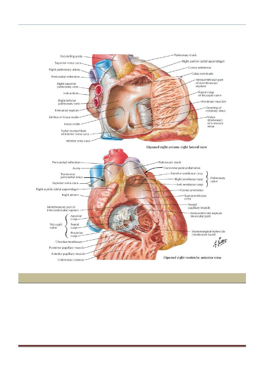
1 . Right Atrium
The right atrium consists of a main cavity and a small out pouching, the auricle.
The main part of the atrium that lies posterior to the ridge is smooth walled and is
derived embryologically from the sinus venosus.
The part of the atrium in front of the ridge is roughened or trabeculated by bundles of
muscle fibers, the musculi pectinati, which run from the crista terminalis to the
auricle.
Openings into the Right Atrium:
1. The superior vena cava opens into the upper part of the right atrium; it has no
valve. It returns the blood to the heart from the upper half of the body.
2. The inferior vena cava (larger than the superior vena cava) opens into the lower
part of the right atrium; it is guarded by a rudimentary, nonfunctioning valve. It
returns the blood to the heart from the lower half of the body.
3. The coronary sinus, which drains most of the blood from the heart wall , opens into
the right atrium between the inferior vena cava and the atrioventricular orifice.
It is guarded by a rudimentary, nonfunctioning valve.
4. The right atrioventricular orifice lies anterior to the inferior vena caval opening
and is guarded by the tricuspid valve.
CHEST ANATOMY THI-QAR UNIVERSITY
COLLEGE OF MEDICINE
LECTURE 4 2019/2020
Dr. Rafid AL-Temimi ; Clinical radiology ( CABM)
Page
4
Dr. Ahmed Abdulameer Daffar ; Thoracic & Vascular Surgeon ( FIBMS )
5. Many small orifices of small veins also drain the wall of the heart and open directly
into the right atrium.
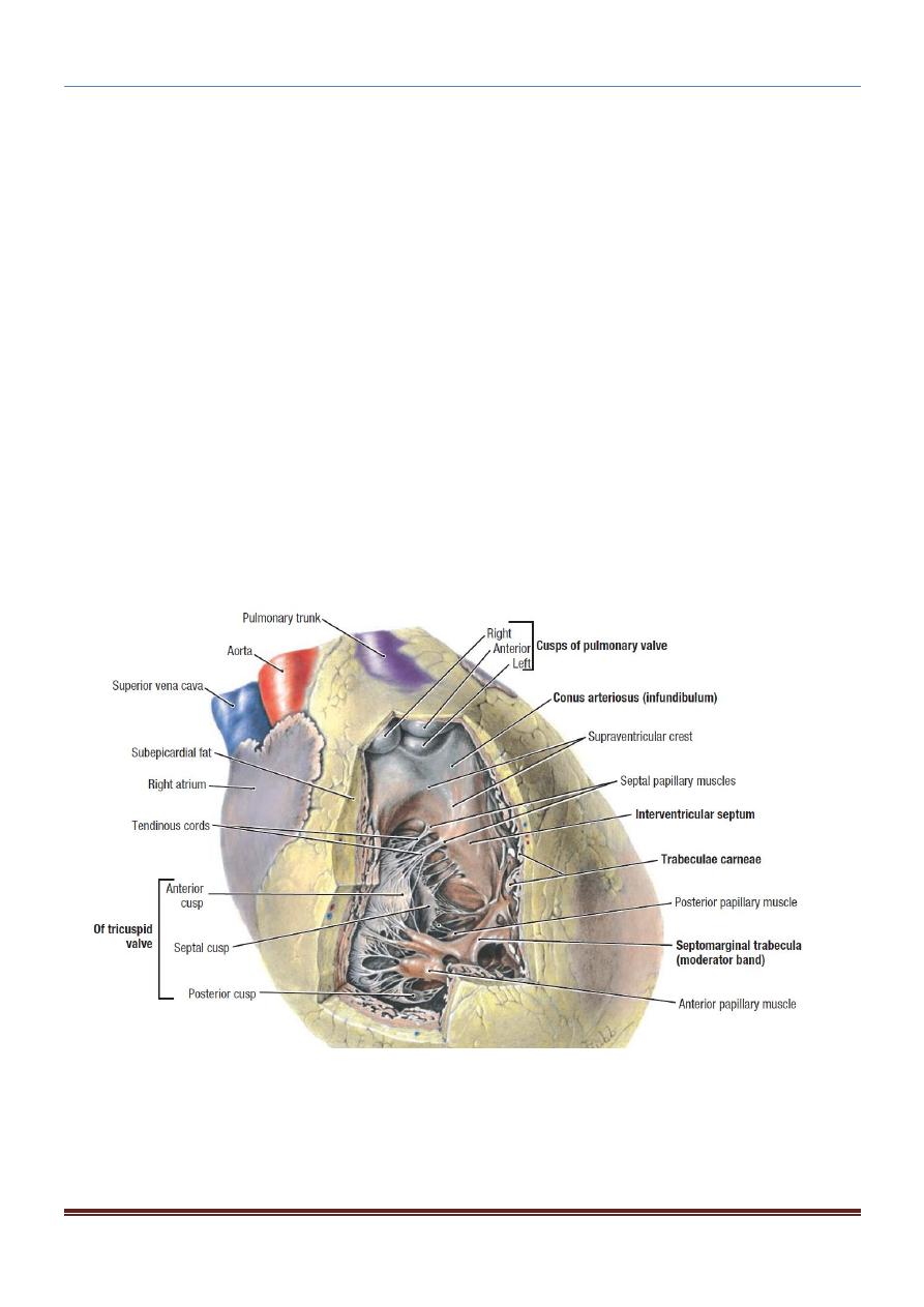
2. Right Ventricle
The right ventricle communicates with the right atrium through the atrioventricular
orifice and with the pulmonary trunk through the pulmonary orifice.
As the cavity approaches the pulmonary orifice, it becomes funnel shaped, at which
point it is referred to as the infundibulum.
CHEST ANATOMY THI-QAR UNIVERSITY
COLLEGE OF MEDICINE
LECTURE 4 2019/2020
Dr. Rafid AL-Temimi ; Clinical radiology ( CABM)
Page
5
Dr. Ahmed Abdulameer Daffar ; Thoracic & Vascular Surgeon ( FIBMS )
The walls of the right ventricle are much thicker than those of the right atrium and
show several internal projecting ridges formed of muscle bundles.
The tricuspid valve: guards the atrioventricular orifice and consists of three cusps
formed by a fold of endocardium with some connective tissue enclosed:
The bases of the cusps are attached to the fibrous ring of the skeleton of the heart ,
whereas their free edges and ventricular surfaces are attached to the chordae
tendineae.
The chordae tendineae connect the cusps to the papillary muscles.
The pulmonary valve guards the pulmonary orifice and consists of three semilunar
cusps formed by folds of endocardium with some connective tissue enclosed.
At the root of the pulmonary trunk are three dilatations called the sinuses, and one is
situated external to each cusp.