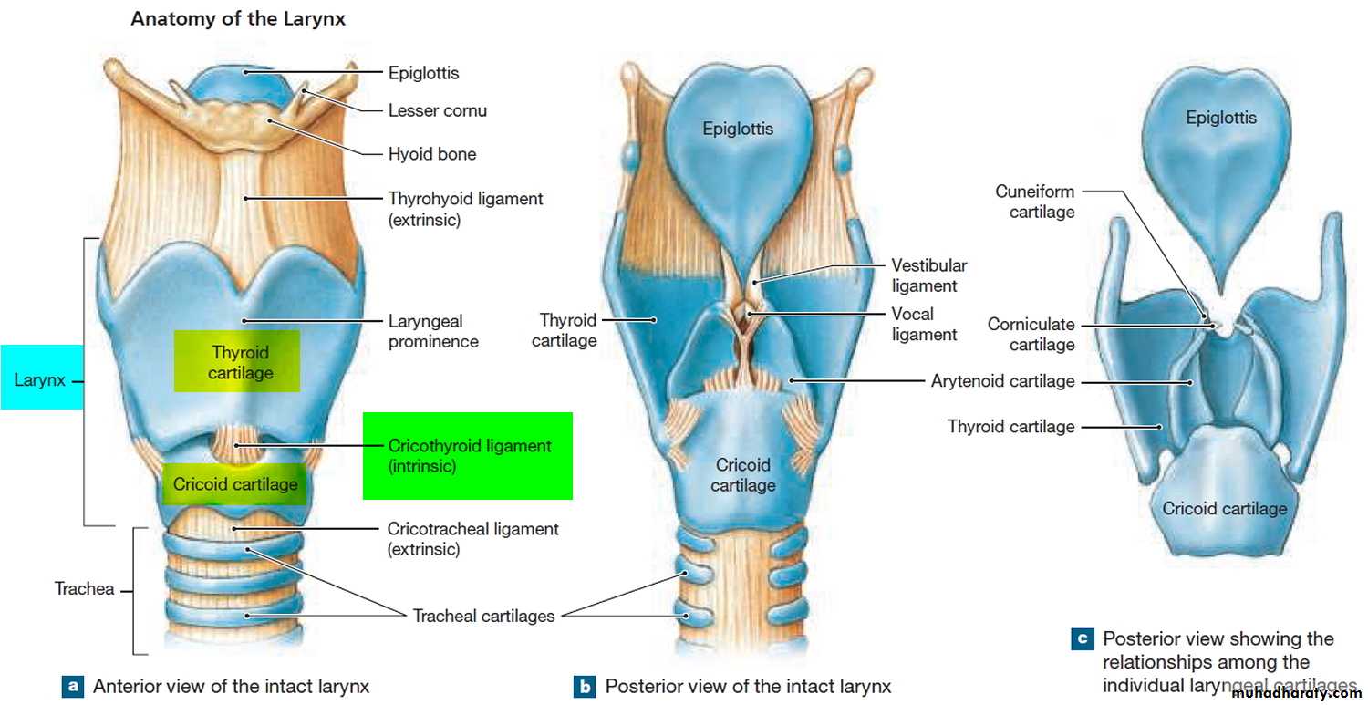
THE LARYNX Larynx is a specialized organ responsible for production of voice. It houses the vocal cords. The wall of the larynx has a complex structure made up of a number of cartilages, membranes and muscles.
1-Mucous Membrane The epithelium lining the mucous membrane of the larynx is predominantly pseudo-stratified ciliated columnar. However, over some parts that come in contact with swallowed food the epithelium is stratified squamous. These parts include the epiglottis (anterior surface and upper part of the posterior surface), and the upper parts of the aryepiglottic folds. The vocal folds do not come in contact with swallowed food, but their lining epithelium is exposed to considerable stress during vibration of the folds. These folds are also covered with stratified squamous epithelium. Serous glands and lymphoid tissue are also present.
2- Cartilages of the Larynx The larynx has a cartilaginous framework which is made of nine cartilages (3 paired and 3 unpaired) that are connected to each other by membranes and ligaments. The cartilages are either hyaline or elastic in nature. These are: 1- Hyaline cartilages a- Thyroid (unpaired) b- Cricoid (unpaired) c- Arytenoid (paired)
2- Elastic cartilages a- Epiglottis (unpaired) b- Cuneiform (paired) c- Corniculate (paired) With advancing age, calcification may occur in hyaline cartilage, but not in elastic cartilage.
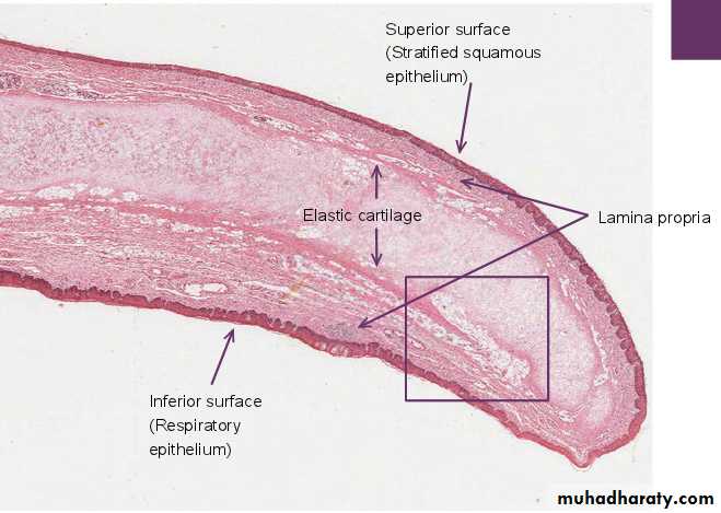
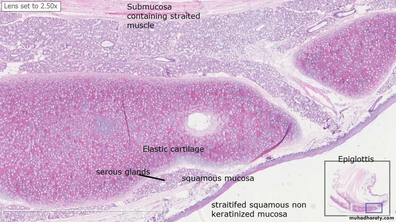
The Epiglottis The epiglottis is considered separately because sections through it are usually included in sets of class slides. The epiglottis has a central core of elastic cartilage. Overlying the cartilage there is mucous membrane. The greater part of the mucous membrane is lined by stratified squamous epithelium (non-keratinizing). The mucous membrane over the lower part of the posterior surface of the epiglottis is lined by pseudo-stratified ciliated columnar epithelium . This part of the epiglottis does not come in contact with swallowed food as it is overlapped by the aryepiglottic folds. Numerous glands, predominantly mucous, are present in the mucosa deep to the epithelium. Some of them lie in depressions present on the epiglottic cartilage.
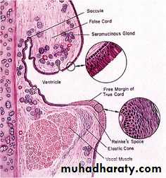
Below the epiglottis and laryngeal vestibule, the mucosa projects into the lumen bilaterally with two pairs of folds separated by a narrow space or ventricle. The upper pair, the immovable vestibular folds, is partly covered with typical respiratory epithelium overlying numerous seromucous glands and occasional lymphoid nodules. The lower pair of folds, the vocal folds (or cords), have features important for phonation or sound production is lined by non keratinizing squamous epithelium .
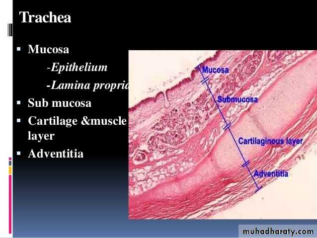
THE TRACHEA AND PRINCIPAL BRONCHI The trachea is a fibroelastic cartilaginous tube. It extends from the lower border of cricoid cartilage to its level of bifurcation into right and left bronchi. The trachea consists of four layers :
1-Mucosa : The lumen of the trachea is lined by mucous membrane that consists of a lining epithelium and an underlying layer of connective tissue. The lining epithelium is pseudo stratified ciliated columnar. It contains numerous goblet cells, and basal cells that lie next to the basement membrane. Numerous lymphocytes are seen in deeper parts of the epithelium.
2- Submucosa The subepithelial connective tissue contains numerous elastic fibres. It contains serous glands that keep the epithelium moist; and mucous glands that provide a covering of mucous in which dust particles get caught. The mucous is continuously moved towards the larynx by ciliary action. Numerous aggregations of lymphoid tissue are present in the subepithelial connective tissue. Eosinophil leucocytes are also present.
3- Cartilage and Smooth Muscle Layer The skeletal basis of the trachea is made up of 16 to 20 tracheal cartilages. Each of these is a C-shaped mass of hyaline cartilage. The open end of the ‘C’ is directed posteriorly. Occasionally, adjoining cartilages may partly fuse with each other or may have Y-shaped ends. The intervals between the cartilages are filled by fibrous tissue that becomes continuous with the perichondrium covering the cartilages. The gaps between the cartilage ends, present on the posterior aspect, are filled in by smooth muscle and fibrous tissue. The connective tissue in the wall of the trachea contains many elastic fibres.
4- Adventitia It is made of fibroelastic connective tissue containing blood vessels and nerves.
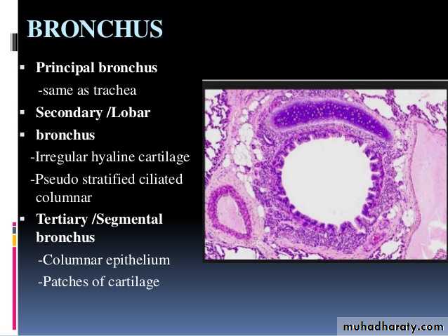
Principal Bronchi The trachea divides at the level of T4 into right and left principal bronchi (primary or main bronchi). They have a structure similar to that of the trachea.