Muscle tissue
and Nervous tissueINTRODUCTION:
All cells are capable of some sort of movement the dominant function of several cell types is to generate motile forces through contraction.In these specialized contractile cells, motile forces are generated by the interaction of the proteins actin and myosin (contractile protein).
Certain forms of contractile unite:
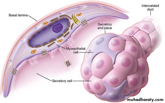
-Myo-epithelial cell: are an important component of certain secretory glands where they function to expel secretions from glandular acini.-pericyte: are smooth muscles –like cells that surround blood vessels
-Myo-fibroblasts: are cells that have contractile role in addition to being able to secret collagen.
This type of cell generally inconspicuous in normal tissues but comes to be a dominant cell type when tissue undergo repair after damage in the formation of a scar.
multicellular contractile units termed muscle such muscle cells can be divided into three types:
Skeletal muscle is responsible for the movement of the skeleton and organs such as the globe of the eye and the tongue.
Skeletal muscle if often referred to as voluntary muscle since it is capable of voluntary (conscious) control.
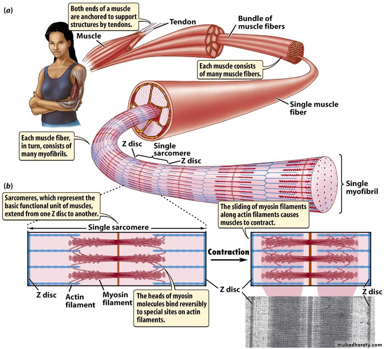
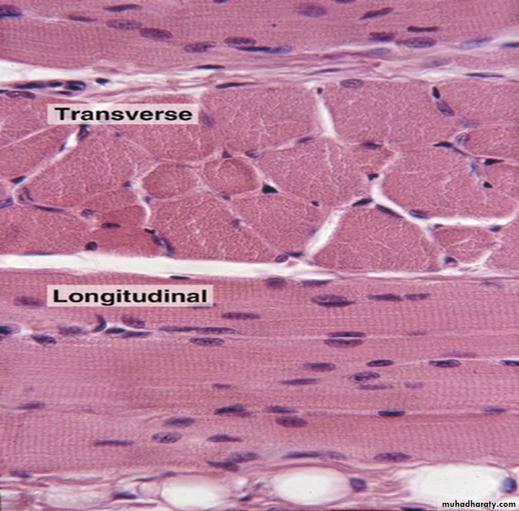
The arrangement of the contractile protein gives rise to the appearance of prominent cross – striation ,the name striated muscle is often applied to skeletal muscle.
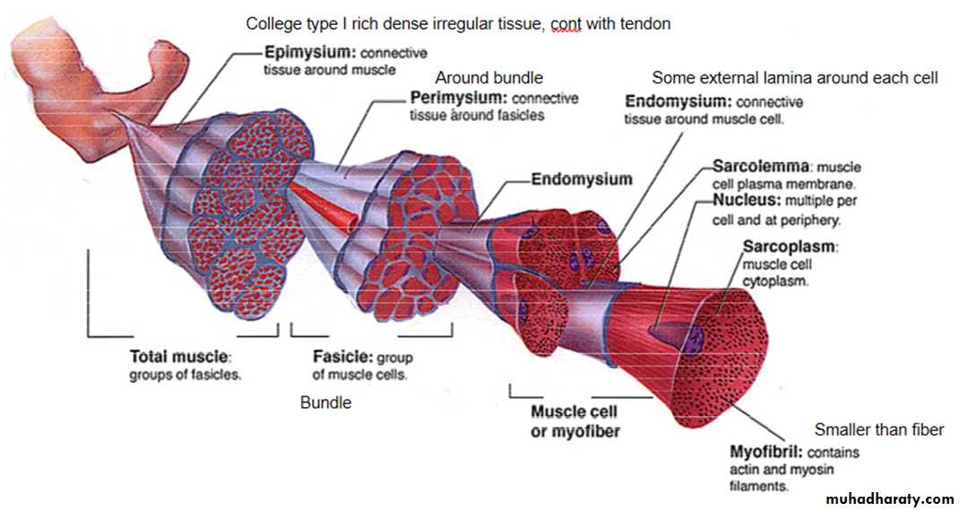
The highly developed functions of the cytoplasmic organelles of muscle cells have led to the use of a special terminology for some muscle cell components:
plasma membrane or plasma lemma ≡ sarcolemma,
cytoplasm ≡ sarcoplasm,
endoplasmic reticulum ≡ sarcoplasmic reticulum.
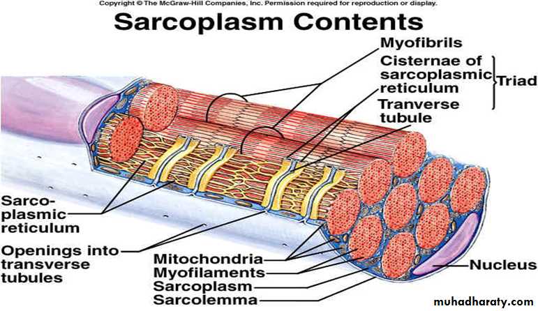
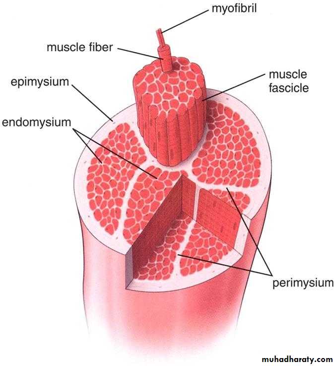
Skeletal muscles is composed of extremely elongated, multinucleated contractile cells often described as individual muscle fibers vary considerably in diameter from 10 to 100 Mm and may extend throughout the whole length of a muscle reaching up to 35 cm in length.
Skeletal muscle contraction is controlled by large motor nerves, individual nerve fibers branching within the muscle to supply muscle fibers, collectively described as a motor unit.
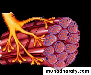
Excitation of motor nerve results in simultaneous contraction of all the muscle fibers of the corresponding motor unit.
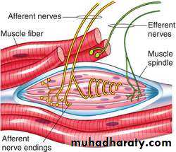
The vitality of skeletal muscle fibers is dependent on the maintenance of their nerve supply. Skeletal muscle contains highly specialized stretch receptors known as neuromuscular spindles.
Smooth muscle:
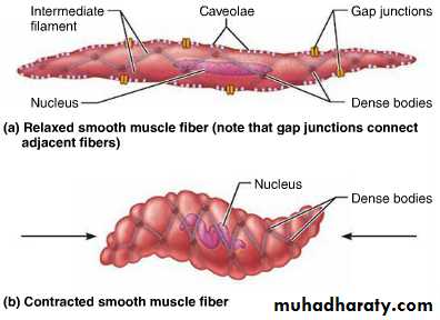
is so named because unlike other forms of muscle, the arrangement of contractile proteins does not give the histological appearance of cross-striations.This type of muscle forms the muscular component of visceral structures such as blood vessels, the gastrointestinal tract, the uterus and the urinary bladder, giving rise to the alternative name of visceral muscle, since smooth muscle is under inherent autonomic and hormonal control, it is described as involuntary muscle.
In contrast to skeletal muscle, which is specialized for relatively forceful contractions of short duration and under fine voluntary control, smooth muscle is specialized for continuous contraction of relatively low force, producing diffuse movements resulting in contraction of the whole muscle mass rather than contraction of individual motor units.
Contractility of smooth muscle occurring independently under influences of the autonomic nervous system, hormones and local metabolites which modulate contractility to accommodate changing functional demands.
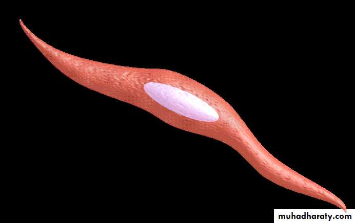
The cells of smooth muscle are relatively small with only a single nucleus.
The fibers are bound together in irregular branching fasciculi, the arrangement varying considerably from one another according to functional requirements.
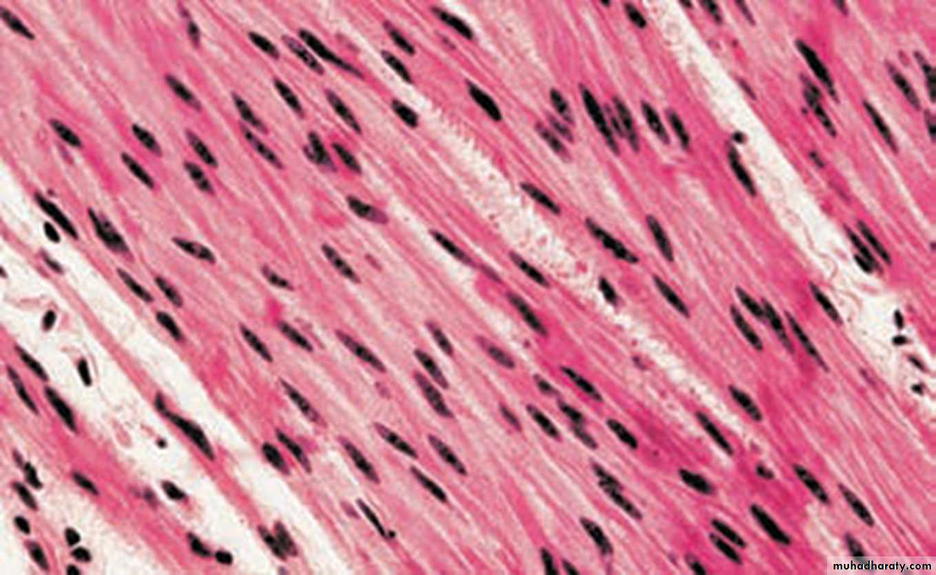
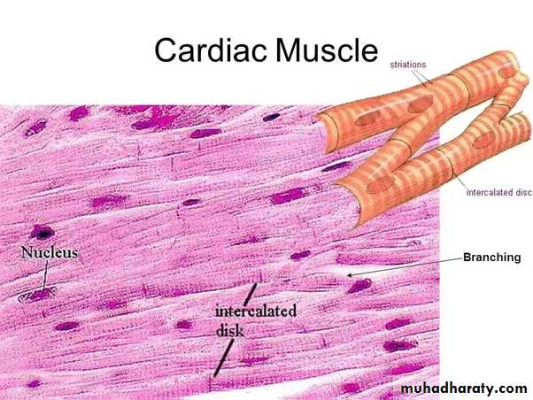
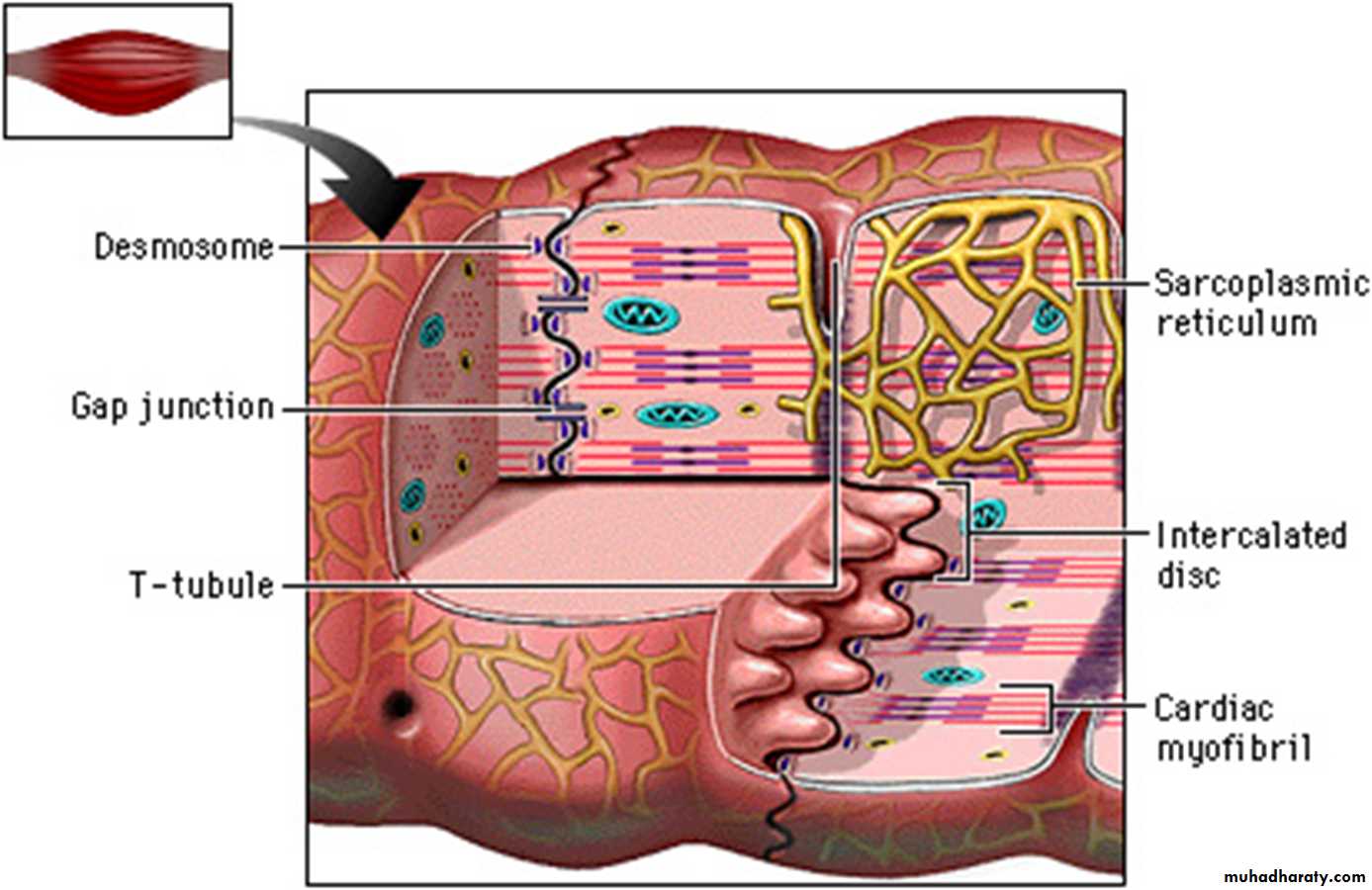
Cardiac muscle:
it has many structural and functional characteristics intermediate between those of skeletal and smooth muscle and provides for the continuous, rhythmic contractility of the heart, although striated in appearance, cardiac muscle is readily distinguishable from skeletal muscle and should not be referred to by the term, striated muscle.Muscle cells of all three are surrounded by an external lamina. In all muscle cell type, contractile forces developed from the internal contractile proteins are transmitted to the external lamina via link proteins which span the muscle cell membrane.
The external lamina binds individual muscle cells into a single functional mass.
Like the skeletal muscle, its contractions are strong and utilize a great deal of energy, and like the smooth the contractions are continuous and initiated by external autonomic and hormonal stimuli.
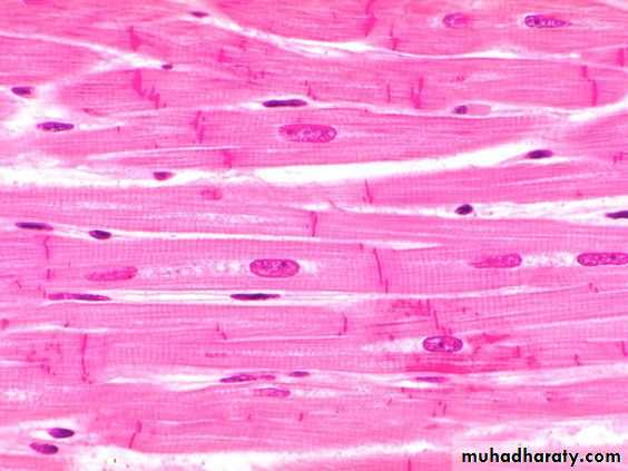
Cardiac muscle fibers are essentially long cylindrical cell with one or at most two nuclei, centrally located within the cell. The ends of the fibers are split longitudinally into a small number of branches, the ends of which giving the impression of a continuous three dimensional cytoplasmic network, this was formerly described as a syncytium before the discrete intercellular boundaries were recognized.
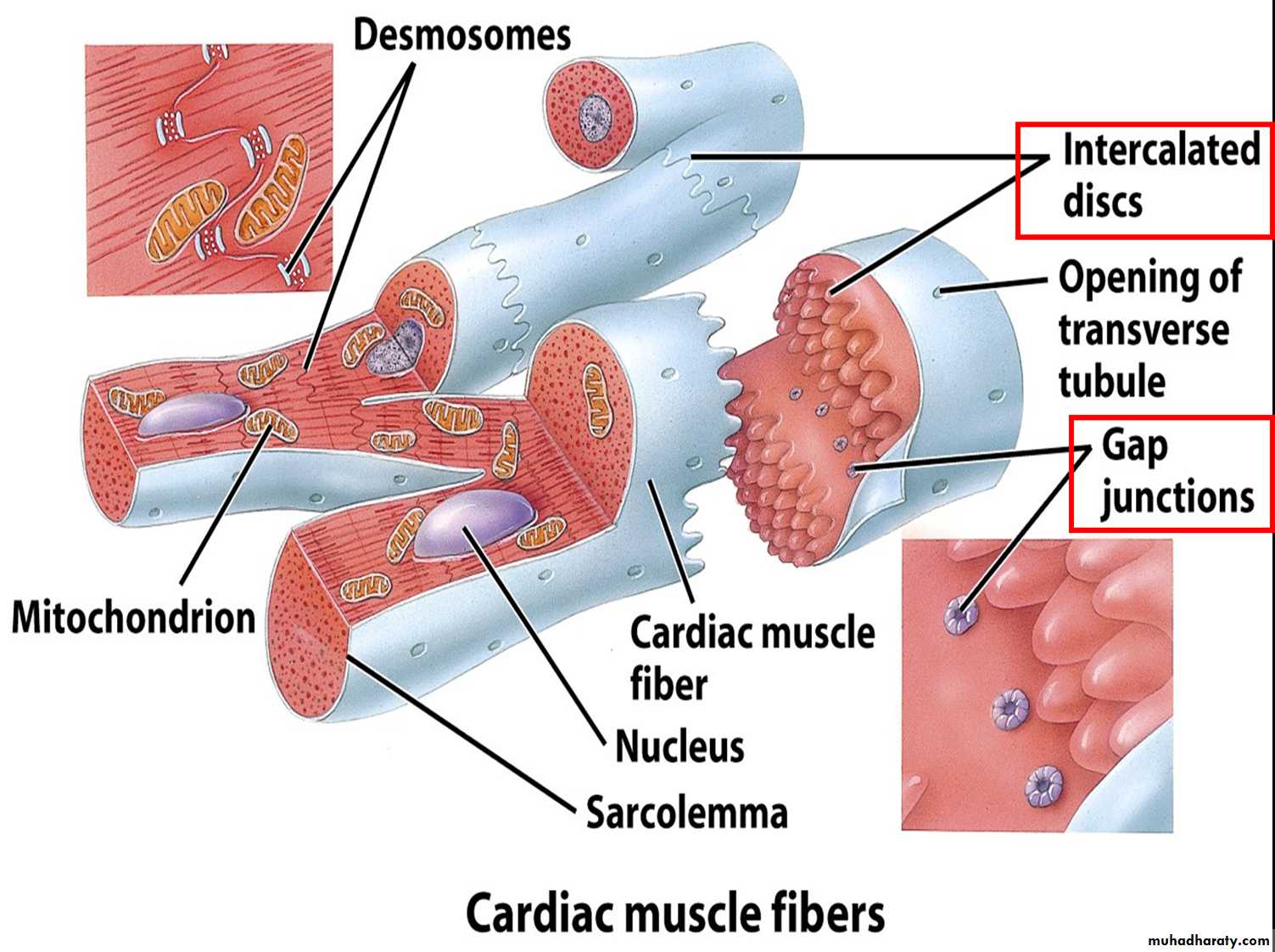
Between the muscle fibers, delicate collagenous tissue analogous to the endomysium of skeletal muscle supports the extremely rich capillary network necessary to meet the high metabolic demands of strong continuous activity.
Cardiac muscle fibers have an arrangement of contractile proteins similar to that of skeletal muscle and are consequently striated in similar manner. However, this is often difficult to visualize with light microscopy due to the irregular branching shape of the cells and their myofibrils.
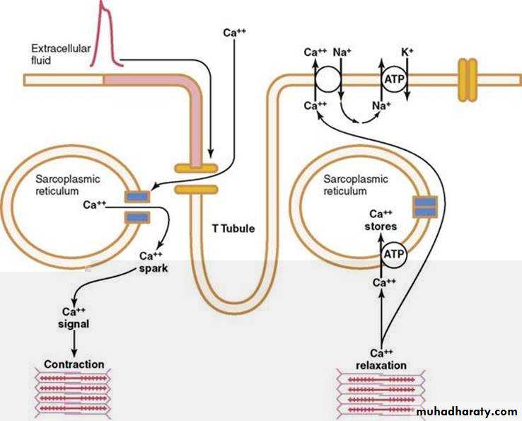
Cardiac muscle fibers have a system of T tubules and sarcoplasmic reticulum analogous to that of skeletal muscle.
In the case of cardiac muscle, however, there is a slow leak of calcium ions into the cytoplasmic from the sarcoplasmic reticulum after recovery from thepreceding contraction; this causes a succession of automatic contraction independent of external stimuli. modulated by external autonomic and hormonal stimuli.
Nerve tissue
The human nervous system is the most complex system in the human body and is formed by a network of more than 100 million nerve cells (neurons). Assisted by many more glial cells, each neuron has, on average, at least a thousand interconnections with other neurons, forming a very complex system for communication.Neurons are grouped as circuits, like electron circuits, neural circuits are highly specific combination of elements that make up system of various sizes and complexities
Although a neural circuit may be single, in most cases it is a combination of two or more circuits that interact to generate a function.
A neural function is a set of coordinated processes intended to produce a definite result. A number of elementary circuits may be combined to form higher-order system.
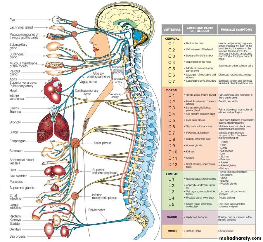
Nerve tissue is distributed throughout the body as on integrated communications network.
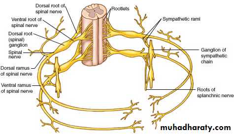
Anatomically the nervous system is divided into:Central nervous system (CNS): consisting of the brain and the spinal cord
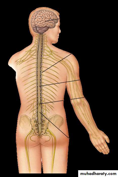
Peripheral nervous system composed of nerve fibers and small aggregates of nerve cells called nerve ganglia.
structurally nerve tissue consists of two cell types:
nerve cells or neurons, which usually show numerous long processes,glial cells which have short processes, support and protect neurons, and participate in neural activity, neural nutrition, and the defense processes of the central nervous system.
Neurons respond to environmental changes (stimuli) by cell outer surfaces membranes are called excitable, or irritable.
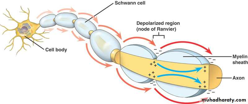
Neuron reacts promptly to stimuli with a modification of electrical potential that may be spread (propagated) throughout the neuron by the plasma membrane. This propagation, called the action potential or nerve impulse, is capable of traveling long distances; it transmits information to the neurons, muscles, and glands.
By creating, analyzing identifying and integrating information, the nervous system generates two great classes of functions:
stabilization of the intrinsic condition (e.g., blood pressure, O2 and CO2 content. PH blood glucose levels, and hormones levels) of the organism within normal ranges;
behavioral patterns (e.g., feeding, reproduction, defense, interaction with other living creatures)
Neurons
Nerve cells or neurons are responsible for
the reception, transmission, and processing of stimuli,
the triggering of certain cell activities
the release of neurotransmitters and other informational molecules.
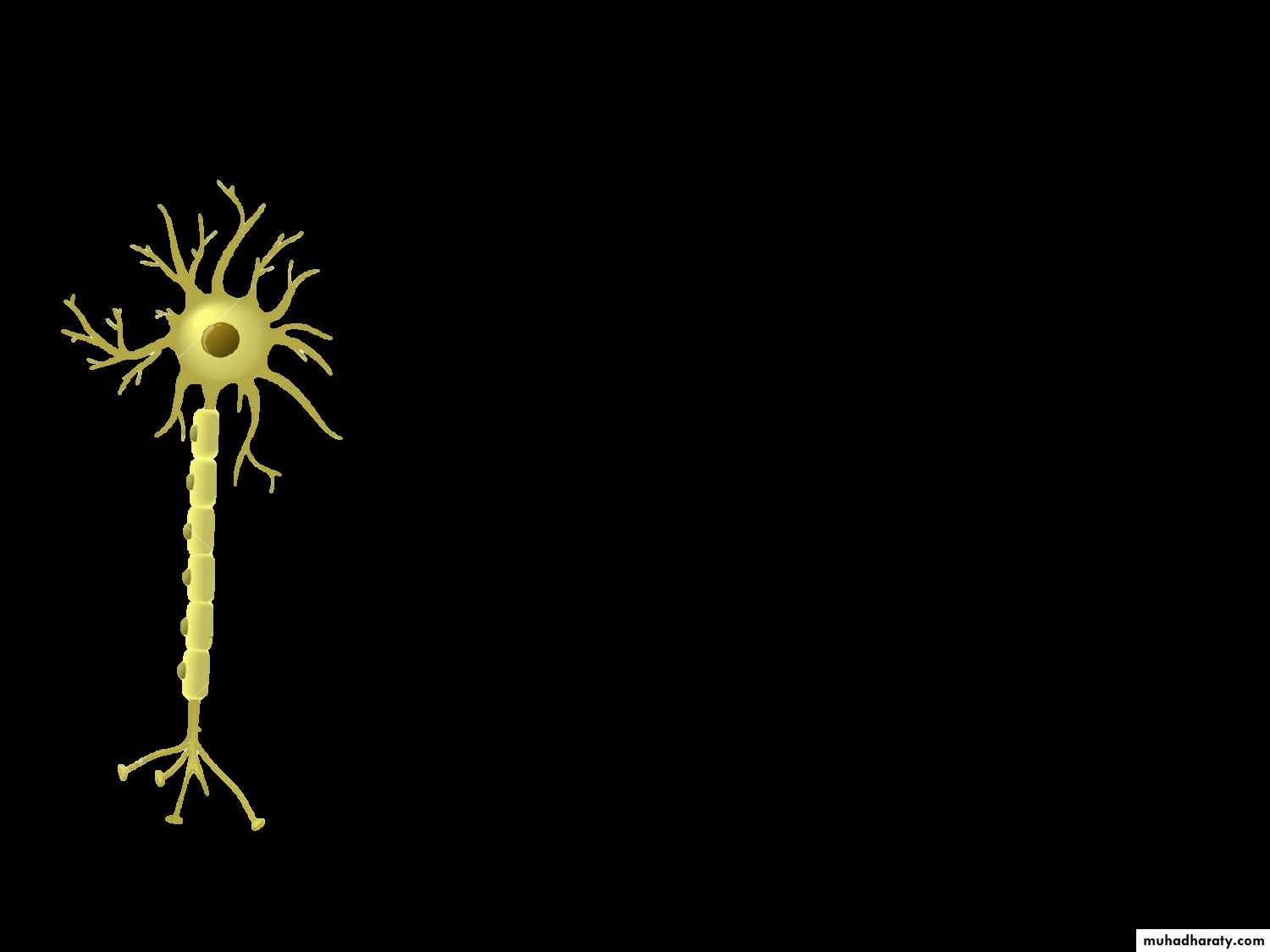
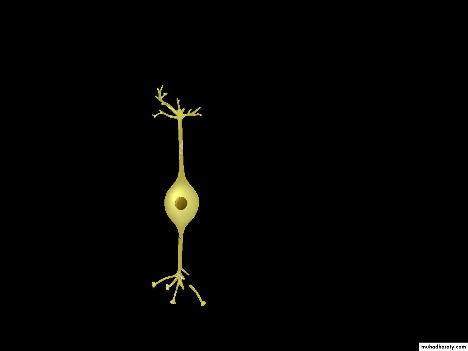
Most neurons consist of 3 parts:
• the dendrites which are multiple elongated processes specialized in receiving stimuli from the environment, sensory epithelial cells , or other neurons,• The cell body, or perikaryon. Which is the trophic center for the whole nerve cell and is also receptive to stimuli,
• The axon, which is a single process specialized in generating or conducting nerve impulses to other cells (nerve, muscle, and gland cells), the distal portion of the axon is usually branched and constitutes the terminal arborization.
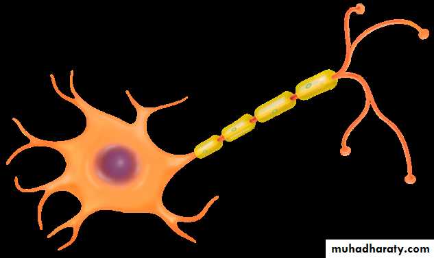
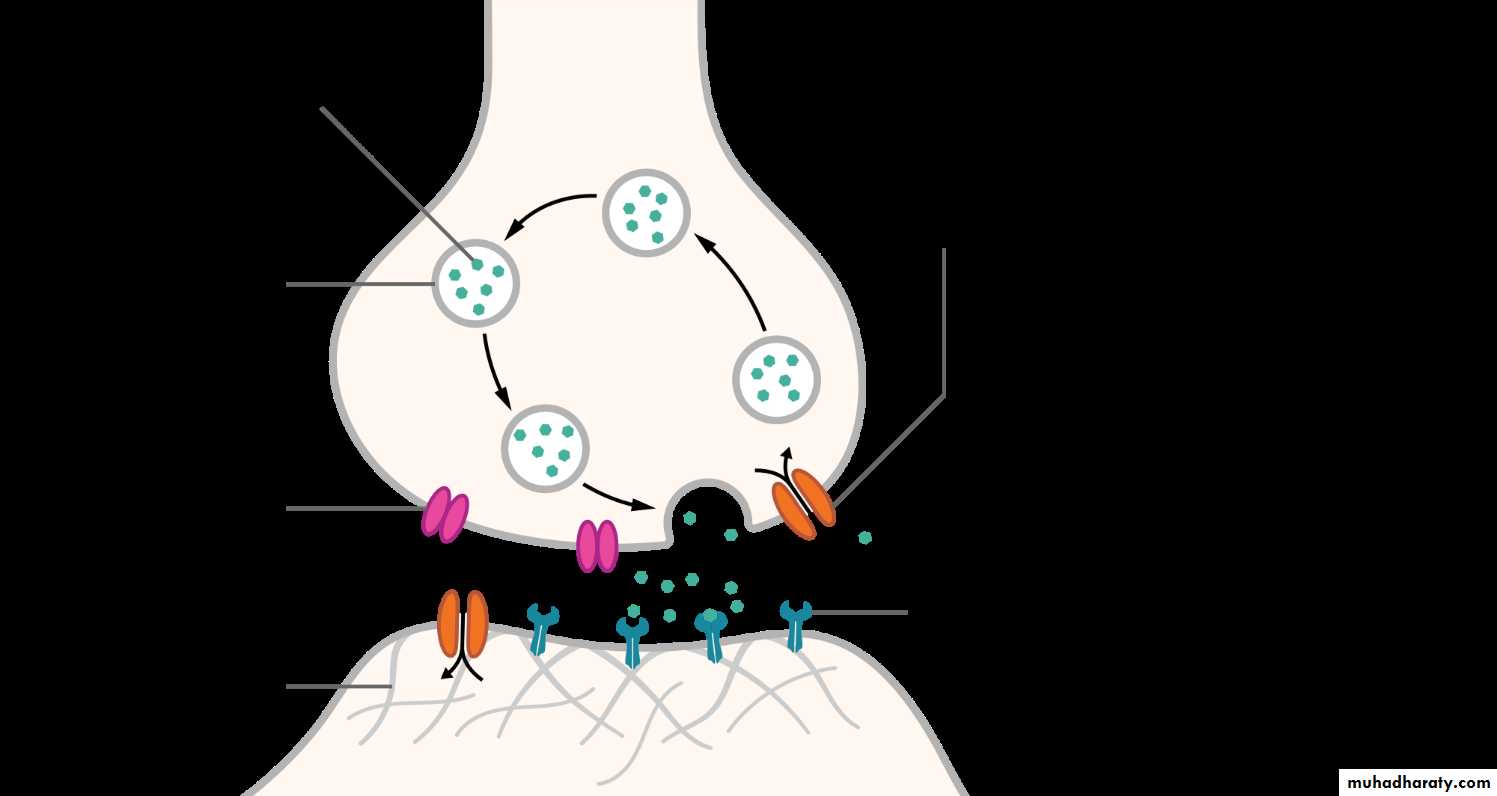
Each branch of this arborization terminates on the next cell in dilatation called bulbs (boutons), which interact with other neurons or non- nerve cells, forming structures called synapses.
Synapses transmit information to the next cell in the circuit.
Neurons and their processes are extremely variable in size and shape. Cell bodies can be spherical, ovoid, or angular.
Some is very large measuring up to 150 Mm in diameter –large enough to be visible to the naked eye.
Other nerve cells are among the smallest cells in the body, for example, the cell bodies of granule cells of the cerebellum are only 4-5 Mm in diameter.
According to the size and shape of their processes, most neurons can be placed in one of the following categories:
Multipolar neurons, which have more than two cell processes, one processes being the axon and the dendrites. Most neurons of the body are multipolar
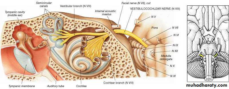
Dipolar neurons, with one dendrite and one axon. Bipolar neurons are found in the cochlear and vestibular ganglia as well as in the retina and the olfactory mucosa
Psuedounipolar neurons, which have a single process that is close to the perikaryon and divides into two branches. The process then forms a T shape, with one branch extending to a peripheral ending and the other toward the central nervous system.
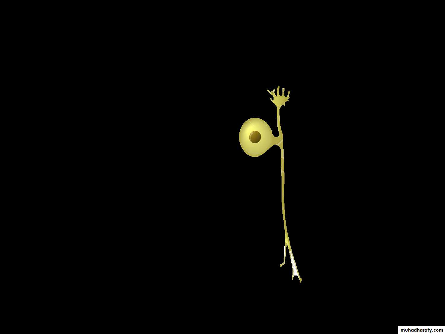
Psuedounipolar neurons are found in the spinal ganglia (the sensory ganglia located in the dorsal roots of the spinal nerves). They are also found is most cranial ganglia.
In pseudounipolar neurons, stimuli that are picked up by the dendrites travel directly to the axons terminal without passing through the perikaryon.
Neurons can also be classified according to their functional roles.
Motor (efferent) neurons control effectors organs such as fibers and exocrine and endocrine glands
Sensory (afferent) neurons are involved in the reception of sensory stimuli from the environment and from within the body.
Interneurons establish relationships among other neurons, forming complex functional networks or circuits (as in the retina).