Done by
Dr.Rafid Remthan Al-Temimi
Clinical Radiology
CAMB,DMRD,M.B.Ch.B.,.
المرحلة
:
الثانية
المادة
:
التشريح
ج
امعة ذي قار
/
كلية الطب
الدكتور
رافد
رمثان التميمي
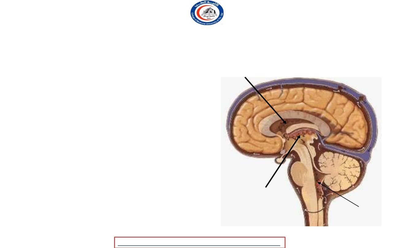
• It is formed by invaginating
of vascular pia mater into the
ventricular cavity
• It becomes highly convoluted &
produce a spongy-like appearance
• It enters the 3rd and 4th
ventricles through their roofs,
and the lateral ventricles
through the choroid fissure
• produces cerebrospinal fluid (CSF)
CHOROID PLEXUS
Lateral ventricle
Third ventricle
Fourth
ventricle
University Of Thi-Qar
College Of medicine
Dr.Rafid Remthan AL-Temimi,Clinical Radiology,CAMB, 2020
Anatomy lecture . 2
nd
stage
Dr.Rafid Al-Temimi
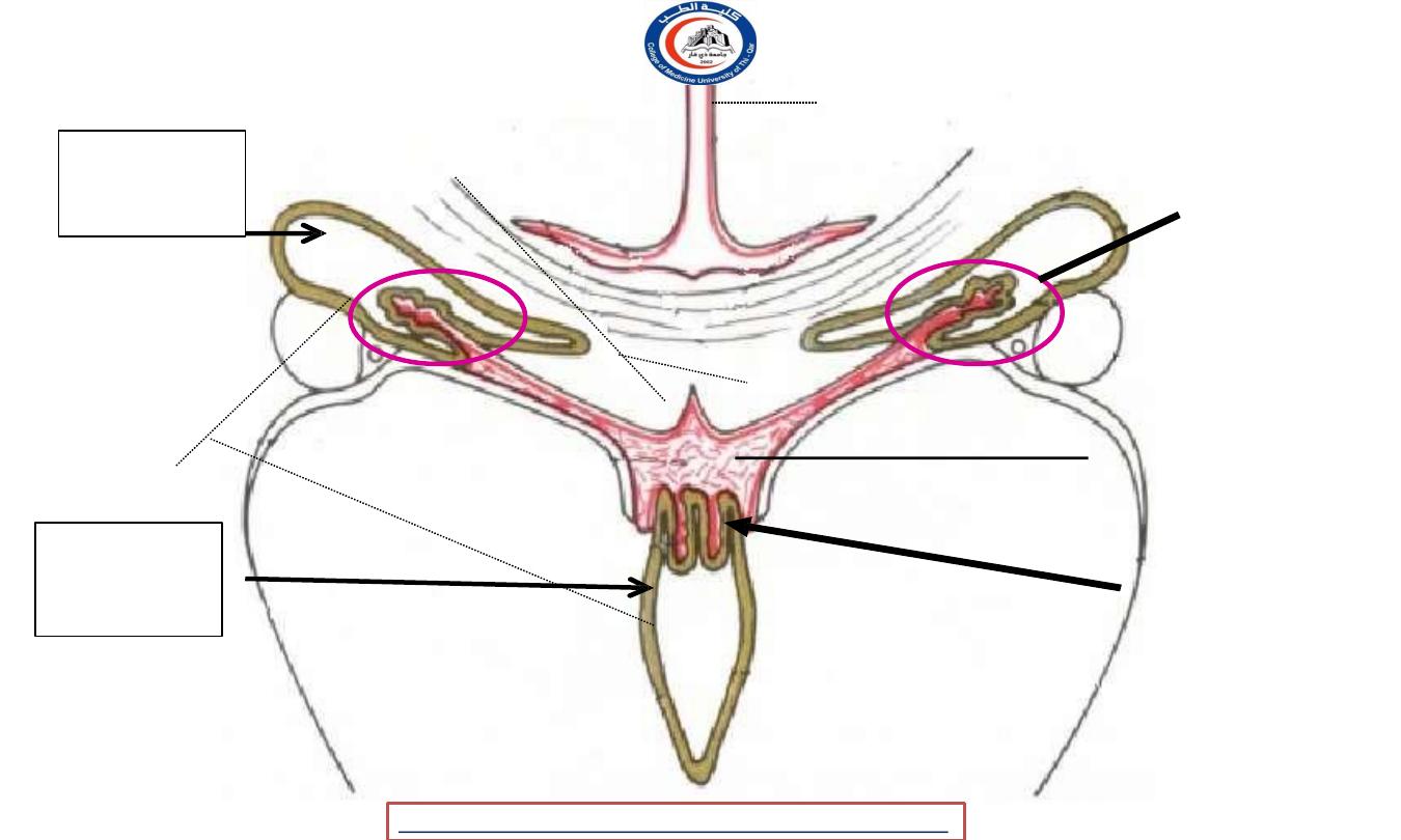
Cavity Of
Lateral
Ventricle
Cavity Of
Third
Ventricle
Pia Mater
Ependyma
CN
THALAMUS
CORPUS
CALLOSUM
Choroid
Plexus of The
Lateral
Ventricle
Pia Mater of
Tela
Choroidae
BODY OF
FORNIX
Choroid
Plexus of
The Third
Ventricle
Blood supply derives from choroidal branches of the internal carotid & basilar arteries
Coronal section of the cavities of the lateral and 3
rd
ventricles
University Of Thi-Qar
College Of medicine
Dr.Rafid Remthan AL-Temimi,Clinical Radiology,CAMB, 2020
Anatomy lecture . 2
nd
stage
Dr.Rafid Al-Temimi
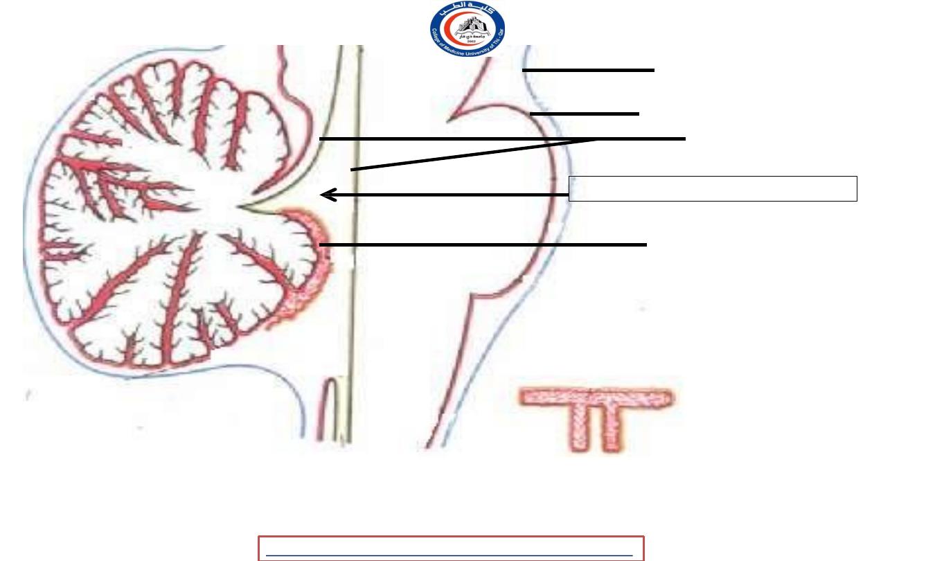
Arachnoid mater
Pia mater
Ependyma
CEREBELLUM
Cavity of fourth ventricle
Choroid plexus of the
fourth ventricle
•
T shaped, vertical part is double
•
Horizontal part extends into lateral recesses of each ventricle (foramina of Luskha)
•
Blood supply ; posterior inferior cerebellar arteries
University Of Thi-Qar
College Of medicine
Dr.Rafid Remthan AL-Temimi,Clinical Radiology,CAMB, 2020
Anatomy lecture . 2
nd
stage
Dr.Rafid Al-Temimi

What is cerebrospinal fluid (CSF) ?
•
Clear, colorless fluid
•
Produced by the choroid plexus
•
Found in the :
Ventricles of the brain
Subarachnoid space (between Arachnoid + Pia mater) around the brain & spinal cord
• The pressure of the CSF is kept remarkably constant.
• Based on the Monro-Kellie doctrine :
•
“Volume of BLOOD, CSF & BRAIN at any time must be relatively
constant”
University Of Thi-Qar
College Of medicine
Dr.Rafid Remthan AL-Temimi,Clinical Radiology,CAMB, 2020
Anatomy lecture . 2
nd
stage
Dr.Rafid Al-Temimi
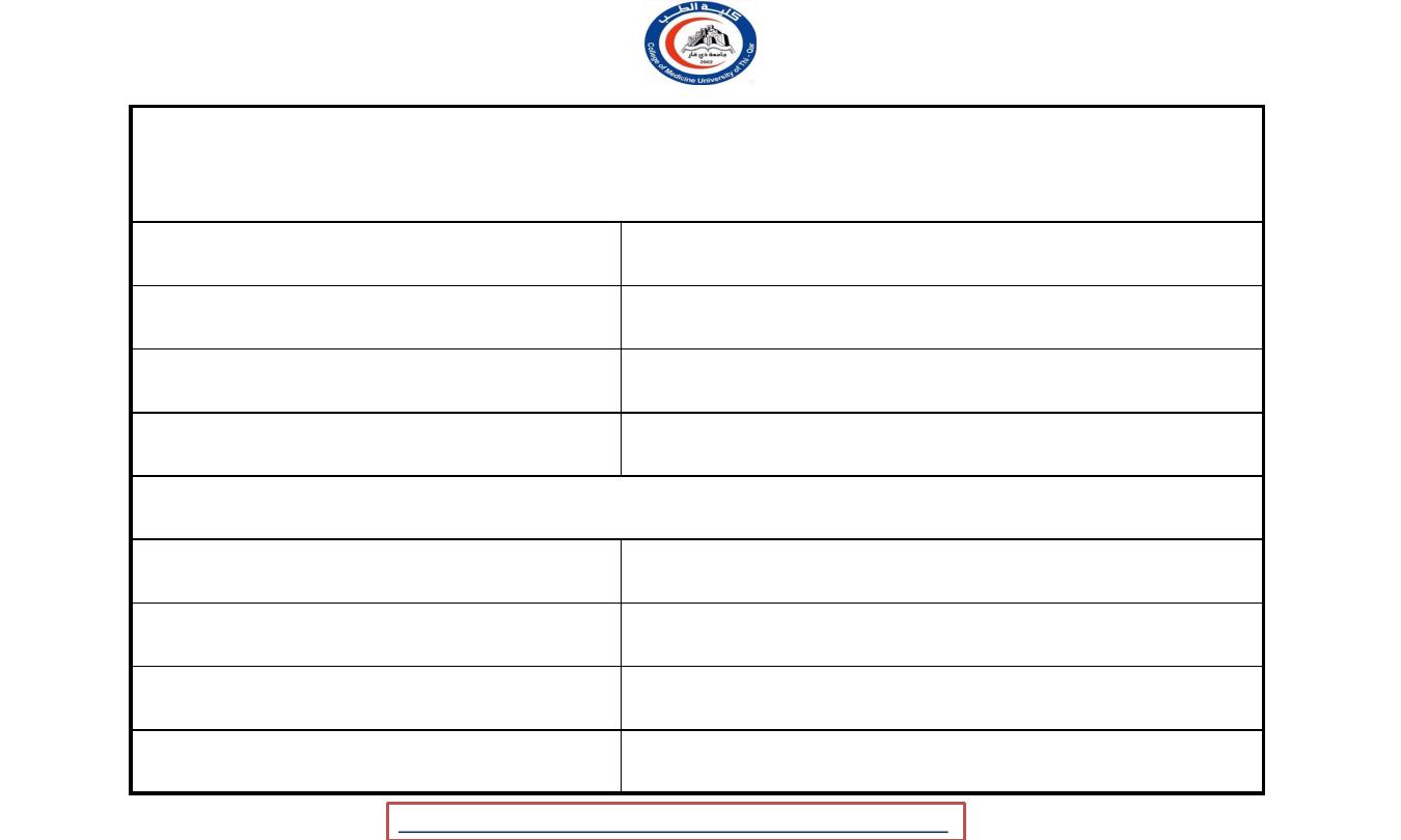
Physical characteristics and composition of the CSF
Appearance
Clear and colourless
Volume
130 ml
Rate of production
0.5 ml/min
Pressure
60-150 mm of water
Composition
protein
15-45 mg/100 ml
glucose
50-85 mg/ 100 ml
chloride
720-750 mg/100 ml
No. of cells
0-3 lymphocytes/cu mm
University Of Thi-Qar
College Of medicine
Dr.Rafid Remthan AL-Temimi,Clinical Radiology,CAMB, 2020
Anatomy lecture . 2
nd
stage
Dr.Rafid Al-Temimi

Function of the CSF :
1. Cushions & protects the CNS from trauma
2. Provides mechanical buoyancy & support for the brain
3. Serves as a reservoir & assists in the regulation of the contents of
the skull
4. Nourishes the CNS
5. Removes metabolites from the CNS
6. Serves as a pathway for pineal secretions to reach the pituitary
gland
University Of Thi-Qar
College Of medicine
Dr.Rafid Remthan AL-Temimi,Clinical Radiology,CAMB, 2020
Anatomy lecture . 2
nd
stage
Dr.Rafid Al-Temimi

Sites of formation :
1.
Choroid plexus of the ventricle cavities, mostly is formed in the LATERAL
VENTRICLES
2.
Some originate from the ependymal cells lining the ventricles
3.
Some from the brain substances through perivascular spaces
Movement of CSF inside the ventricle is controlled by the:
1. Pulsation of the artery in the choroid plexus
2. By the aid of the cilia & microvilli of the ependymal cells
University Of Thi-Qar
College Of medicine
Dr.Rafid Remthan AL-Temimi,Clinical Radiology,CAMB, 2020
Anatomy lecture . 2
nd
stage
Dr.Rafid Al-Temimi
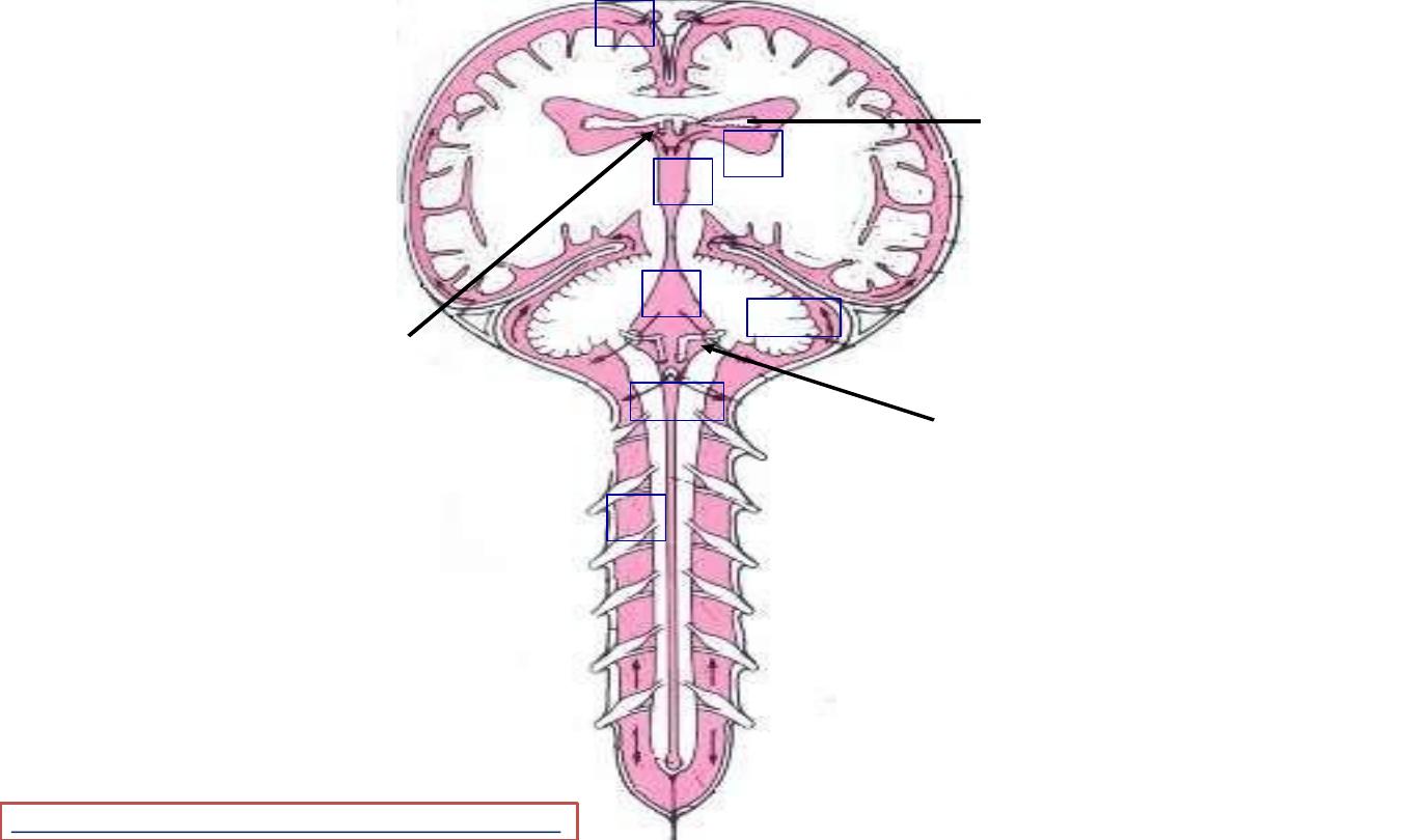
Choroid plexus of the
4
th
ventricle
Choroid plexus of the
3
rd
ventricle
1
2
3
5
3.2
3.1
4
Superiorly = lateral
aspect of each
cerebral
hemisphere
Inferiorly =
subarachnoid space
around the brain &
spinal cord
Choroid plexus of the
lateral ventricle
University Of Thi-Qar
College Of medicine
Dr.Rafid Remthan AL-Temimi,Clinical Radiology,CAMB, 2020
Anatomy lecture . 2
nd
stage
Dr.Rafid Al-Temimi
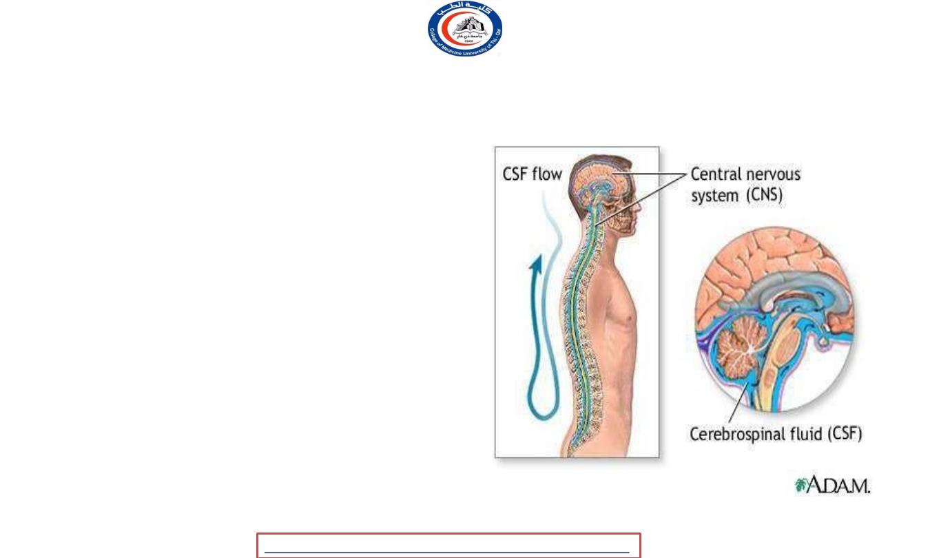
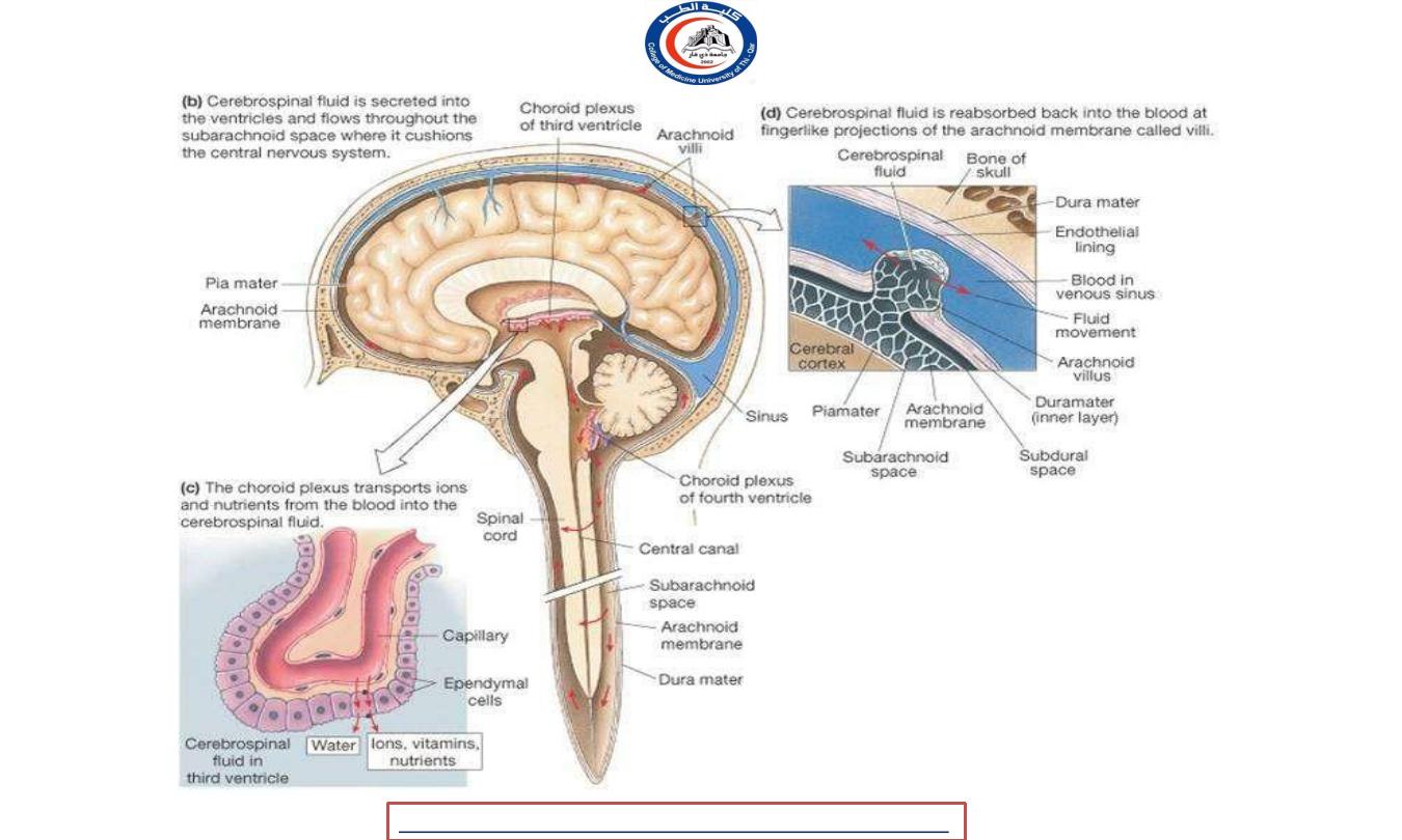
cerebrospinal fluid (CSF)
•
The CSF is formed in the lateral ventricles
escapes by the foramen of monro into the
third ventricle
•
From the third ventricle by the
aqueduct into the fourth ventricle.
•
Then from the fourth ventricle the fluid is
poured into the subarachnoid spaces through
the medial foramen of majendie and the two
lateral foramina of luschka.
•
There is no evidence that functional
communications between the cerebral
ventricles and the subarachnoid spaces exist
in any region except from the fourth
ventricle.
University Of Thi-Qar
College Of medicine
Dr.Rafid Remthan AL-Temimi,Clinical Radiology,CAMB, 2020
Anatomy lecture . 2
nd
stage
Dr.Rafid Al-Temimi
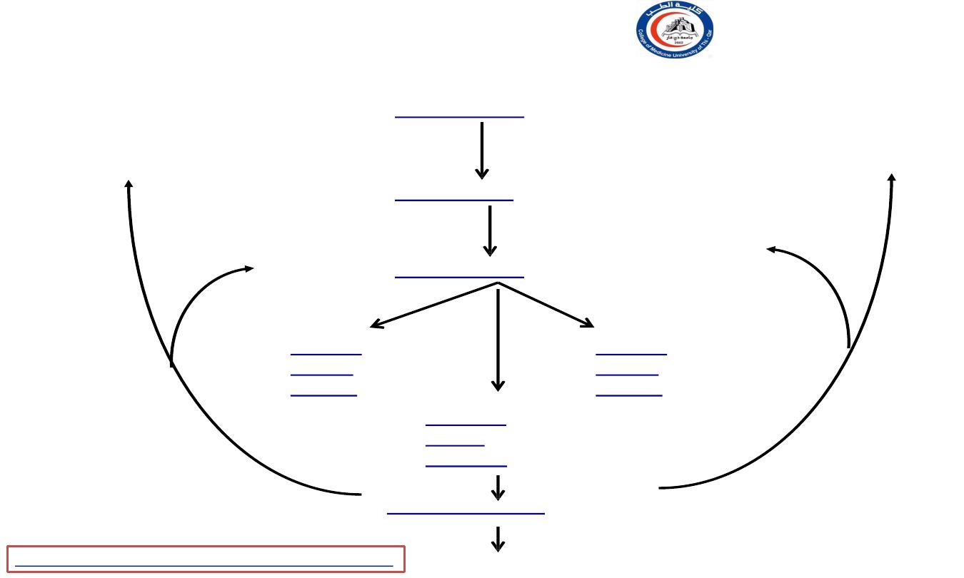
Choroid plexus of the
lateral ventricle
1. Lateral ventricle
Site of formation
.2Third ventricle
Interventricular foramina
3. Fourth ventricle
Cerebral aqueduct
3.2 Lateral
foramina
(Luschka)
3.2Lateral
foramina
(Luschka)
3.1 Median
foramen
(Magendie)
4. Subarachnoid space
Inferiorly
Superiorly
Absorbed
Superiorly
Absorbed
University Of Thi-Qar
College Of medicine
Dr.Rafid Remthan AL-Temimi,Clinical Radiology,CAMB, 2020
Anatomy lecture . 2
nd
stage
Dr.Rafid Al-Temimi
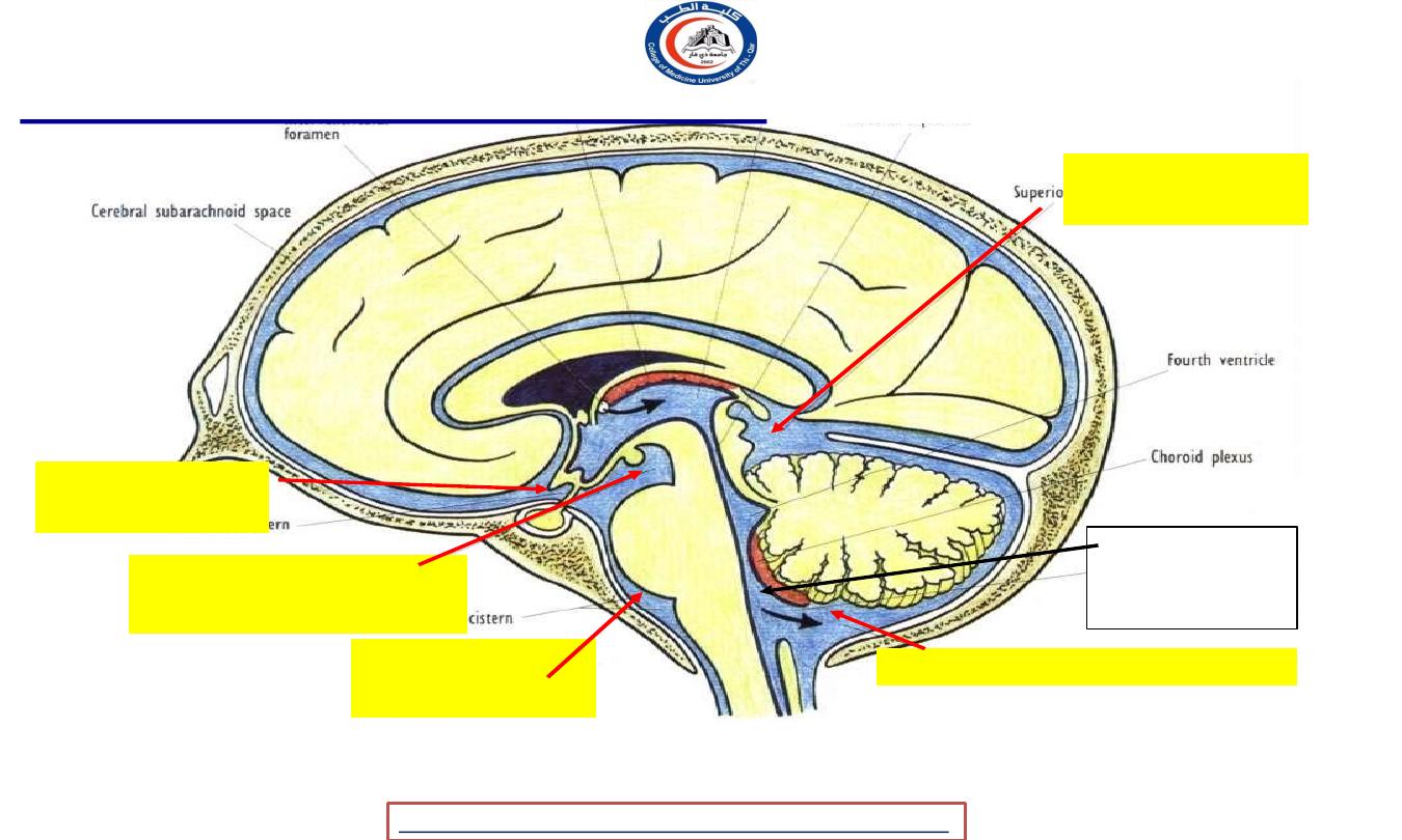
Median sagittal section to show the subarachnoid cisterns & circulation of CSF
Superior
cistern
Interpeduncular
cistern
Cerebellomedullary cistern
Chiasmatic
cistern
Pontine
cistern
Circulation of CSF in subarachnoid space :
Median
foramen of 4
th
ventricle
University Of Thi-Qar
College Of medicine
Dr.Rafid Remthan AL-Temimi,Clinical Radiology,CAMB, 2020
Anatomy lecture . 2
nd
stage
Dr.Rafid Al-Temimi

Factors that facilitate the flow of CSF in
subarachnoid space ;
1. Pulsation of the cerebral & spinal arteries
2. Movements of the vertebral column
3. Respiration & coughing
4. Changing of the positions
University Of Thi-Qar
College Of medicine
Dr.Rafid Remthan AL-Temimi,Clinical Radiology,CAMB, 2020
Anatomy lecture . 2
nd
stage
Dr.Rafid Al-Temimi
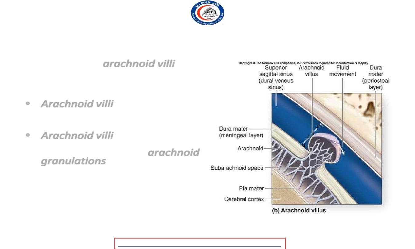
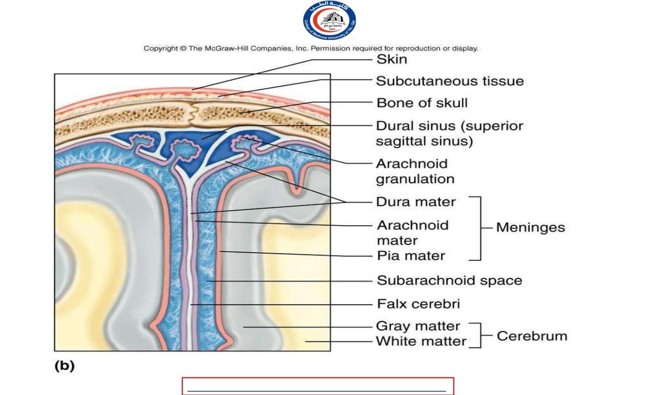
Absorption of CSF into dural venous sinuses
• Main sites -
arachnoid villi
(project
into dural venous sinuses,
especially, superior sagittal sinus)
• Arachnoid villi
are covered by
endothelium of the venous sinus
• Arachnoid villi
tend to be grouped
together & form elevations known as
arachnoid granulations
• CSF pressure >> the pressure in the
sinus
•
The rate of absorption of CSF
through the arachnoid villi controls
the CSF pressure
University Of Thi-Qar
College Of medicine
Dr.Rafid Remthan AL-Temimi,Clinical Radiology,CAMB, 2020
Anatomy lecture . 2
nd
stage
Dr.Rafid Al-Temimi
University Of Thi-Qar
College Of medicine
Dr.Rafid Remthan AL-Temimi,Clinical Radiology,CAMB, 2020
Anatomy lecture . 2
nd
stage
Dr.Rafid Al-Temimi
University Of Thi-Qar
College Of medicine
Dr.Rafid Remthan AL-Temimi,Clinical Radiology,CAMB, 2020
Anatomy lecture . 2
nd
stage
Dr.Rafid Al-Temimi
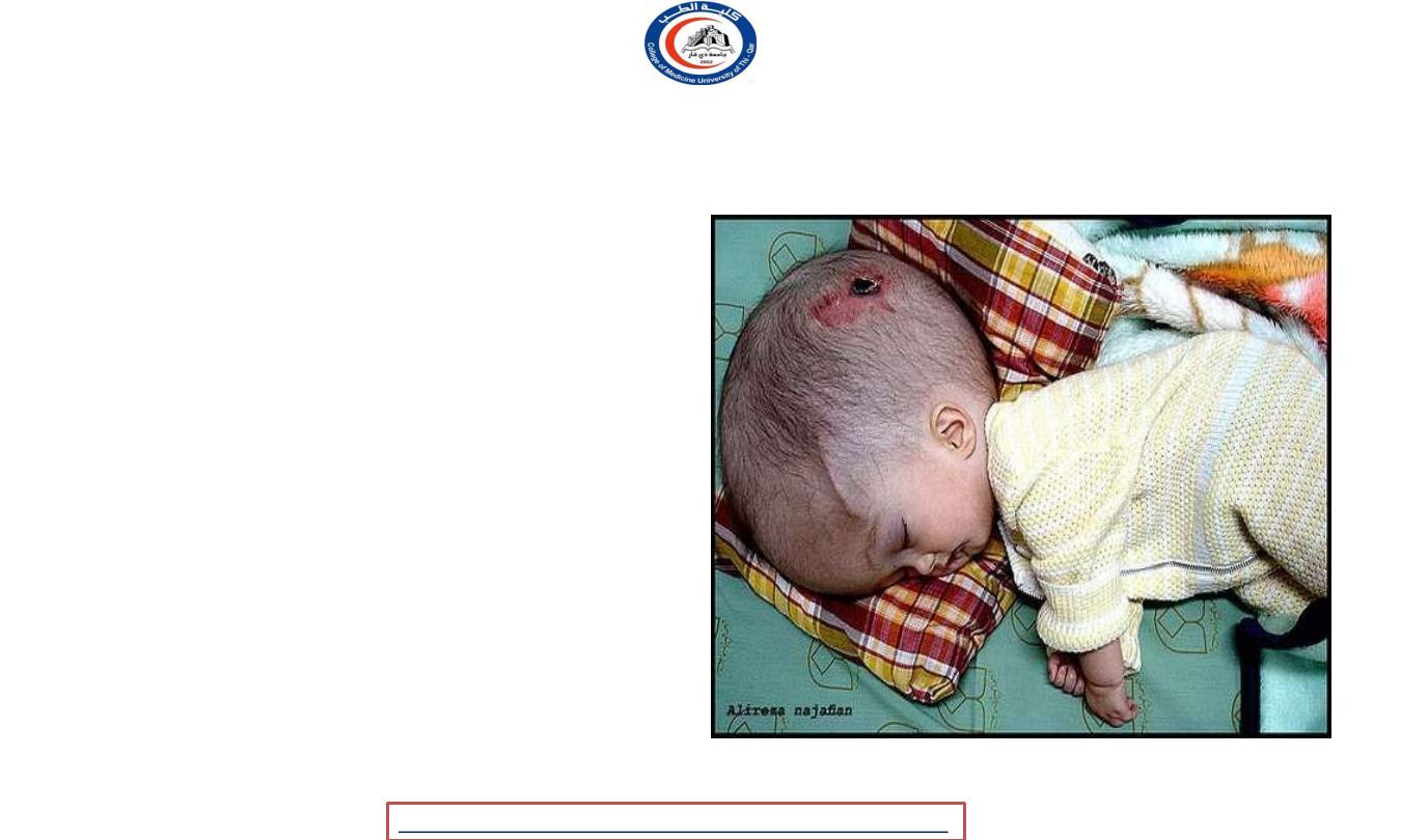
CLINICAL APPLICATION
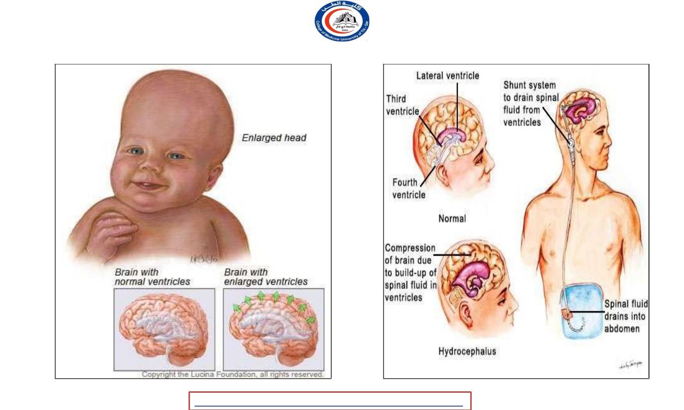
Hydrocephalus
• The term hydrocephalus is
derived from the Greek words
"hydro" meaning water and
"cephalus" meaning head.
• It is excessive accumulation of
fluid in the brain.
University Of Thi-Qar
College Of medicine
Dr.Rafid Remthan AL-Temimi,Clinical Radiology,CAMB, 2020
Anatomy lecture . 2
nd
stage
Dr.Rafid Al-Temimi
University Of Thi-Qar
College Of medicine
Dr.Rafid Remthan AL-Temimi,Clinical Radiology,CAMB, 2020
Anatomy lecture . 2
nd
stage
Dr.Rafid Al-Temimi

University Of Thi-Qar
College Of medicine
Dr.Rafid Remthan AL-Temimi,Clinical Radiology,CAMB, 2020
Anatomy lecture . 2
nd
stage
Dr.Rafid Al-Temimi
What is the blood brain barrier (BBB)?
•The brain is a privileged site, sheltered from the systemic
circulation by the blood-brain barrier (BBB).
.
Semipermeable barrier.
• Highly specialised brain endothelial structure.
• Helps maintain stable environment for brain.
• Separates neurons from some blood borne substances.

University Of Thi-Qar
College Of medicine
Dr.Rafid Remthan AL-Temimi,Clinical Radiology,CAMB, 2020
Anatomy lecture . 2
nd
stage
Dr.Rafid Al-Temimi
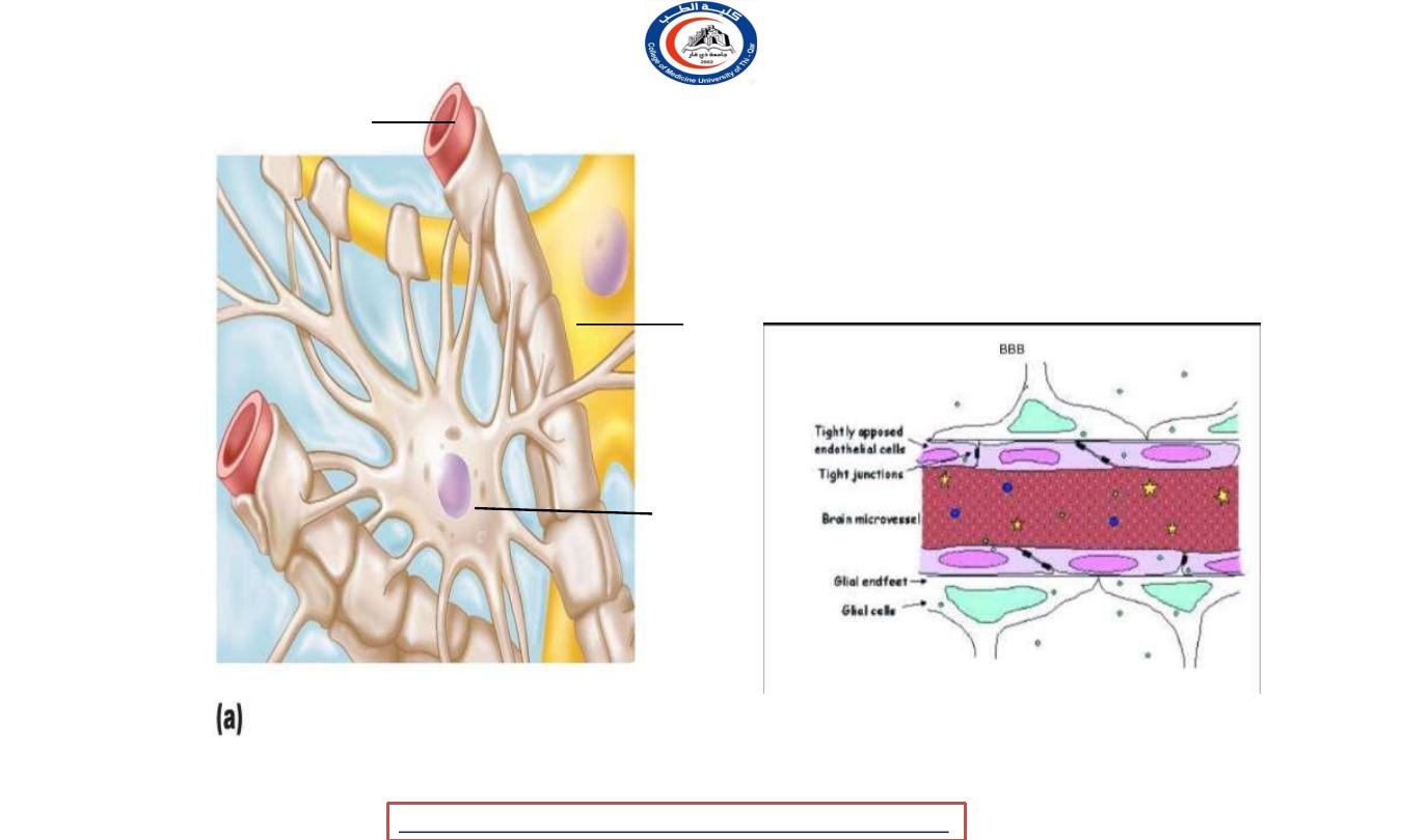
• It intervene between the blood in the capillaries
and extracellular spaces surrounding neurons.
• Principal components:
1. Capillary endothelial cells
and
tight junctions
between
them.
2. A
basement membrane
on which capillary endothelial
cells are arranged.
3. Foot process of astrocytes
.
University Of Thi-Qar
College Of medicine
Dr.Rafid Remthan AL-Temimi,Clinical Radiology,CAMB, 2020
Anatomy lecture . 2
nd
stage
Dr.Rafid Al-Temimi
Capillary
Neuron
Astrocyte
Astrocytes are the most abundant CNS neuroglia.

University Of Thi-Qar
College Of medicine
Dr.Rafid Remthan AL-Temimi,Clinical Radiology,CAMB, 2020
Anatomy lecture . 2
nd
stage
Dr.Rafid Al-Temimi
Blood Brain Barrier: Functions
• Selective barrier
– Allows nutrients to move by facilitated diffusion
– Metabolic wastes, proteins, toxins, most drugs, small
nonessential amino acids, K
+
denied
– Allows any fat-soluble substances to pass, including
alcohol, nicotine, and anesthetics
• Absent in some areas, e.g., vomiting center and
hypothalamus, where necessary to monitor chemical
composition of blood

University Of Thi-Qar
College Of medicine
Dr.Rafid Remthan AL-Temimi,Clinical Radiology,CAMB, 2020
Anatomy lecture . 2
nd
stage
Dr.Rafid Al-Temimi
• Areas that are devoid of:
1. Pineal gland
2. Neurohypophysis
3. Area postrema (at lower end of floor of fourth
ventricle.
4. Wall of supraoptic recess of third ventricle.

University Of Thi-Qar
College Of medicine
Dr.Rafid Remthan AL-Temimi,Clinical Radiology,CAMB, 2020
Anatomy lecture . 2
nd
stage
Dr.Rafid Al-Temimi
BLOOD-CEREBROSPINAL FLUID BARRIER
It is the barrier between the blood and cerebrospinal fluid that exists
at the choroid plexus.
The function of this barrier is same as of BBB.
It doest not allow the movement of following substances like
chemical agents,pathogens,bile pigments
etc.
It allows the movement of only those substances which are allowed by
BBB like Oxygen,Carbon Dioxide,Water etc

University Of Thi-Qar
College Of medicine
Anatomy lecture . 2
nd
stage
Dr.Rafid Al-Temimi
Dr.Rafid Remthan AL-Temimi,Clinical Radiology,CAMB, 2020
