
Pneumonia
Tikrit University
College of Medicine
Department of Radiology
Chest Series

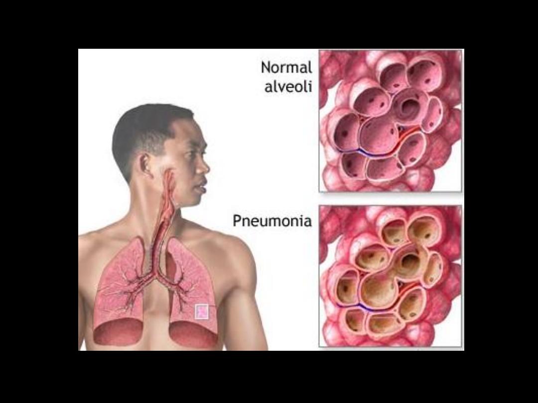
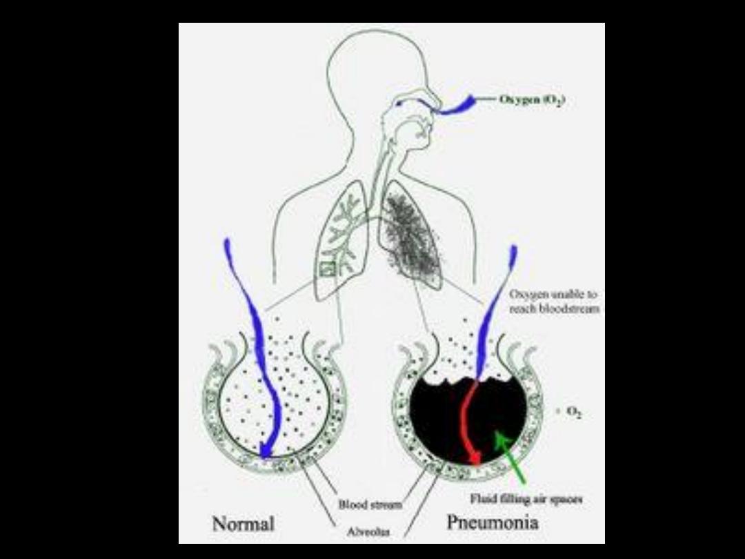
Pneumonia
is a lung parenchyma
infection caused by bacteria, a virus
or fungi, with a consolidation on
radiological examination.
Pneumonitis
is an inflammation of
the lungs caused by chemical or
radiation therapy but not with
infectious agents.
Definition

– Inhalation
– Aspiration of oropharingeal secretion
– Hematogenic spread
– Direct spread (thorax wall, mediastinum)
Spread of lung infections

Predisposing factors of pneumonia
• Airways mechanical barrier damage
• Specific and/or nonspecific immune defense
mechanisms injury
• Bronchial obstruction
• Micro aspiration of upper respiratory truck
secretion.
• Lung edema
• Viral infections.

Diagnostic Methods
• History, physical examination
• Chest X-Ray
• Sputum examination (gram stained)
• Sputum , blood cultures
• Serological tests
• Peripheral blood analysis

Symptoms
fever, shaking chills,
cough, sputum (expectoration),
pleuritic pain.
Others:
(dispnea, fatigue, sweating, loss of appetite...)
Diagnosis

Radiology:
lobar opacities – Lobar pn.
interstitial shadows – interstitial pn.
Patchy opacities – bronchopn.
Others
(absea, pneumatocele)
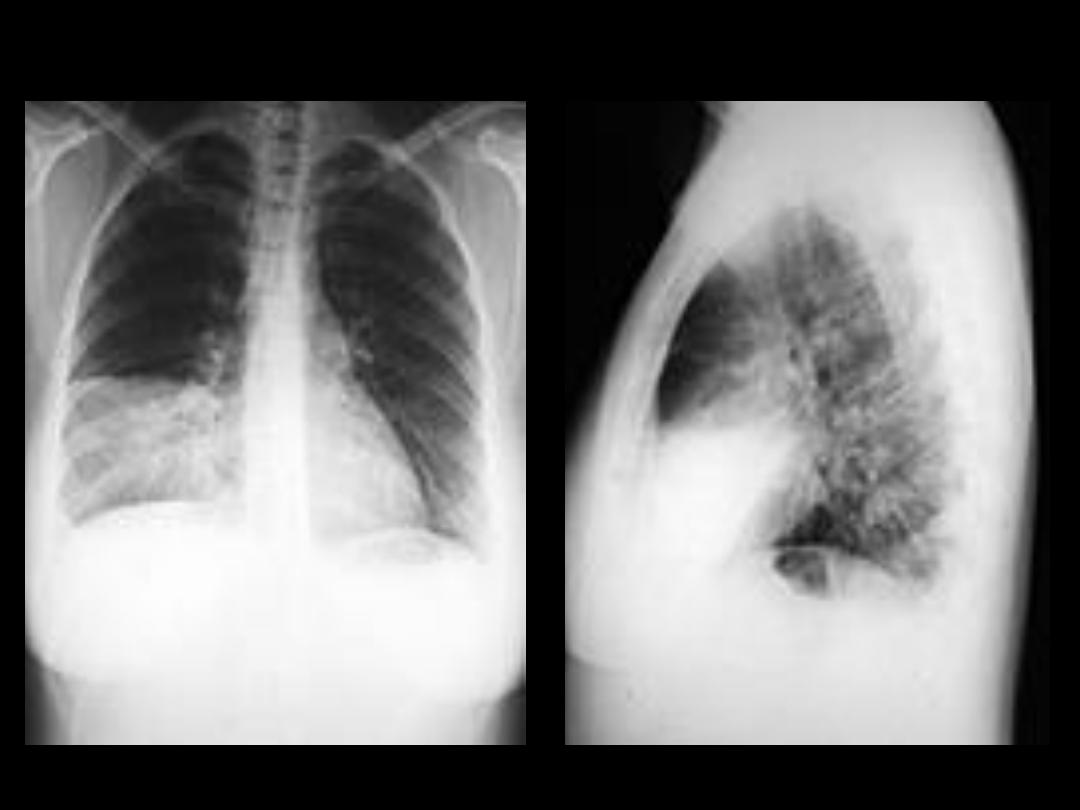
Diagnosis

Chest X-Ray
• Gold standart test for pneumonia
• For differencial diagnosis
• For grading pneumonia severity
• For examining complications
• For Follow up

Radiological classification
Classification with anatomical localization
• Lobar consolidation
• Bronchopneumonia
• Interstitial pneumonia
• Round pneumonia
Classification with targeting therapy
• Community acquired pneumonia
• Hospital acquired pneumonia (Nosocomial)
• Immunosuppresed (immunocompromised)
patients pneumonia

Just to Know
Community acquired
pneumonia is the:
Pneumonia acquired outside
hospital, and frequently seen in
healthy persons

Lobar pneumonia
– Focal Opacity
– Lobar
Lobular pneumonia (bronchopneumonia)
– Multiple patchy opacities
– If Bilateral , Asymmetrical
Interstitial pneumonia
– Bilateral symmetrical dirty shadow.
Round pneumonia
– Seen in pediatrics
In general - Pleural effusion could be seen with pneumonia.

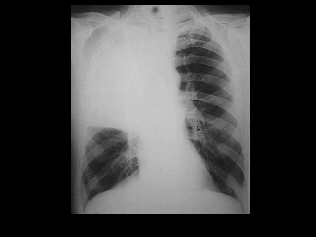
Lobar pneumonia
– Focal Opacity
• Infection primarily involves alveolus.
• Spread through pores of Kohn and canals of Lambert
throughout a segment and ultimately an entire lobe.
• The larger bronchi remain patent, causing
– Air bronchogram
– No volume loss because airways are open
• Chx by Patchy consolidation of a single lobe.
• It is usually bacterial in origin and is the most common
presentation of community acquired pneumonia.
• S. pneumoniae
• K. pneumoniae
• Others:
• S. aureus
• H. influenzae
• Fungal
• Nowadays uncommon due to early treatment

Lobular pneumonia (bronchopneumonia)
– Multiple
• Primarily affects the bronchi and adjacent
alveoli.
• Volume loss may be present as bronchi fill
with exudate.
• Bronchial spread results in multifocal patchy
Opacities
• Manifests as patchy consolidatin with poorly
defied airspace opacitis, usually involving
several lobes, and most commonly due to:
– S. pneumoniae
– K. pneumoniae
– Others:
• S. aureus
• H. influenzae

Interstitial pneumonia
Chx by Bilateral symmetrical dirty shadow.
It is caused by inflammatory cells located
predominantly in the
interstiail tissue
of
the alveolar septa causing:
– diffuse or patchy ground glass opacification.
• It can be caused by:
• Viral pneumonia
• Mycoplasma
• Chlamydia
• Pneumocysti.

Round pneumonia
• It is an infectious mass-like opacity seen
only in children, most commonly due to
Streptococcus pneumonia.
• Infection remains somewhat confined due
to incomplete formation of pores of Kohn.
Lobar pneumonia
Lobar pneumonia
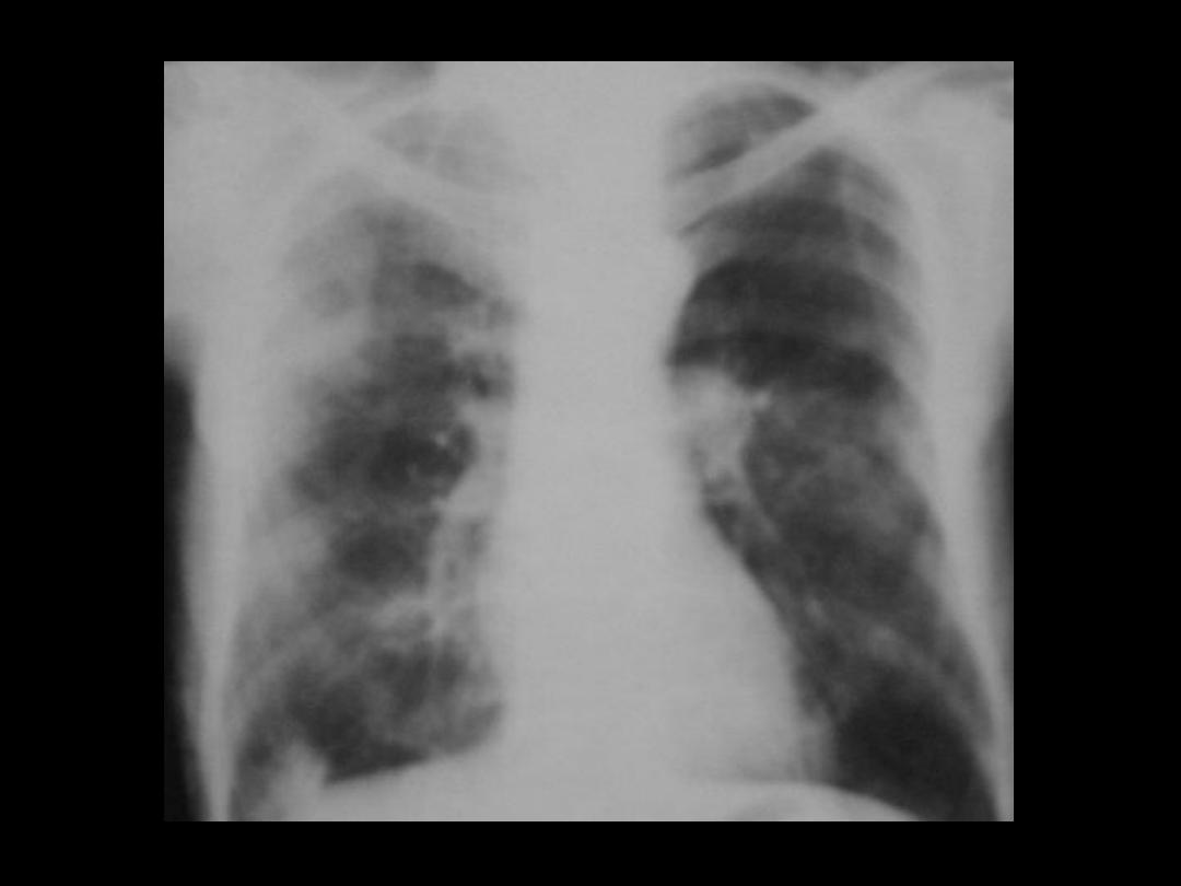
Bronchopneumonia
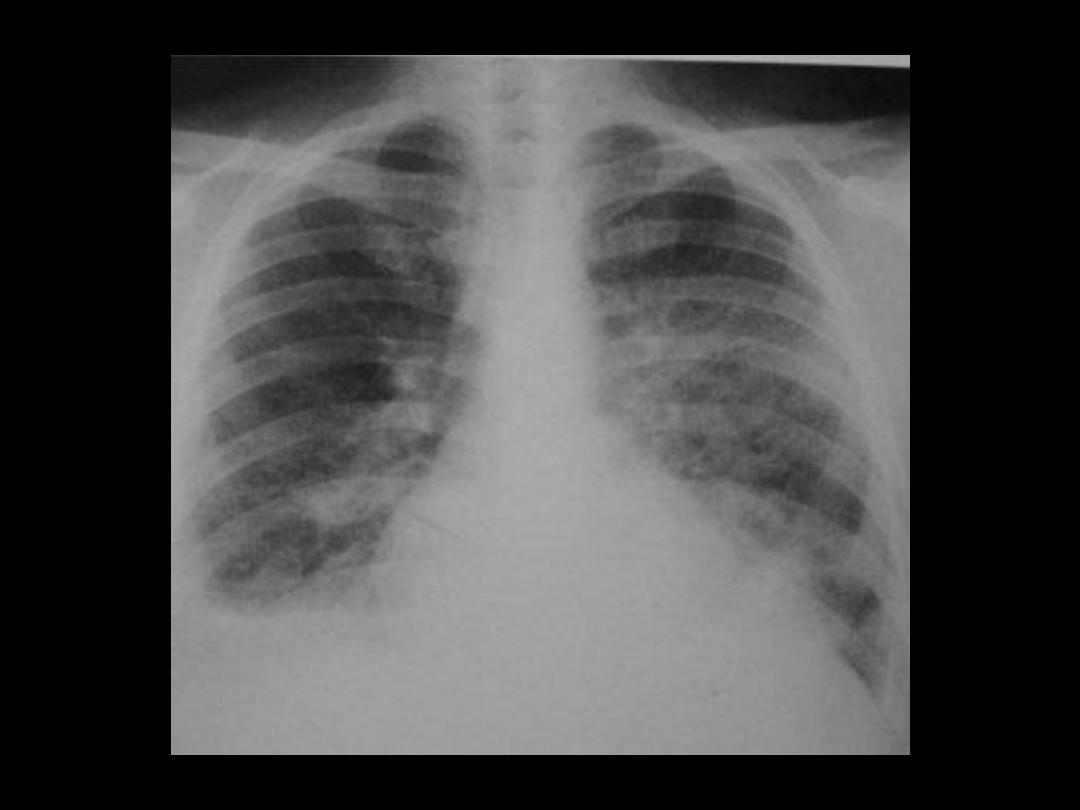
Interstitial pneumonia