STRUCTURE AND FUNCTION OF THE SKIN
The skin – the interface between humans and their environmentSkin is your passport into the world

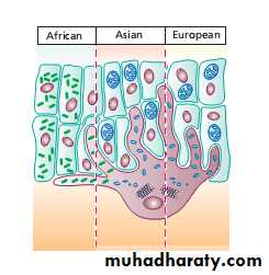
Skin can decide if you are : or
criminal
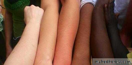
innocentSkin can decide your race
qqqqqqqqqqq
Skin is the largest organ in the body

Skin weighs an average
of
Skin covers an area of 2m2
It acts as a barrier, protecting the body from harsh external conditions and preventing the loss of important body constituents, especially water.
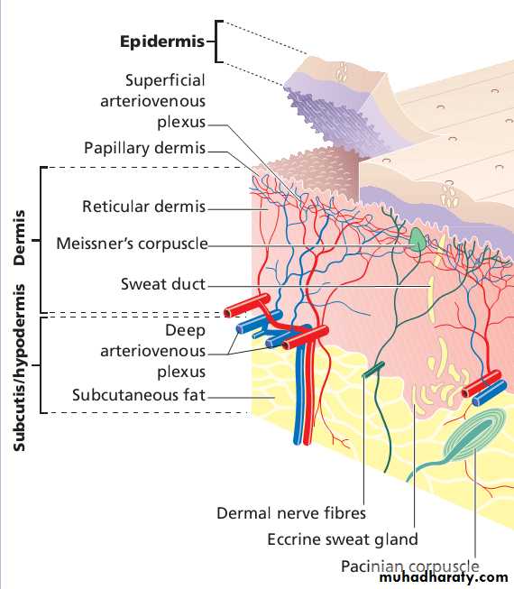
Structure of the skin
The skin has three layers :
The outer one is the epidermis, which is firmly attached to, and supported by connective tissue in the underlying dermis.
Beneath the dermis is loose connective tissue, the subcutis/hypodermis, usually contains abundant fat.
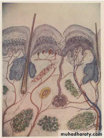
Structure of the skin, from WS Gottheil, Skin Diseases
(1897).Epidermis
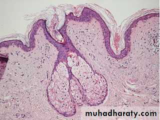
The epidermis consists of many layers of closely packed cells, the most superficial of which are flattened and filled with keratins; it is therefore a stratified squamous epithelium.It adheres to the dermis partly by the interlocking of its downward projections (epidermal ridges) with upward projections of the dermis (dermal papillae) . The epidermis contains no blood vessels.
It varies in thickness from less than 0.1 mm on the eyelids to nearly 1 mm on the palms and soles.
from basal layer to the surface takes 30 to 60 days.

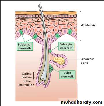
Stem cells reside amongst interfollicular basal cells and amongst the cells of the external root sheath at the bulge in the hair follicle at the level of attachment of the arrector pili muscle.
The basal layer, the deepest layer, rests on a basement membrane.
It is a single layer of columnar cells, whose basal surfaces sprout many fine processes and hemidesmosomes, anchoring them to lamina densa of basement membrane.
The spinous or prickle cell layer is composed of keratinocytes.
These differentiating cells, which synthesize keratins, are larger than basal cells.Keratinocytes are firmly attached to each other by small interlocking cytoplasmic processes, by abundant desmosomes and by other cadherins .
Under the light microscope, the desmosomes look like ‘prickles’.
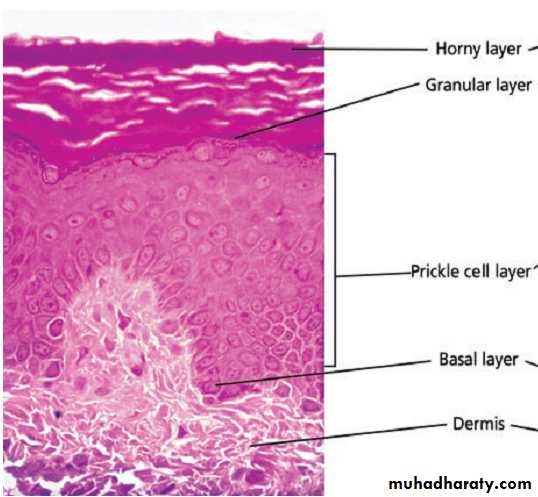
The granular layer, consists of two or three layers cells are flatter than those in the spinous layer,have more tonofibrils.
cells contain large irregular basophilic granules of keratohyalin, merge with tonofibrils.
keratohyalin granules break up and their contents are dispersed throughout the cytoplasm.
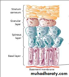
The horny layer (stratum corneum) is made of piled-up layers of flattened dead cells (corneocytes)– the bricks – separated by lipids – the mortar – in the intercellular space.
The corneocyte cytoplasm is packed with keratin filaments, embedded in a matrix and enclosed by an envelope derived from keratohyalin granules.
Keratinization
All cells have an internal skeleton made up of microfilaments (actin), microtubules (tubulin) and intermediate filaments .Keratins ( meaning ‘horn’) are the main intermediate filaments in epithelial cells
During differentiation, the keratin fibrils in the cells of the horny layer align and aggregate, under the influence of filaggrin. Cysteine, found in keratins of the horny layer, allows cross-linking of fibrils to give the epidermis strength to withstand injury.
The typical ‘basket weave’ appearance of the horny layer in routine histological sections is artefactual.
Other cells in the epidermis
MelanocytesThey migrate from the neural crest into the basal layer.
also found in hair bulbs, retina and pia arachnoid.
Each dendritic melanocyte associates with a number of keratinocytes, forming an ‘epidermal melanin unit’ .
The dendritic processes wind between the epidermal cells and end as discs in contact with them.
cytoplasm contains discrete organelles,melanosomes, containing varying amounts of the pigment melanin.
Langerhans cells
dendritic cell like the melanocyte.lacks desmosomes,tonofibrils, has a lobulated nucleus.
their dendritic processes fan out as striking network
highly specialized macrophages. They take up exogenous antigen, process it and present it to T lymphocytes either in skin or in local lymph nodes.
immunosurveillance for viral and tumour antigens.
Topical or systemic glucocorticoids reduce their density
The Langerhans cell is principal cell in skin allografts to which T lymphocytes of the host react during rejection
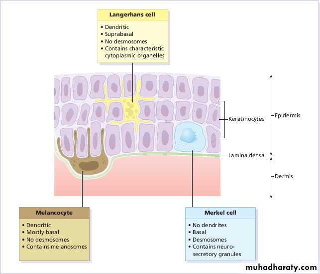
Merkel cells
act as transducers for fine touch.non-dendritic cells, lying in or near the basal layer.
concentrated in localized thickenings of the epidermis near hair follicles (hair discs), and contain membrane-bound spherical granules.
Sparse desmosomes connect these cells to neighbouring keratinocytes.
Fine unmyelinated nerve endings are often associated with Merkel cells
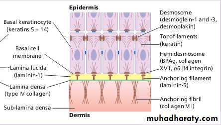
Dermo-epidermal junction
lies at the interface between epidermis and dermis.
Electron microscopy shows that the lamina densa is separated from the basal cells by an electron-lucent area, the lamina lucida.
The plasma membrane of basal cells has hemidesmosomes.
lamina lucida contains the adhesive macromolecules, laminin. Fine anchoring filaments cross the lamina lucida and connect the lamina densa to the plasma membrane of the basal cells.
Anchoring fibrils extend from papillary dermis to lamina densa.
provide mechanical support, growth, differentiation and migration of the overlying basal cells
semi-permeable filter
Dermis
lies between the epidermis and the subcutaneous fat.It supports the epidermis structurally and nutritionally.
Its thickness varies, being greatest in the palms and soles and least in the eyelids and penis.
The dermis interdigitates with the epidermis so that upward projections of the dermis, the dermal papillae, interlock with downward ridges of the epidermis, the rete pegs. This interdigitation is responsible for the ridges seen most readily on fingertips (as fingerprints).
the dermis has three components: cells, fibres and amorphous ground substance.
Cells of the dermis
main cells are fibroblasts, resident and transitory phagocytes, lymphocytes, Langerhans and mast cells.Fibres of the dermis
dermis is largely made up of interwoven fibres, principally of collagen, packed in bundles.70 – 80% .
individual collagen molecules consist of three polypeptide chains. The alignment of the chains is stabilized by covalent cross-links
Reticulin fibres are fine collagen fibres, seen in foetal skin,around the blood vessels,appendages of adult skin.
Elastic fibres: two distinct protein components: elastin core and a surrounding microfibrillar component.
Ground substance of the dermis
consists largely of two glycosaminoglycans (hyaluronic acid and dermatan sulphate) with smaller amounts of heparan sulphate and chondroitin sulphate.
It binds water, allowing nutrients, hormones and waste products to pass through the dermis.
It acts as a lubricant between the collagen and elastic fibre networks during skin movement
it provides bulk to act as a shock absorber.
Muscles
Both smooth and striated muscle found in skin.smooth arrector pili muscles used by animals to raise their fur, so protect them from cold. They are vestigial in humans, may help to express sebum. Smooth muscle is also responsible for ‘goose pimples’ (bumps) from cold, nipple erection, and the raising of the scrotum by the dartos muscle.
Striated fibers : platysma and some of the muscles of facial expression are also found in the dermis.
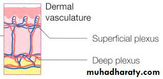
Blood vessels
The blood vessels lie in two main horizontal layers:The deep plexus is just above the subcutaneous fat, and its arterioles supply the sweat glands and hair papillae.
The superficial plexus is in the papillary dermis and arterioles from it become capillary loops in the dermal papillae.
Cutaneous lymphatics
Afferent lymphatics begin as blind-ended capillaries in the dermal papilla, pass to superficial lymphatic plexus in papillary dermis then two deeper horizontal plexuses, run with the veinsNerves
1 million nerve fibres. Most are found in the face and extremities. Both myelinated and non-myelinated fibres. Most free sensory nerves end in dermis.
Free nerve endings detect the potentially damaging stimuli of heat and pain (nocioceptors), Pacinian and Meissner corpuscles, register deformation of skin caused by pressure (mechanoreceptors), vibration , touch.
Autonomic nerves supply blood vessels, sweat glands arrector pili muscles.
Sebaceous glands
hair germs, few free glands arise from epidermis.
with hairs lie in the obtuse angle between the follicle and the epidermis. multilobed and contain cells full of lipid, which are shed whole (holocrine secretion) during secretion so that sebum contains their remnants.
It lubricates and waterproofs the skin, protects it from drying; mildly bactericidal and fungistatic.
Free sebaceous glands in the eyelid (meibomian glands),mucous membranes (Fordyce spots), nipple, perianalregion and genitalia.
Sweat glands
Eccrine sweat glands2–3 million sweat glands,most numerous on the palms, soles and axillae.
The tightly coiled glands lie deep in the dermis, and the emerging duct passes to the surface by penetrating the epidermis in a corkscrew fashion.
Initially, sweat is isotonic with plasma but, under normal conditions, it becomes hypotonic by the time it is discharged at the surface.
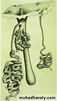
Apocrine sweat glands
Apocrine glands are limited to the axillae, nipples,periumbilical area, perineum and genitalia.The coiled tubular glands (larger than eccrine glands) lie deep in the dermis, and during sweating the luminal part of their cells is lost (decapitation secretion).
Apocrine sweat passes via the duct into the mid-portion of the hair follicle. The action of bacteria on apocrine sweat is responsible for body odour.
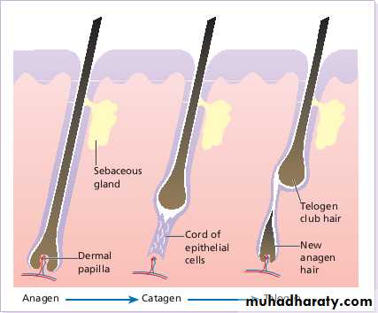
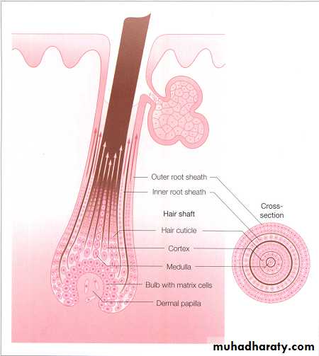
Hair follicle
Hair is the keratinized product of the hair follicle, a tube-like structure continuous with the epidermis at its upper end.
The follicles are sloped in the dermis, and longer follicles extend into the subcutaneous layer. An oblique muscle, the arrector pili, runs from the mid-region of the follicle wall to a point in the papillary dermis.
Above the muscle, one or more sebaceous glands.
The hair fibre is made up of three cell layers: an outer cuticle, the cortex and a variable central medulla
the inner root sheath which surrounds the hair fibre disintegrates before the hair emerges from the skin.
The inner root sheath is itself enclosed by the outer root sheath, which forms a continuous structure extending from the hair bulb to the epidermis.
Hairs are classified into three main types.
1.Lanugo hairs: Fine long hairs covering the foetus2.Vellus hairs: Fine short unmedullated hairs covering much of body, replace lanugo hairs before birth.
3.Terminal hairs: Long coarse medullated hairs scalp or pubic regions. growth influenced by androgen