
Thrombo-embolism in Pregnancy:

Thrombo-embolism include all vascular
occlusive processes like:
thrombophlebitis.
phlebothrombosis.
septic thrombophlebitis.
embolization of venous clots to the
lungs.

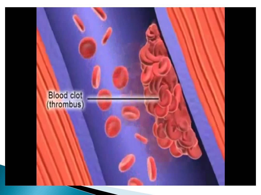
the formation of a thrombus within the
veins, can occur anywhere in the venous
system but the clinically predominant
sites are in the vessels of the legs
giving rise to deep venous thrombosis
in the lungs resulting in pulmonary
embolism
.

The patho-physiology of venous
thrombosis in pregnancy
related to
increased venous stasis but
alterations in the balance of proteins
of the coagulation and fibrinolytic
systems have a role.

Blood stasis .
Hypercoagulability .
Blood vessel damage.

It’s 3- 5 times common in pregnancy than
non pregnant.
VTE is increased 4-6 times antenatal and 20
fold postnatal.
Absolute risk 1in1000 pregnancies.
Cesarean Section increases the incidence to
1-2%.

Risk factors
Pregnancy is hyper-coagulable state due
to Increased clotting factors like Factor
XI,X,VII, VIII,II.
Decreased fibrinolytic activity
(decrease protein S)
Obesity, Operative delivery.
Restricted activities.

Acquired thrombophilia
associated with antiphospholipid
syndrome, the combination of
lupus anticoagulant with or
without, anticardiolipin
antibodies, with a history of
recurrent miscarriage and or
thrombosis.

Inherited thrombophilias
Protein-C, protein-S, and
Antithrombin III deficiency

Maternal age > 35 years.
Pre-pregnancy weight > 80 kg.
Pre-existing Thrombophilia.
Previous DVT.
Severe varicose veins .

Prolonged bed rest.
Multi foetal pregnancies.
Severe pre-eclampsia.
Caesarean section delivery.
Sepsis, especially pelvic.

Diagnosis:
History:
Clinical presentation.
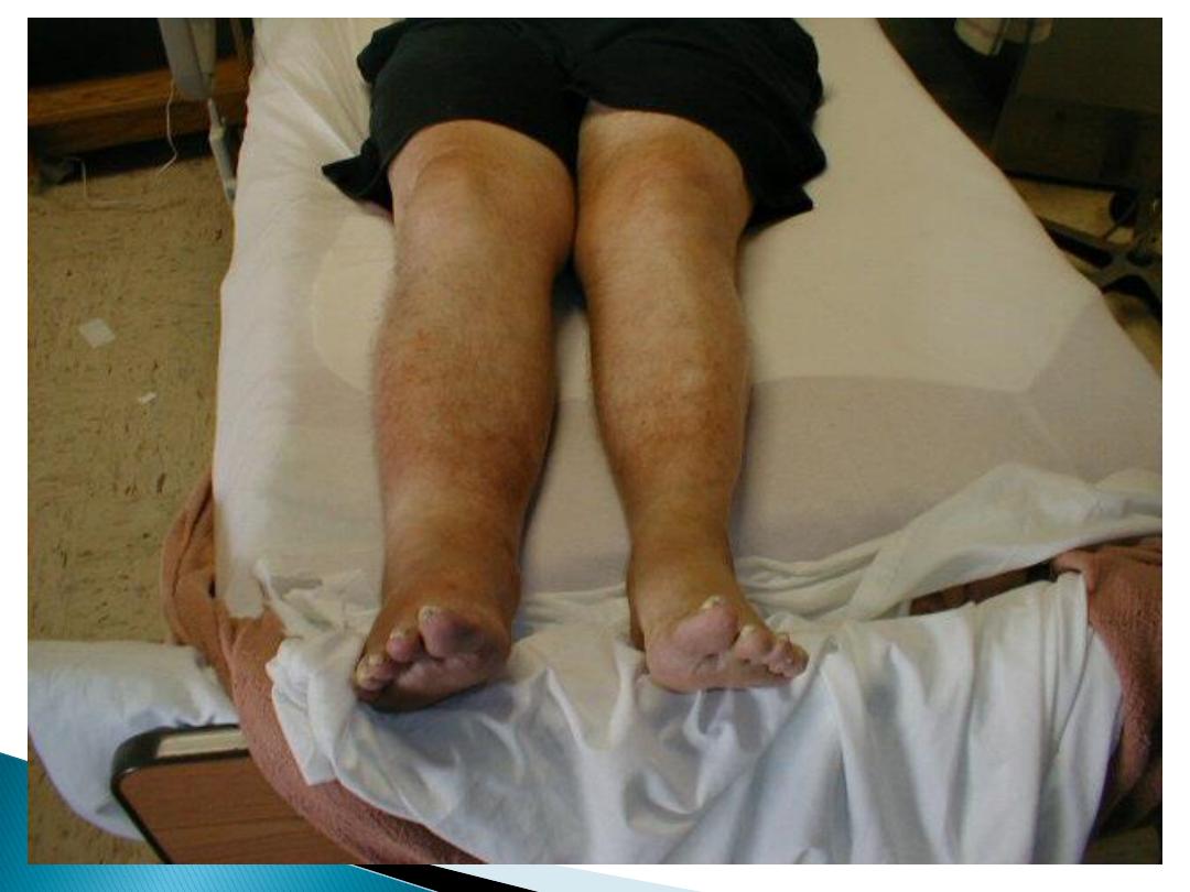
The symptoms and signs of VTE include leg
pain and swelling (usually unilateral), lower
abdominal pain, low-grade pyrexia,
For PE.dyspnoea, chest pain, haemoptysis
and collapse .
Risk factors.
Medical history
Exam.
Investigations

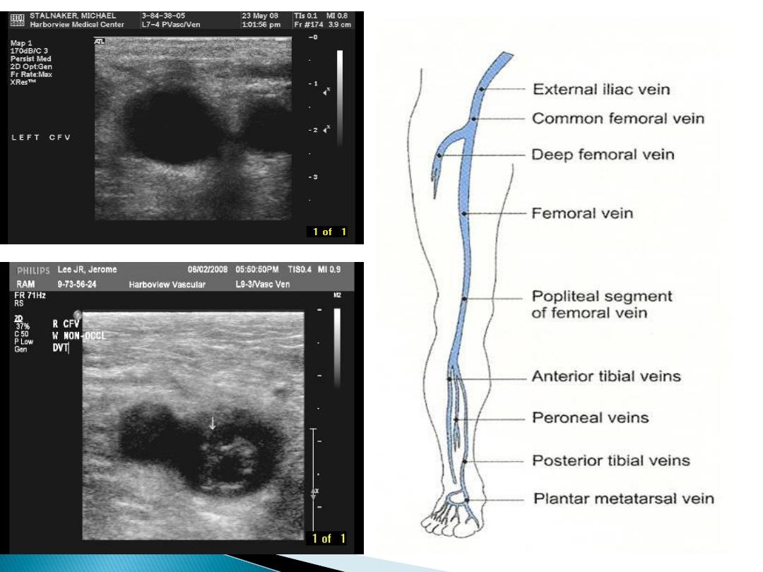
Compression duplex ultrasound:
It is the primary diagnostic test for
DVT should be undertaken where
there is clinical suspicion of DVT or
PE.
If it is negative and there is a low level
of clinical suspicion, anticoagulant
can be discontinue
If it confirms the diagnosis of DVT no
further investigation anticoagulant
treatment should be continued.

If it is negative and a high level of
clinical suspicion exists, anti-
coagulation continue and
ultrasound repeated in Day 7 or an
alternative diagnostic test
employed. If repeat testing is
negative, anticoagulant should be
discontinued.
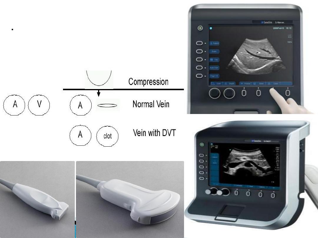
High resolution B-mode
ultrasound system
l Linear array transducers:
3-5 MHz - Large legs
5-10 MHz – Small legs

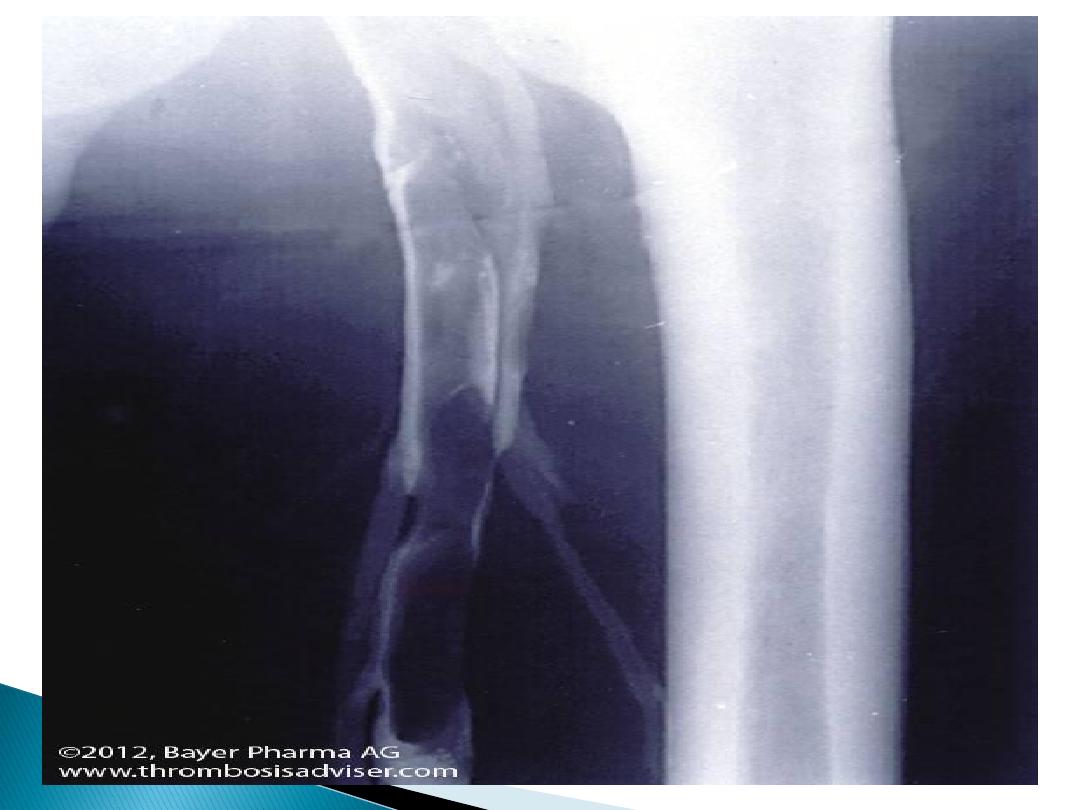
Venography:
it is an invasive procedure, requiring the
injection of a contrast medium and the
use of X-ray, is not preferable during
pregnancy.
It’s best when iliac vein thrombosis is
suspected (if back pain and swelling of
the entire limb).
Magnetic resonance venography or
Conventional contrast venography may
be considered.

Full blood count.
Coagulation screen.
D- dimer levels are not recommended in
pregnancy
Blood urea and electrolytes
Liver function test
Performing a thrombophilia screen prior to
therapy is not routinely recommended

For hemodynamic stable patient
suspected PE: with symp.&signs :
First line investigations:
CXR ,ECG.
ECG :T wave inversion ,right bundle
branch block

abnormal features caused by PE
include atelectasis, effusion, focal
opacities,
It also identify other pulmonary
disease as pneumonia, pneumothorax
or lobar Collapse. regional oligemia or
pulmonary edema.
If CXR normal V/Q can be interpreted.
If CXR abnormal V/Q can not be
interpreted

If no symptoms do ,CT Pulmonary
angiography ,Ventilation –perfusion
scan
:
V/Q carries a increased risk of childhood
cancer .
CTPA has higher risk for maternal breast
cancer (lifetime risk increased 13%).
The average fetal radiation dose with
CTPA is less than 10% of that with V/Q
scanning

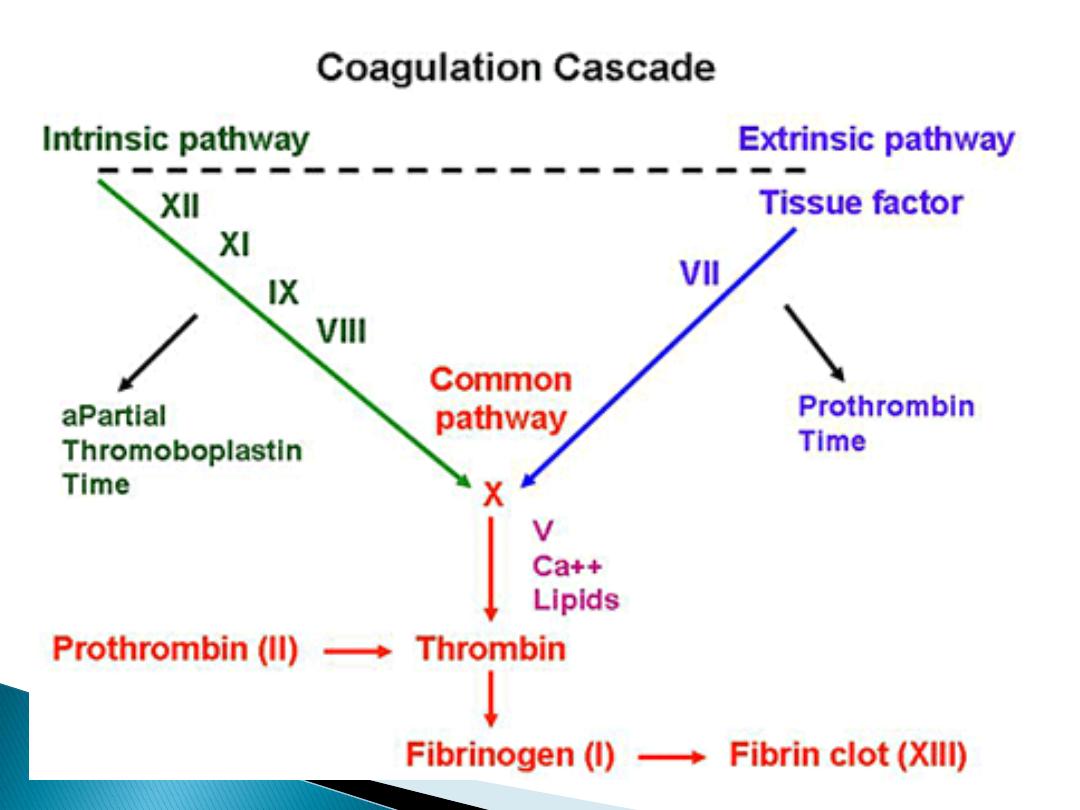
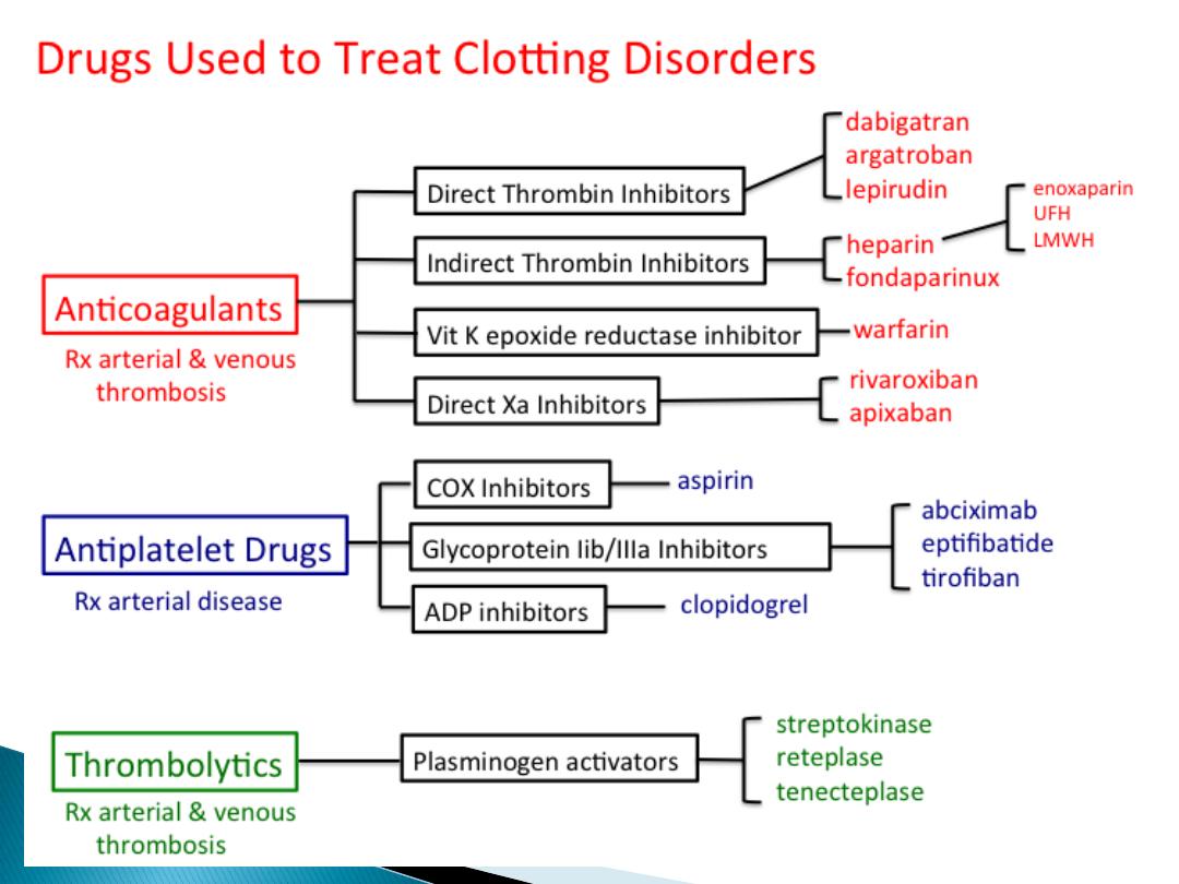
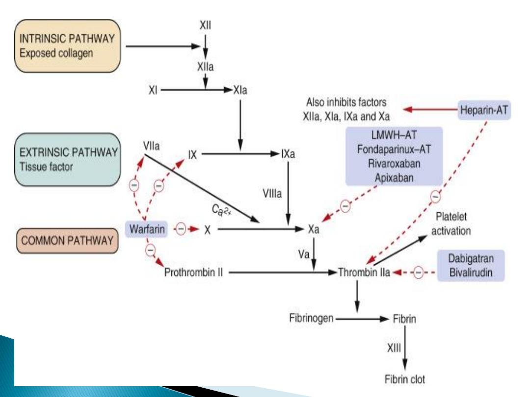
Anticoagulant therapy
:
1- Heparin;
It does not cross the placenta, not
teratogenic.
.Un-fractioned Heparin prolongs the
activated partial thromboplastin time
(APTT).
LMWH affect factor X activity.
it’s effect can be stopped within hours .
It may be associated with idiosyncratic
reaction, thrombocytopenia and higher
risk of osteoporosis
.

In clinically suspected DVT or PE,
treatment with LMWH should be
given until the diagnosis is excluded
by testing.
Treatment of VTE in pregnancy:
Dose: (enoxaparin 1 mg/kg once
daily; dalteparin 100 units /kg once
daily).

When delivery is planned, LMWH
maintenance therapy should be
discontinued 24 hours before planned
delivery, or when labor established.
Regional anesthetics or analgesic
techniques should not be undertaken
until at least 24 hours after the last
dose of therapeutic LMWH or 12 hrs
after prophylactic.

A thromboprophylactic dose of
LMWH should be given by 3 hours
after a caesarean section (more
than4 hours after removal of the
epidural catheter).
The epidural catheter should not be
removed within 12 hours of the
most recent injection.

Anticoagulant prophylaxis
Women had high risk ,previous DVT
on ante-natal prophylaxis LMWH&
for 6weeks postnatal.
Prophylaxis for low risk 10 days
postnatal if had two or more risk
factors

Women with a history:
of recurrent DVT .
or DVT occurring in non-pregnant
state
thrombophilias,or +ve family
history
or multiple risk factors
offered anticoagulant prophylaxis
ante-natal and post-natal for 6
weeks.

Women with artificial heart valves
definite history of previous
pulmonary embolism require full
anticoagulation throughout
pregnancy and post-natal.

2- Oral Anticoagulant,
Warfarin
teratogenic:
It prolongs the prothrombin time (PT).
It crosses the placenta and can cause limb
and facial defects characteristic embryo-
pathy in the first trimester, central nervous
system abnormalities at any trimester, fetal
hemorrhage and neonatal haemorrhage
Neither heparin (un-fractionated or LMWH)
nor
warfarin is contraindicated in breastfeeding.

Massive PE , Once suspected, early
intervention by full I.V. anticoagulant
therapy may be life saving.
Full I.V. un fractioned heparin is
immediately started, with supportive
oxygen therapy.
Management should involve a
multidisciplinary resuscitation team
including senior physicians,
obstetricians and radiologists.
.