


1
CEPHALOMETRIC RADIOGRAPHY
Cephalometric radiography is a standardized method of production of skull
radiographs, which are useful in making measurements of the cranium and the
orofacial complex. The radiograph thus obtained is called a cephalogram.
Uses of cephalometrics
1. Study of craniofacial growth: Serial cephalogram studies have helped in providing
information regarding:
• The various growth patterns.
• The formation of standards, against which other cephalograms can be compared.
• Prediction of future growth.
• Predicting the consequences of a particular treatment plan.
2. Diagnosis of craniofacial deformity: Cephalograms help in identifying, locating and
quantifying the nature of the problem, the most important result being a
differentiation between skeletal and dental mal-relationships.
3. Treatment planning: By helping in diagnosis and prediction of craniofacial
morphology and future growth, cephalometries help in developing a clear treatment
plan. Even prior to starting orthodontic treatment an orthodontist can predict the final
position of each tooth within a given patient's craniofacial skeleton to achieve
aesthetic and more stable results. it helps in distinguishing cases which can be treated
with growth modification appliances or which may require orthognathic surgery in
future.

2
4. Evaluation of treated cases: Serial cephalograms permit the orthodontist to evaluate
and assess the progress of treatment and also helps in guiding any desired change.
5. Study of relapse in orthodontics: Cephalometries also helps in identifying causes of
orthodontic relapse and stability of treated malocclusions. It helps in establishing
positions of individual teeth within the maxilla or the mandible, which can be
considered to be relatively stable.
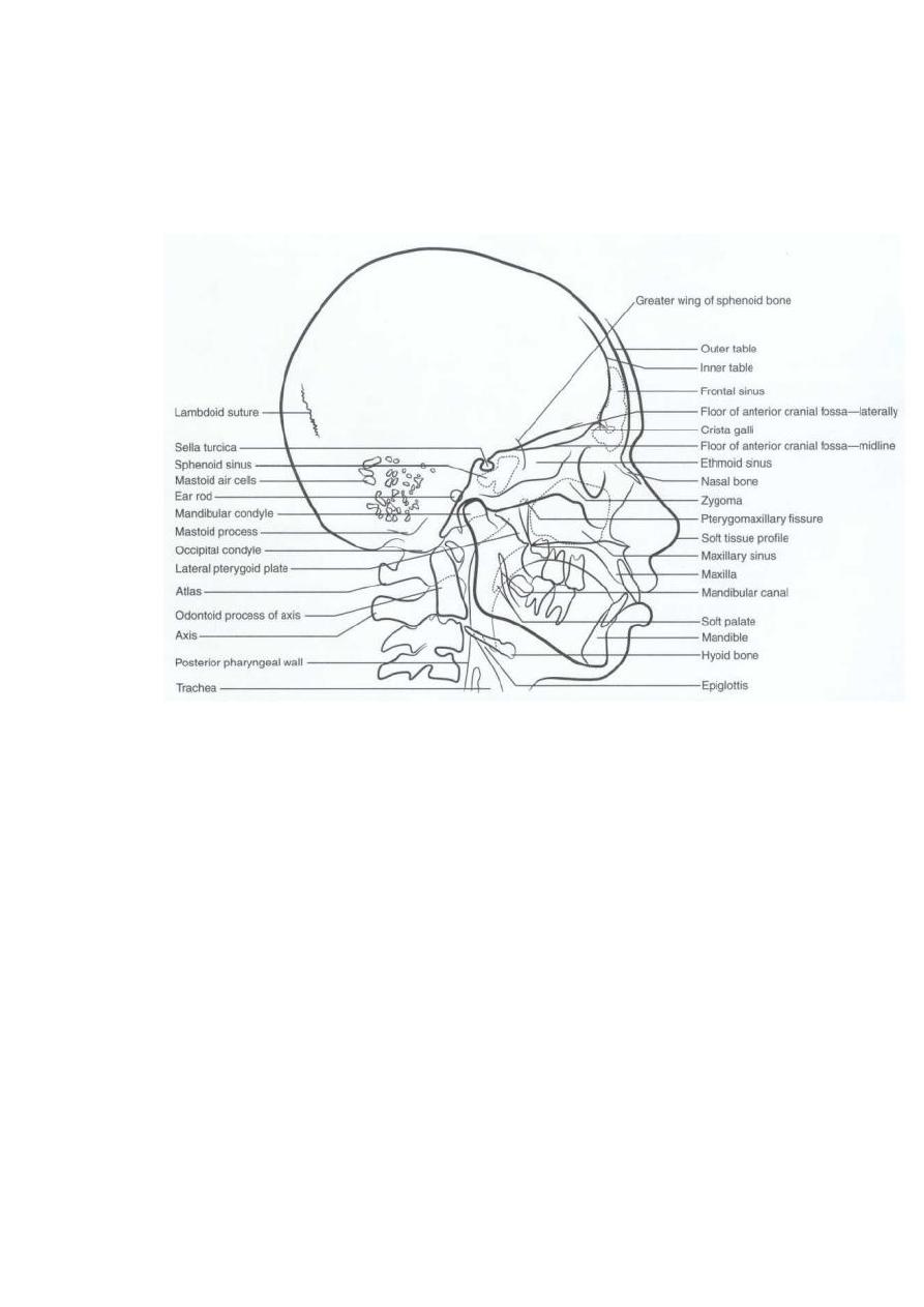
Cephalometric views
Lateral projection: The mid-sagittal plane of the subject's head is conventionally
placed at 60 inches (152.4 cm) from the target of the X-ray tube with the left side
(European convention is the right side) of the subject towards the film.
Postero-anterior projection: The head is rotated by 90 degrees so that the central ray
perpendicularly bisects the trans-metal axis.
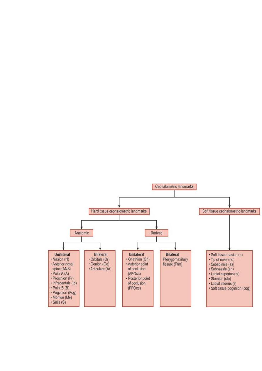
Cephalometry makes use of certain landmarks or points on the skull, which are used
for quantitative analysis and measurements. The landmarks used in cephalometrics
are of two broad categories: hard tissue landmarks and soft tissue landmarks. Some of
the hard tissue landmarks are unilateral while others are bilateral.
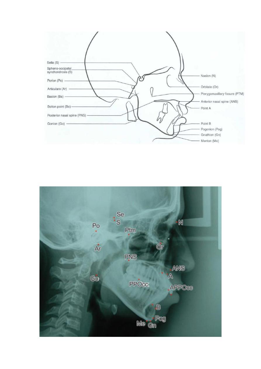
Hard Tissue Landmarks
Nasion (N)
Code/Abbreviation: Nasion is abbreviated as N (Capital alphabet).
Defnition: Nasion is the most anterior point of frontonasal outline in the midline.
Orbitale (Or)
Code/Abbreviation: Orbitale is abbreviated as Or (Capital alphabet O followed by
small r) Definition: The lowest point on the inferior bony margin of the orbit.
Anterior Nasal Spine (ANS)
Code: Anterior nasal spine is abbreviated as ANS (all capital letters).
Definition: Anterior nasal spine is the tip of bony anterior nasal spine in the midline
or median plane.
Point A (A)
Code: Point A is abbreviated as point A itself. Definition: Point A is the deepest
point on the curved bony outline between the anterior nasal spine (ANS) and
prosthion.
Prosthion (Pr)
Code: Prosthion is abbreviated as Pr (Capital P followed by small r). Definition: It is
the lower most anterior point of alveolar process of pre-maxilla in the midline
between two central incisors.
Anterior Point of Occlusion (APOcc)
Code: APOcc (Capital APO followed by double small c). Definition: Anterior point
of occlusal plane is the midpoint in the incisors overbite in the occlusion.

3
Infradentale (Id)
Code: Infradentale is abbreviated as Id (Capital I followed by small d).
Definition: Infradentale is the highest anterior point of the alveolar process of
mandible between two central incisors in midline.
Point (B)
Code: Point B is abbreviated as point B itself. Definition: It is the deepest point of
curvature on the contour of mandibular alveolar process in the middle between
infradentale and pogonion.
Pogonion (Pog)
Code: Pogonion is abbreviated as Pog (Capital P followed by small o and g).
Definition: Pogonion is the most anterior point of the bony chin in the midline.
Gnathion (Gn)
Code: Gnathion is abbreviated as Gn (Capital G followed by small n). Definition : It
is the most anterior and inferior point of the bony chin. It is constructed by
intersecting a line drawn perpendicularly to the line connecting menton and pogonion
with bony outline.
Menton (Me)
Code: Menton is abbreviated as Me (Capital M followed by small e).
Definition: According to Krogman and Sassouni, Menton is the most caudal point in
the outline of the symphysis. It is regarded as the lowest point of the mandible.
Sella (S)
Code: Sella is abbreviated as S (capital letter ‘S’). Definition: Sella is the midpoint of
sella turcica or hypophyseal fossa or pituitary fossa.
Se (Se)
Code: Se is abbreviated as Se (Capital S followed by small e). Definition: Se is the
mid-entrance point of sella turcica or pituitary fossa.
Pterygomaxillary Fissure (Ptm)
Code: Pterygomaxillary fssure is abbreviated as Ptm (Capital P followed by small t
and m). Definition: It is the intersection of the inferior border of the foramen
rotundum with the posterior wall of the pterygomaxillary fissure.
Posterior Nasal Spine (PNS)
Code: Posterior nasal spine is abbreviated as PNS (all capital letters). Definition: It
is the intersection of a continuation of the anterior wall of the pterygopalatine fossa
and the floor of the nose.
Posterior Point of Occlusion (PPOcc)
Code: Posterior point of occlusion is abbreviated as PPOcc (capital PPO followed by
small double c). Definition: Posterior point for the occlusal plane is the most distal
point of contact between the most posterior molar in occlusion.
4
Articulare (Ar)
Code: Articulare is abbreviated as Ar (capital A followed by small r). It was
introduced by Bjorio (1947). Definition: Articulare is the point of intersection of the
posterior margin of the ascending ramus and the outer margin of the cranial base.
Condylion (Cd)
Code: Condylion is abbreviated as Cd (Capital C followed by small d). Definition: It
is the superior most point of the head of the condyle of the mandible.
Gonion (Go)
Code: Gonion is abbreviated as Go (Capital G followed by small o).
Definition: Gonion is the intersection of the lines tangent to the posterior margin of
the ascending ramus and the mandibular base.
Porion (Po)
Code: Porion is abbreviated as Po (Capital P and small o)\ Definition: It is the
midpoint on the upper edge of the porus acusticus externus located by means of the
metal rods on the cephalometer.
Basion (Ba)
Code: Basion is abbreviated as Ba (Capital B followed by small a).
Definition: Basion is the lowest point on the anterior margin of the foramen magnum
in the midline.
5
6
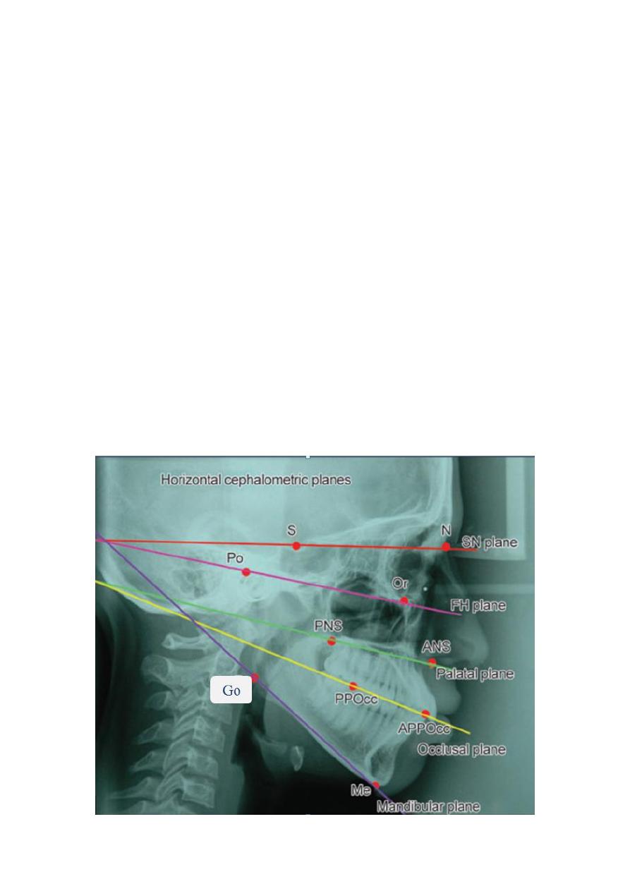
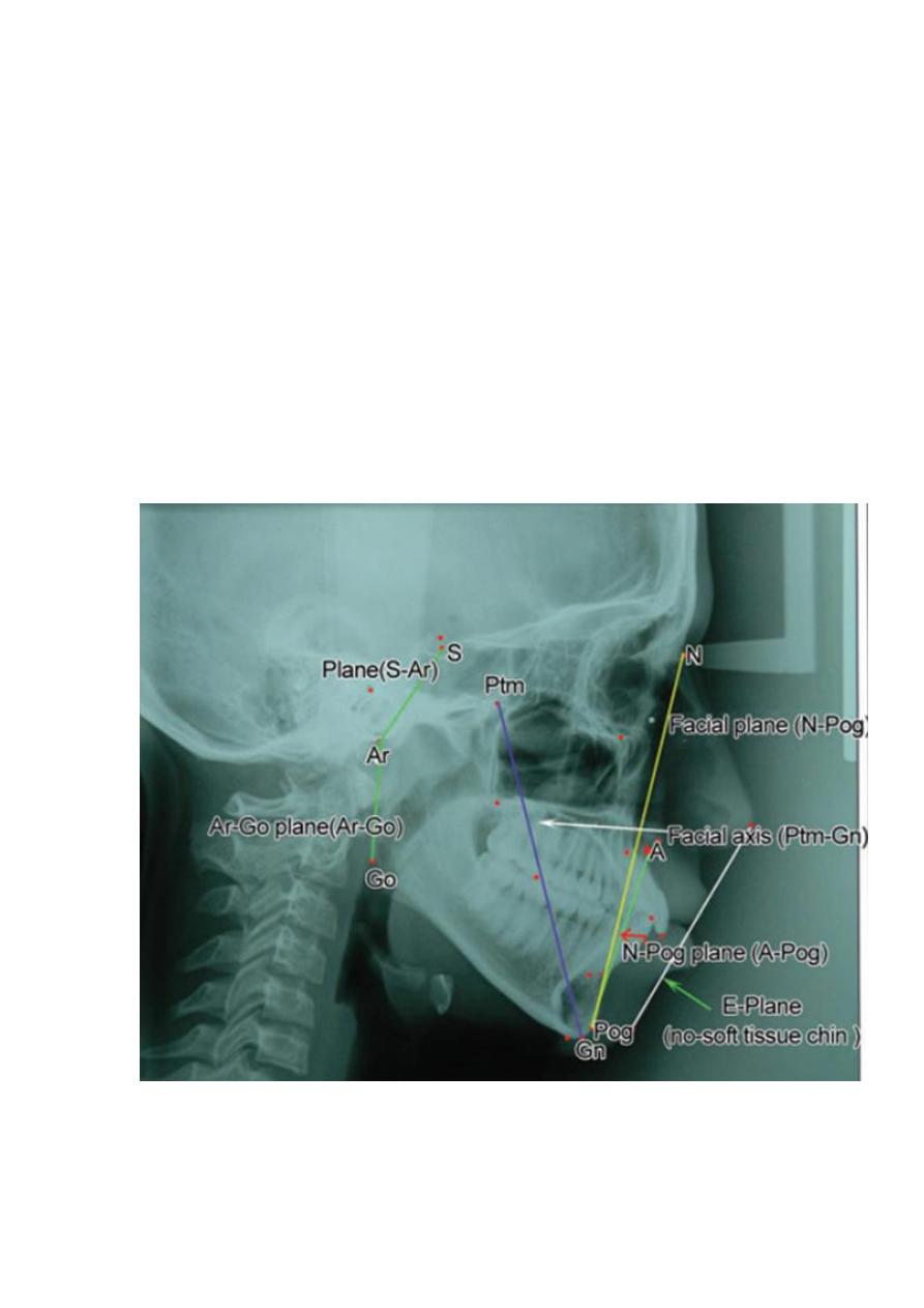
Cephalometric planes:
Reference planes are classifed into the following two groups:
1. Horizontal cephalometric reference planes
2. Vertical cephalometric reference planes.
Horizontal Cephalometric Reference Planes
1. Frankfort Horizontal Plane: It is drawn from the point orbitale to the
superior most point on the external auditory meatus (Porion).
2. S-N Line- The S-N line represents the anterior cranial base. It is constructed
by connecting the points sella turcica and the Nasion.
3. Palatal Plane: The palatal plane is drawn by extending a line from the
anterior nasal spine (ANS) to posterior nasal spine(PNS).
4. Occlusal Plane (Functional OP, Anatomic OP): It was originally described
as the line connecting the molars in occlusion to the bisector of the overbite
(vertical overlap of the incisors anteriorly), also known as the anatomic
occlusal plane. It was later modified to be represented by the line passing
through the occlusion of the premolars and the molars, also known as the
functional occlusal plane.
5. Mandibular Plane: Mandibular planes have been defined by various authors
based upon their clinical experience and use in their cephalometric analyses.
Tweed described the mandibular plane as a line that is a tangent to the inferior
border of the mandible. Down considered the mandibular plane to represent a
line connecting the points gonion and menton. Steiner drew the mandibular
plane by joining the points Gonion and Gnathion.
7
Vertical Cephalometric Reference Planes
1. A-Pog Line: It is a line from point A on the maxilla to pogonion on the
mandible.
1. Facial Plane:It is a line from the anterior point of the frontonasal suture
(nasion) to the most anterior point of the mandible (pogonion).
2. Facial Axis: A line from Ptm point to cephalometric gnathion.
3. E-Plane: E-plane is also called esthetic plane and it is a line between the most
anterior point of the soft tissue nose and chin.
4. S-Ar Plane: It is the plane between the sella point (center of sella turcica) and
the Ar (articulare) point.This plane represents the lateral extent of cranial base.
5. Ar-Go Plane: Ar-Go plane is formed by the line connecting from articulare
(Ar) to the gonion (Go). This plane is important in the determination of length
of ramus.

8
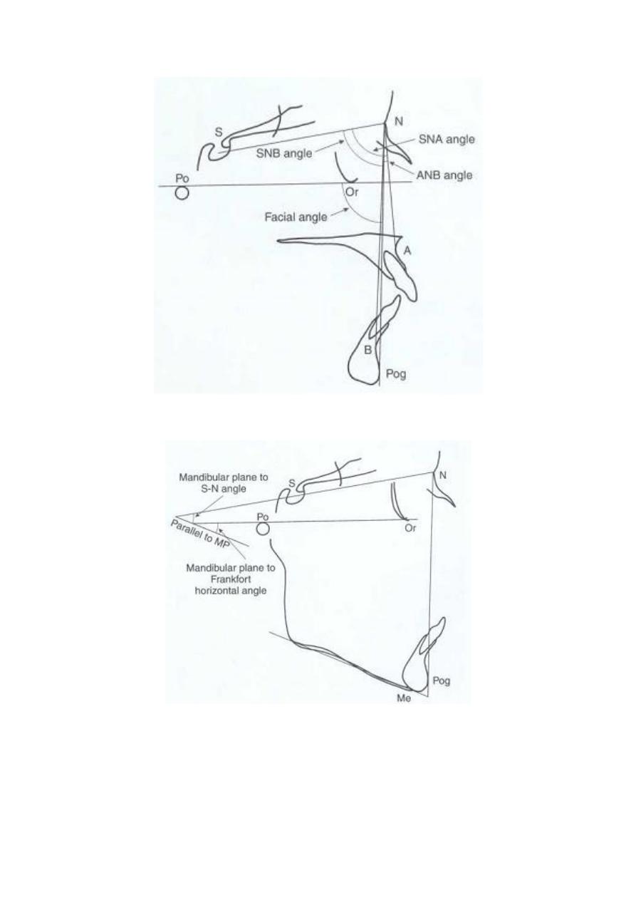
The Skeletal Analysis
Relating the Maxilla to the Skull
The angle SNA is formed by joining the lines S-N and N-A. The mean reading for
this angle is 82°. If the angular reading is more than 82°, it would indicate a relative
forward positioning or protrusion of the maxilla. Conversely, should the reading be
less than 82°, it would indicate a relative backward or recessive location of the
maxilla.
Relating the Mandible to the Skull
To assess whether the mandible is protrusive or recessive relative to the cranial base,
the SNB angle is read. The mean for this angle is 80°. If the angle is less than 80°, it
is indicative of a retruded mandible. An angle greater than 80°degrees suggests a
prognathic or forwardly positioned mandible.
Relating the Maxilla to the Mandible
The angle ANB, provides information on the relative positions of the jaws to each
other. The ANB angle provides a general idea of the antero-posterior discrepancy of
the maxillary to the mandibular apical bases. The mean reading for this angle is 2°. A
reading greater than 2° indicates a Class II skeletal tendency. As a rule, the larger the
number, the greater the antero-posterior jaw discrepancy, and hence the greater the
difficulty in correcting a malocclusion. Angles less than 2° and readings of below
zero (e.g. -1°,-2°) indicate that the mandible is located ahead of the maxilla,
suggesting a Class III skeletal relationship.
Occlusal Plane Angle
The occlusal plane is drawn through the region of the overlapping cusps of the first
prernolars and first molars. The angle of the occlusal plane to S-N plane is measured.
The mean reading for normal occlusions is 14°. The angle is increased in long face or
vertically growing individuals and also skeletal open bite cases. It may be decreased
in horizontally growing individuals or cases with a skeletal deep bite.
Mandibular Plane Angle
The mandibular plane is drawn between Gonion (Go) and Gnathion (Gn). The
mandibular plane angle is formed by joining the mandibular plane to the anterior
cranial base (S-N plane). The mean reading for this angle is 32°. Excessively high
(vertical growers) or low (horizontal growers) mandibular plane angles are suggestive
of unfavourable growth patterns and these may complicate treatment results.
9
Angles relating the anterior cranial base to the maxilla and mandible.
Different mandibular plane to other plane angles.
11
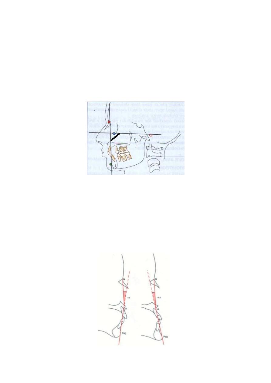
Facial Angle
The facial angle is used to measure the degree of retrusion or protrusion of the lower
jaw. The facial angle provides an indication of the degree of recession or protrusion of
the mandible in relation to the upper face. Facial angle is the inferior inside angle
formed by the intersection of the facial line (Nasion-Pogonion) to the Frankfort
Horizontal (FH) Plane. The mean reading for this angle is 87.8° (± 3.6°) with a range
of 82° to 95°. A prominent chin increases this angle, whereas a smaller than average
angular reading suggests a retrusive or retro-positioned chin.
Angle of Convexity
The angle of convexity is formed by the intersection of line N-point A to point A-
Pogonion. This angle measures the placement of the maxillary basal arch at its
anterior limit (point A) relative to the total facial profile (Nasion-Pogonion). This
angle is read in plus or minus degrees starting from zero. If the line Pogonion-point A
is extended and located anterior to the N-A line, the angle is read as positive. A
positive angle suggests prominence of the maxillary denture base relative to the
mandible. A negative angle of convexity is associated with prognathic profile or in
other words a Class III profile. The range extends from -8.5° to +10°, with a mean
of 0°.