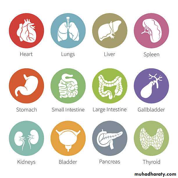
Anatomy of Spleen


Introduction:
Lymphatic organ connected to vascular system.Site: it lies obliquely in the post. Part of the Lt. hypochondrium wedged between the fundus of stomach and the diaphragm.
Surface anatomy: it lies opposite the ribs 9,10, and 11.
N.B: normally, the spleen is not palpable as it does not extend below costal margin.
The Spleen:Size and Weight: (easy to remember by the add numbers 1, 3, 5, 7, 9, and 11) The average spleen is 1” thick, 3” broad, 5” long, 7 ounces (200gm) in weight, and is related to the 9-11 ribs.
Shape and Features: it is shaped as cupped hand:
2 ends: med. (post.) and lat. (ant.)
2 borders: upper and lower
2 surfaces: diaphragmatic and visceral.
Shape and Features
• Medial (post.) end: is rounded and directed upwards, backwards and medially.• Lateral (ant.) end: is expanded and directed downwards, forwards.
• Upper border: is sharp and notched near the ant. End and terminates laterally by the angle.
• Lower border: is thick, rounded, and smooth (no notches)
• Diaphragmatic surface: is convex, smooth and related to the diaphragm.
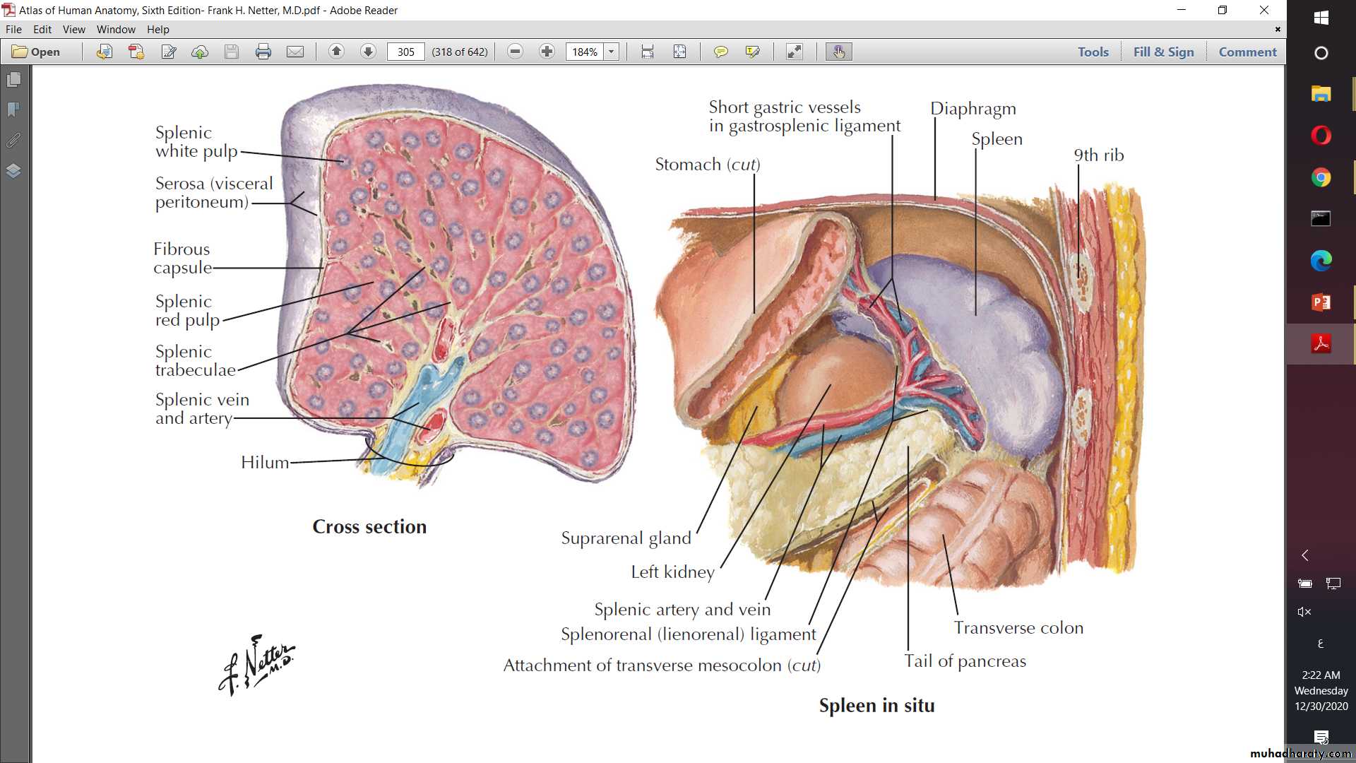
• Visceral surface: concave, irregular and related to abdominal viscera. It presents the hilum.
Relations of the Spleen:
• The Diaphragmatic surface: is related to diaphragm which separates the spleen from Lt. Pleura and Lt. Lung.• The Visceral surface:
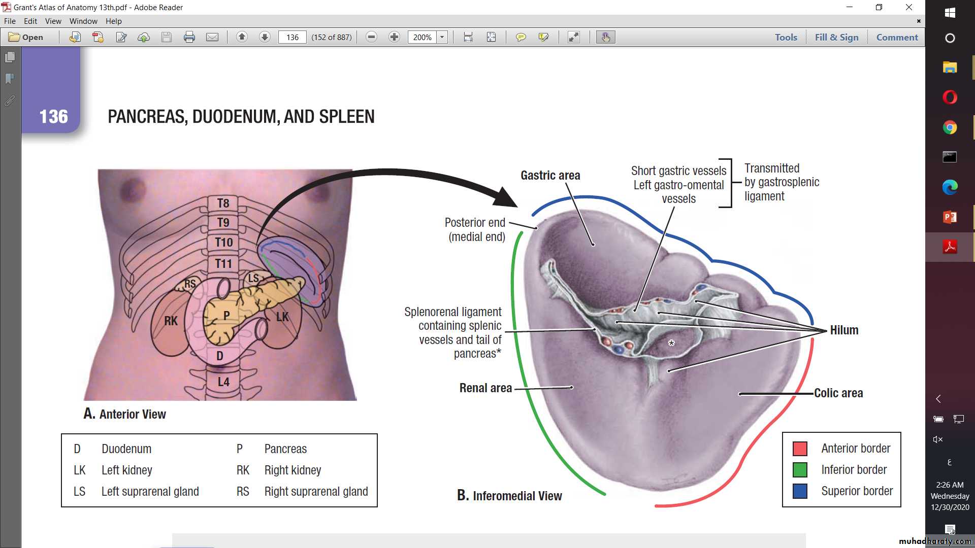
• Gastric impression: it is a large concave area between the hilum and the upper border (related to post. Wall of the fundus of stomach).
• Renal impression: a small shallow impression between the hilum and the lower border (related to the front of the left kidney).
• Pancreatic impression: s small impression below lateral end of hilum (related to tail of pancreas)
• Colic impression: a flat area close to lat. end of the spleen (related to the Lt. colic flexure).
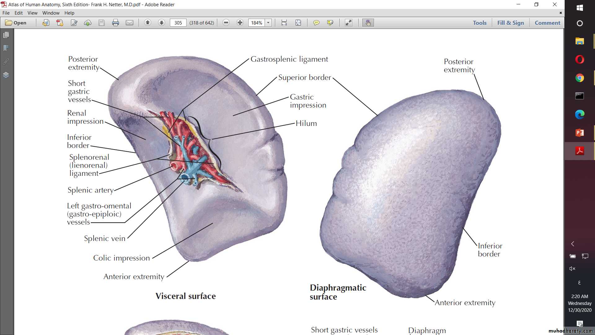
Hilum of the Spleen:
It is a longitudinal slit in the visceral surface between the gastric and colic impressions.It transmits:
• Terminal branches of splenic artery.
• Tributaries of splenic vein.
• Automatic nerves and lymphatics.
It gives attachment to 2 ligaments:
• Linorenal lig.
• Gastrosplenic lig.
Peritoneal Relations of the Spleen:
The spleen is completely covered with peritoneum of the greater sac except the pancreatic impression.Stability of the Spleen:
Depend on:
• Intra-abdominal pressure.
• Position of the surrounding organs.
• Ligaments: As mentioned previously.
Peritoneal ligaments of the Spleen:
Linorenal Ligament:Attachments: it extend between lower part of hilum of spleen and the front of upper half of the Lt. kidney.
Contents:
• Splenic vessels.
• Tail of pancreas.
• Lymphatics and pancreatico-splenic L.Ns.
• Extraperitoneal fatty tissue.
• Autonomic nerve fibers.
Peritoneal ligaments of the Spleen:
Gastrosplenic Ligament:Attachments: it extend between upper part of hilum of spleen and the front of upper 1\3 of the greater curvature of the stomach.
Contents:
• Short gastric vessels.
• Lt. gastric-epiploic vessels.
• Lymphatics and pancreatico-splenic L.Ns.
• Extraperitoneal fatty tissue.
• Autonomic nerve fibers.
Arterial Supply of the Spleen:
Splenic artery:Arises as the largest branch of the coeliac trunk.
It runs a tortuous course along the upper border of the body of the pancreas.
It enters the linorenal lig. To reach the hilum of the spleen to end by dividing into 5-6 branches.
Venous Drainage of the Spleen:
Splenic Vein:Aries at the hilum of the spleen then enters the linorenal ligament.
It runs a straight course behind the pancreas.
It ends by joining the superior mesenteric v. to form the portal v.
How to place the spleen in the correct anatomical position?
Hold the spleen in your left hand with its convex (Diaphragmatic) surface applied to the palm, the rounded post. end towards the wrist, the broad ant. end towards the tips of fingers and the notched upper border applied to the thumb.Thanks You