RESPIRATORY SYSTEM
LUNG DEVELOPMENT(1).Pre-natal lung development :-
The lung bud arises as a pouch from the primitive foregut at 22-26 days gestation. The bronchial tree develops between 5 and 16 wks gestation by continuous budding and branching of the airways . airways branching ends at 16 wks . further growth occurs by an increase in diameter and length but not by increase in air ways number . Insults to lung before 16 wks gestation decrease both air ways number and subsequent alveolar growth and number and the insult after 16 wks affect only alveolar number and growth.
(2).post-natal lung development :---
Approximately 60 million primitive alveoli exist at birth ,the lung grows most rapidly in alveolar number during the 1st 2 years . the growth rates decrease thereafter ,until the adult number of approximately 375 million alveoli is reached at age 8-12 yrs .
Common pathologic features of pulmonary :-
(1) underlying pathologic process : Most lung diseases in children are classified as
(a) .Obstruction (airway narrowing ) may be caused by intraluminal
secretion , edema or inflammation , hyper trophy or contraction of the
bronchial smooth muscles or extrinsic compression
(b) Restriction (impaired lung expansion) may be caused by decrease lung
compliance ., atelactasis or pneumothorax causing lung collapse
,neuromuscular diseases and disorder of chest wall .
(2). Pathophysiology :
(a) .hypoxemia (deficient oxygenation of blood ) most commonly caused by ventilation – perfusion abnormalities but may be the result of intra cardiac or intra pulmonary shunt or hypoventilation .
(b) .hypercapnia (excess carbon dioxide in the blood ) caused commonly by hypoventilation due to upper air way obstruction , neuromuscular weakness or CNS depression
(3) Pathogenic factors :
(a) The small airway of the child result in high airway resistance and put the child at great risk for develop of obstructive lung diseases .boys are affected more than and more severely than girls in part because the peripheral airway in boys younger than 5 yrs are smaller than those in girls .
(b) the young child lack specific immunity
( c) in children most pulmonary diseases has a single cause, whereas in adult ,pulmonary diseases is apt to be multi factorial in etiology .
CONGENITAL MALFORMATION
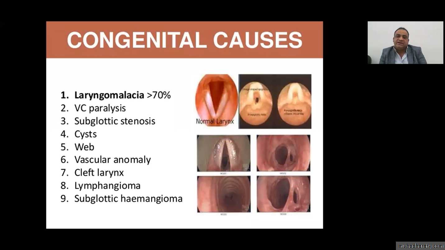
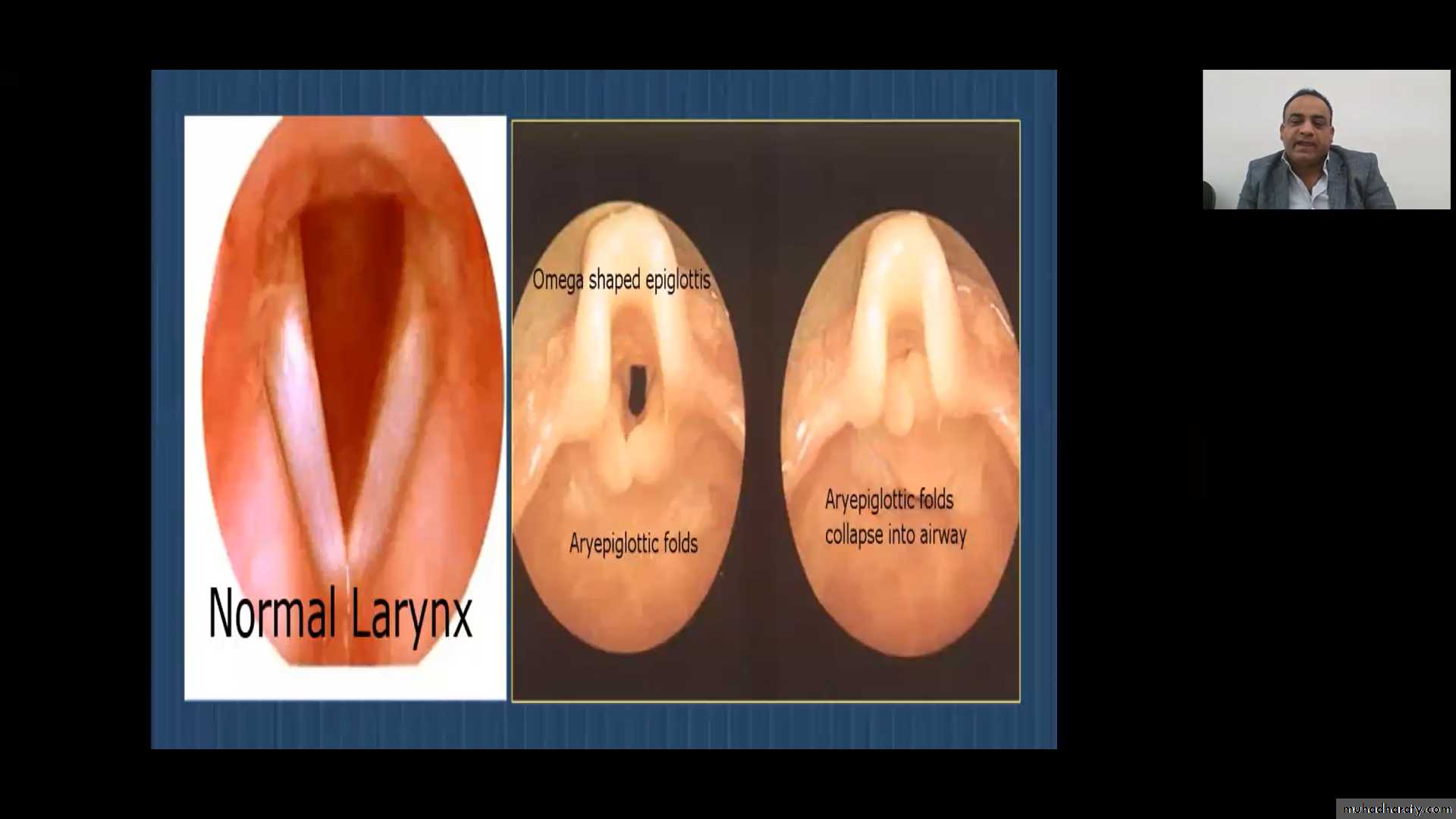
LARYNGOMALACIA AND TRACHEOMALACIA (congenital strider )Strider persisting or appearing after the first few days of life usually result from disturbance to the larynx . The most common of these are laryngomalacia and tracheomalacia due to flabbinees of the epiglottis and subglottic and weakness of the airway walls leading to collapse and sometime air way obstruction with inspiration .
Clinical features :
Noisy respiration sound usually associated with inspiration are relatively common during the neonatal period and the first years of life ,strider may present at birth or may appear at two months ,male affected more than female ,symptom can be intermittent and are worse when affected infant lie on the back . Some infant develop dyspnea and inspiratory retraction in the supraclavicular ,intercostals and subcostal spaces . The strider typically is loudest when the child is feeding or quietly relaxing or in neck flexion position ,and the strider diminished during sleeping or when the child is cryingThe strider usually resolves by the age 2 months but may recur with respiratory infection until about three years of age .
Diagnosis :- Is diagnosed by fibroptic bronchoscopy or direct laryngoscopy .
Therapy :- Is usually not needed ,parent should be reassured and provide slow careful feeding ,sometime need small nipple .
Rarely tracheostomy is required when the strider occur in failure to thrive or with life threatening apnea or obstruction .
SUBGOTTIC STENOSIS
Is a common problem that may be congenital or iatrogenic ,aggressive management of premature infant with intubation and mechanical ventilation may produce residual damage to the larynx .
Infant with down syndrome appear to have a small larynx and more susceptible or sub glottic stenosis .
Subglottic stenosis will produce strider and evidence of obstruction will be present on expiration and inspiration with increasing degrees of respiratory effort ,the strider will worsen .Viral infection may increase strider .
Definitive diagnoses requires endoscopic evaluation .
Treatment may necessitate tracheostomy ,however milder congenital cases improve with age as the larynx grows .

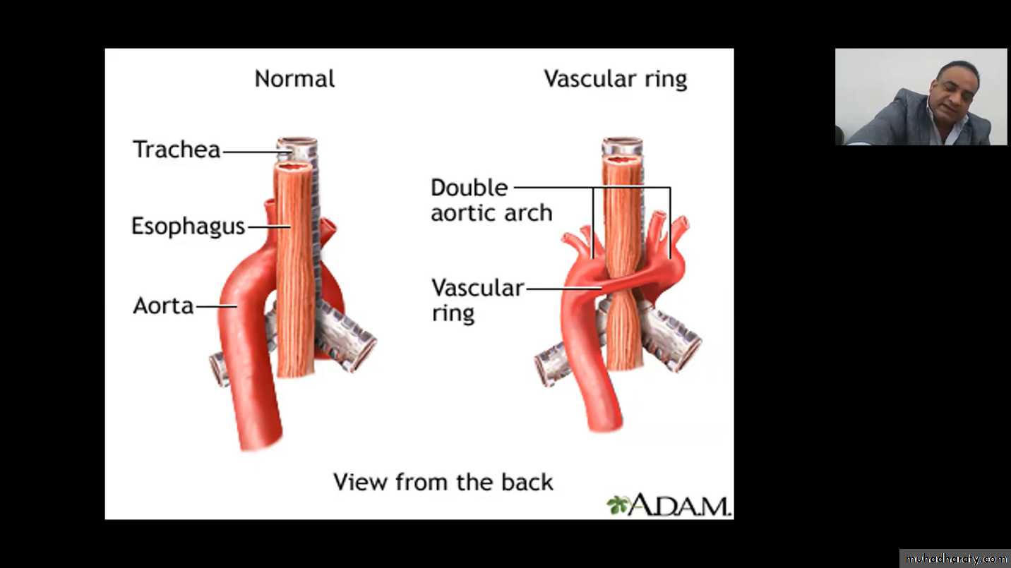
VASCULAR RINGS
Congenital anomalies of the aortic arch or its branches can create a ring around the airway that compromises respiration .Clinical features :
If the vascular ring produce compression of trachea and esophagus symptoms are frequently present at infancy , chronic strider and wheezing is exacerbated by crying , feeding and flexion of the neck . Extension of the neck tends to relieves the noisy respiration . Vomiting is frequent .
Diagnosis :
X-ray examination of the barium filled esophagus , arteriography , two dimensional ECHO , MRI ,CT or angiography . Bronchoscopy may be used to assess air way narrowing . THE CONDITION NOT IMPROVE WITH AGE
Treatment with surgical correction of the defect .
HEMANGIOMA
A number of mass lesion affect the larynx ,but the most common is hemangioma ,that usually found in the subglottic space . Infant with strider should be examined head to toe for cotaneous hemangioma because The presence of such lesion greatly increase the likelihood that the strider due to subglottic hemangioma .The airway obstruction usually worsen with cry and may produce pulmonary hypertension . The diagnosis depend on endoscopy and may be by AP x-ray of the larynx .
Treatments are controversial ,may need trachiostomy , others laser therapy or steroid. As with cutaneous hemangioma ,spontaneous regression is the rule ,but may need years .
PULMONARY SEQUESRATION
A cyst like mass of non-functioning lung tissue which lack normal communications with tracheo –bronchial tree .sometime develops at the embryo .
Most often within the lower lobe , the non functioning part is nourished by systemic arteries .
Clinical features :
Infection can result if a fistula develop between the sequestration . Children usually present with history of recurrent persistent progressive pulmonary sepsis or lung abscess .
Diagnosis of lung sequestration :
(a) . x-ray usually shows the sequestration as area of density with displacement of the bronchovascular marking .
(b) Contrast bronchography shows sequestration as area fail to fill outlined by bronchi that are filled
(c) Arteriography will delineate the anomalous artery supply from the aorta .
The treatment is surgical removal of the sequestration
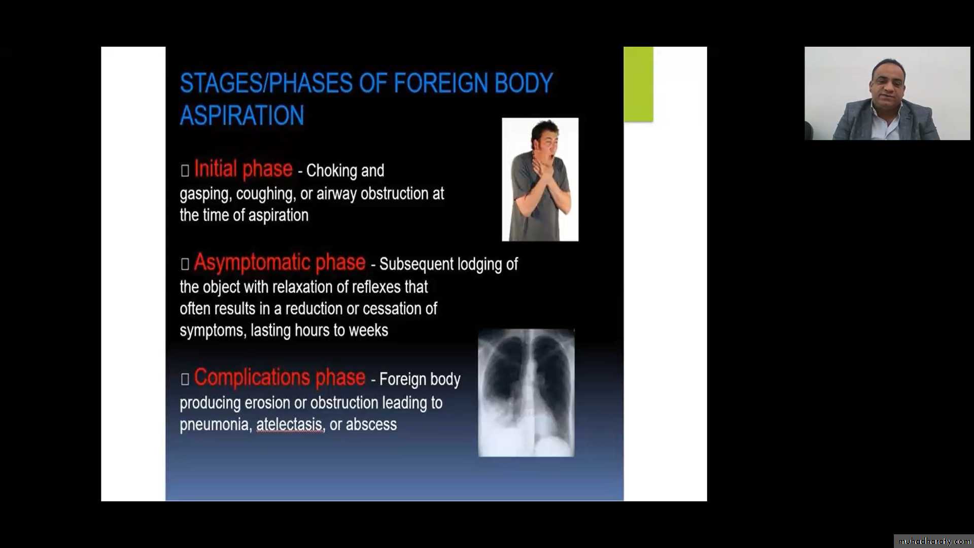
FOREIGN BODY ASPIRATION
The majority of patient are less than 4 yrs and most of death also occur in this age . Younger children most commonly aspirate foods ,toys .
Clinical features :
A high number of children who aspirate foreign bodies will present with either a clear –cut history of choking or physical or x-ray evidence of foreign body ,however a small percentage of patients with foreign body aspirate will have negative history because un observed or unrecognized events .
Physical finding consistent with acute foreign body aspirate include unilateral absence of breath sound ,localized wheeze ,strider or bloody sputum . X-ray may reveal the presence of radiopaque , when aspiration suspected ,expiratory films should be requested . Because the right main bronchus is a more direct continuation of the trachea than the left main bronchus ,foreign bodies tend to enter the right lung more . Some foreign bodies especially nuts ro seeds may migrate from place to place in the air way and even lodge in the larynx on coughing totally occluding the air way . Foreign bodies also may lodge in the esophagus and compress the trachea producing respiratory symptoms .

Diagnosis :-
The majority of foreign bodies are small and quickly coughed out ,but many may remain in the lung for long period before diagnosis and may come to medical attention because of symptoms of fever ,cough sputum production or chest pain . Patient with persistent with wheeze unresponsive to bronchodilators therapy, persistent atelactasis ,recurrent or persistent pneumonia or persistent cough without other explanation should be suspected foreign body aspiration . If there is good evidence in history ,physical examination or by film for bronchialforeign body ,the patient should undergo rigid bronchoscopy . Flexible bronchoscopy may be very useful diagnosis when presentation is not straight forward .
Prevention :-
The best approach to foreign body aspirate is to educate parents . Infant should not have food that must be easily broken into small pieces . Toys should be free of small parts that may be aspirate .

L2-
INFECTIONS OF RESPIRATORY TRACT
The peak incidence of respiratory infections in general is during the months of December to February . The attack rate is higher among the children than the adult and especially severe on those under 3 yrs and more than one affected among the family. The causal agents are a wide variety of viruses and bacteria , usually start with viral infection followed by secondary bacterial invasion .A wide range of clinical pattern from the trivial to the fatal can produce by many viruses , each of which may produce essentially similar clinical illness , Classification of acute respiratory diseases is difficult because the isolation of the viruses is slow and requiring laboratory facilities which are not universally available . On anther hand an anatomical classification can not provide a satisfactory basis because a single virus can affect the respiratory mucous membrane which is continuous from the nose to the alveoli . Further more some of the viruses do not confined to respiratory tract alone ,but also may invade the GIT ,mesenteric lymph nodes and CNS .
There are 3 main groups of viruses that affect the respiratory tract .
( 1) . Myxo viruses include the influenza A and B , respiratory syncytial virus and para influenza virus .
(2 ) . the adenoviruses contain a large number of different types ,some endemic or others epidemic .
( 3) . the picorna viruses are two types ,entero virus such as coxcacke , ECHO virus and rhinoviruses of many serological types .
There is no doubt that RSV is the commonest virus and most severe one ,causing infection of the infant and young children and can cause death in absence of bacterial infection .
UPPER RESPIRATORY TRACT INFECTIONS
The vast majority of cases including these in which tonsillitis is present are due to the virus and few cases of tonsillitis and pharyngitis are due to bacteria of Beta – hemolytic streptococci .
Clinical features :
Upper respiratory infections vary widely in their clinical features
Depend on :---
(1 ) . the area of maximum involvement
( 2 ) . the age of the patient .
( 3) . the causative agent .
Rhinitis with nasal obstruction and mucopurelent discharge may seriously interfere with feeding in the infant whereas a similar common cold in toddler may result only in a mild malaise and low grade fever and nasal discharge . In most cases however the pharynx shows acute hyperaemia and some edema ,may with cervical adenitis that present with brisk fever ,irritability and anorexia . When the cause adenovirus type 3 ,conjunctivitis and posterior cervical lymphodenitis are often present .
The direct inspection of the throat is important because the young child rarely complained of sore throat . When the larynx affected can cause strider When trachea affected the symptoms ,harsh cough ,sometime with retrosternal pain ,often worsen during night . In other cases the prominent complain are abdominal pain and vomiting due to mesenteric lymph nodes , so the different diagnosis from appendicitis may be difficult , but the viral infection are associated with high fever and flushed hot skin and often absence of rigidity and muscle tenderness . In few cases headache and neck stiffness may arouse the suspicion of meningitis
IT IS WISER TO PERFORM A LUMBER PUNCTURE THAN TO RISK LEAVING A PYOGENIC MENINGITIS INADEQUATELY TREATED
If the tonsils affected they are grossly hyperemic and swollen with yellow to white Exudates may appear in crypts with enlarged tender lymph nodes .
TREATMENT OF UPPER RESPIRATORY INFECTIONS
1. The room of the patient should be airy with temp. around 18-20 centigrade .
2. good intake of fluid and glucose in form sweetened drinks must be encouraged
3. diet should be light and palatable and the intake of calories is un important in a short illness.
4. tepid sponging is indicated if the rectal temp. rises above 39 c .
5. pain and high temp. ca relieved by paracetamol .
6. nasal obstruction may be temporarily relieved by the use in each nostril before each reed of 2 drops of normal saline .
7. throat swab should be always be taken for culture when the pharynx is inflamed and when the hemolytic streptococcus are isolated antibiotic should be given such as benzyl penicillin 5oo,ooo unites I .m. twice daily for 3 days and followed by oral penicillin 250 four times daily for a further four days .
CROUP
The most common syndrome of infectious upper obstruction is croup or acute infectious laryngo tracheobronchitis . Croup is of viral etiology para-influenza type 1 ,2 viruses are the most common .
CLINICAL FEATYRES :-
The typical attack begin in a child between 6 mon .-3 yrs. Having symptoms of upper respiratory infections (common cold ) and lasts less than five days . A brassy cough ,inspiratory strider and respiratory distress may develop slowly or acutely . signs of upper air way obstruction such as labored breathing and marked supra sternal ,inter costal and sub costal retractions are evident on examination . Associated lower air way diseases accompanied by wheeze and productive cough may present ,although the majority of such children are not seriously ill. The air way may becomes more severe . The subglotic space is the major site of obstructive which is caused by edema resulting fro the viral inflammation .
Indication of admission to hospital :-
1 . suspected epiglottitis .
2 . progressive strider .
3 . severe distress at rest .
4 . hypoxeamia .
5 . restlessness with pallor or cyanosis .
6 . decrease sensorium .
7 . high fever .
8 . respiratory distress .
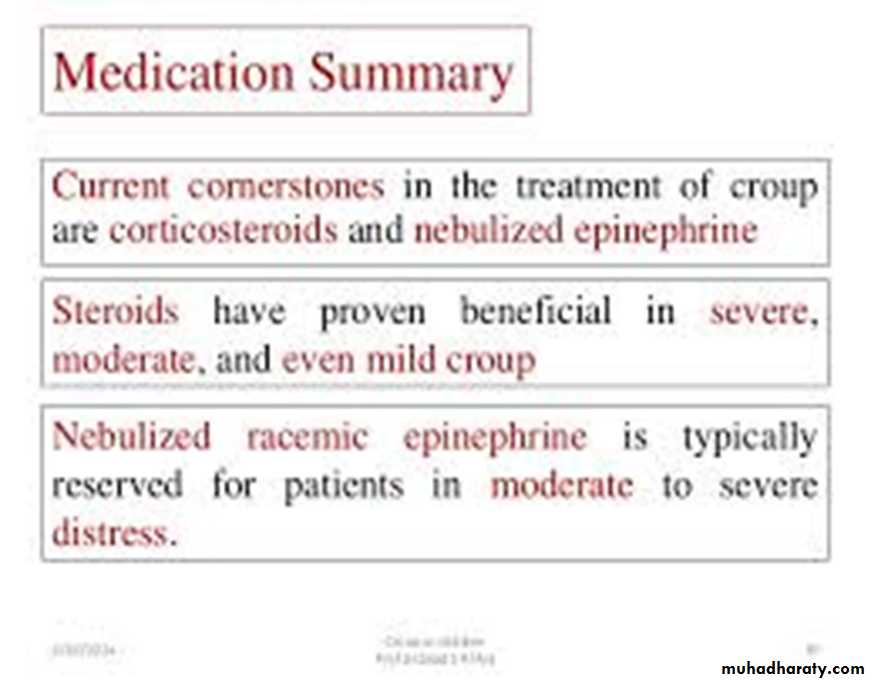
Trearment of croup
1. keep the child quite as possible and the best calm method for a child with croup is to sit in mother lap.
2. racemic epinephrine inhaler may reduce the edema temporarily producing marked clinical improvement ,but the edema and obstruction soon return and the disease run its course over several days . In severe case epinephrine may be repeated every 20 min .
3 . cool mist adminsterd by tent or face mask may help to prevent drying of the Secretion around larynx
4 . sedation should be avoided .
5 . systemic administered corticosteroid is beneficial in treating croup ,but Generally is reserved for ill patient .
If the patient is very young ( less than 4 months ) or if symptoms continued for more than one week ,the patient should undergo care full laryngoscopy, because there is increased possibility that another lesion may present such as subglottic or hemangioma . sudden worsening ( fever ,respiratory distress , increased ) this suggests complicating bacterial tracheitis
6 . Intravenous fluid may be needed in severely distressed child .
EPIGLOTTITIS
Another syndrome o upper air way obstruction typically occur in older children ( 2-7) yrs and the causative agent H . INFLUENZA type b .CLINICAL FEAT URES

Epiglottitis is characterized by sudden onset with high fever ,respiratory distress ,fulminant progression ,severe dysphagia and muffled voice . The patient prefer erect position to breath easily with drooling due to dysphagia . Epiglottitis is true pediatric emergency because the inflamed airway suddenly May become totally obstructed leading to death , Examination of the pharynx should be avoided .
DIFFERNTIAL DIAGNOSIS OF EPIGLOTTITIS
1 . severe croup .
2 . becterial tracheitis .
3 . foreign body aspiration .
4 . Retropharyngeal and peritonsillar abscess .
DIAGNOSIS :-
1 . High suspicion from clinical picture
2. Confirmation depend on direct observation of the inflamed and swollen supraglottic structures and redness of enlarged epiglottis of course examination done at operation room to place nasotracheal
or perform tracheostomy .
3 . Isolation of H. influenza room the surface of epiglottis or from Blood culture .
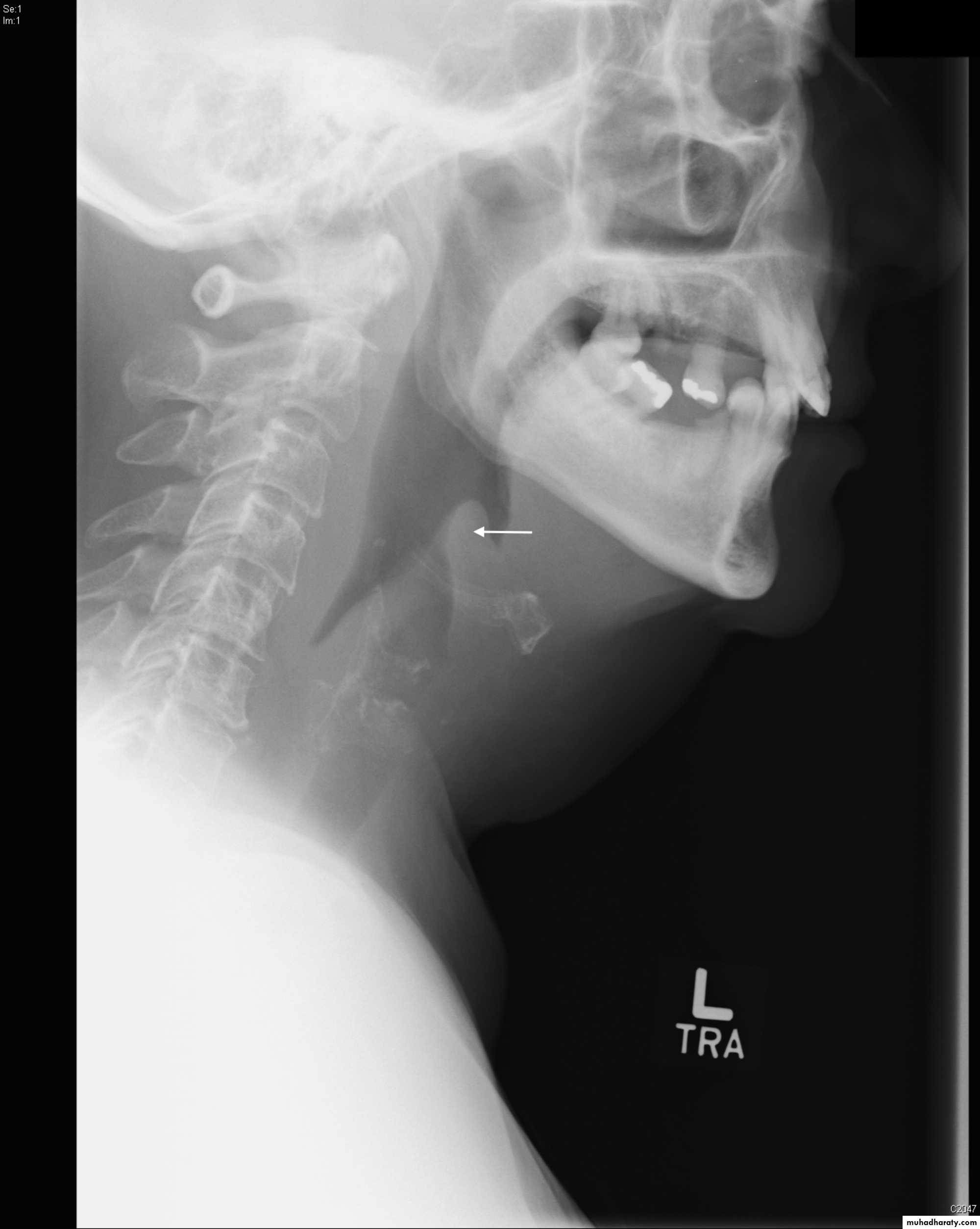
4 . X-ray of lateral neck give us thumb sign of swollen epiglottis .
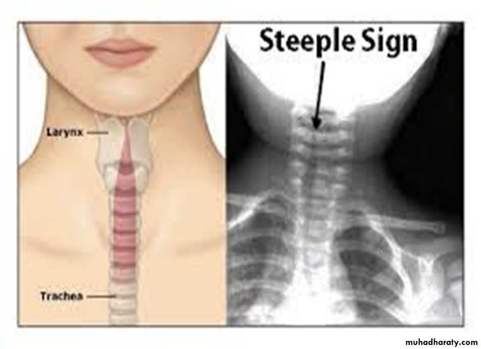
X-ray in croup give us steeple sign due to narrowed subglottic space.
TREATMENT :-
1 .Admission to the respiratory care unit .
2 . nasotracheal intubations with closed observation to prevent Extubation.
3 .tracheostomy .
4 oxygen
5 .antibiotics like ceftriaxone 50 – 75 mg /kg x 2 suitable for H influenzas hould be given and continued for 7-10 days . Or ampicillin +sulbactam combination should be given Parentrally if H influenza sensitive .
6 . Intravenous fluid may be needed during hospitalization .
7 . the epiglottitis resolve after a few days of antibiotics and the Patient can be weaned from the tracheostomy .