1
Fasciolopsis buski
Fasciolopsis buski (Lankester, 1875) Odhner, 102, the giant intestinal fluke, was
first observed by busk in the duodenum of a Laskar sailor at autopsy in London. Its
natural geographical distribution is limited to Oriental countries.
Morphology, Biology, and Life Cycle
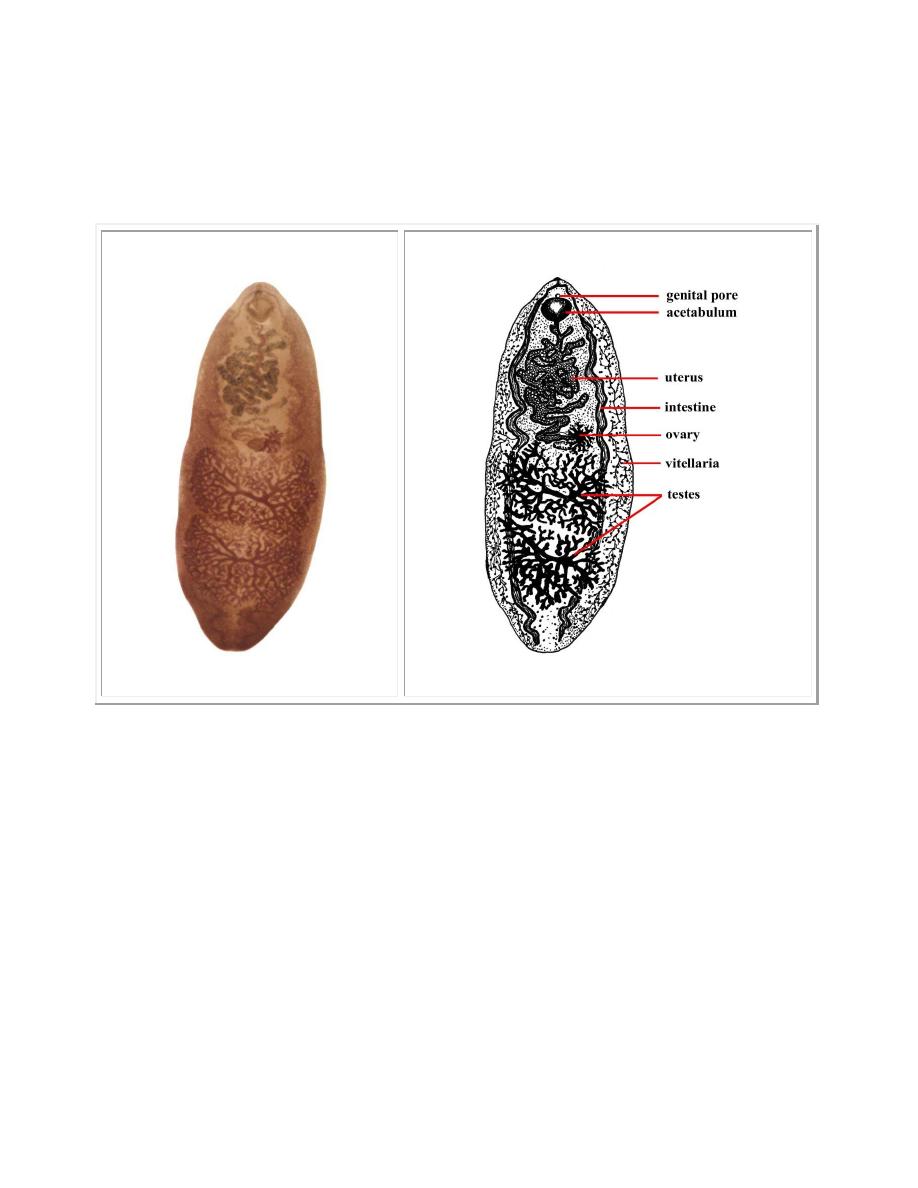
Fasciolopsis buski is a large fleshy worm, broadly ovate or elongate-ovoidal,
attached to the wall of the duodenum or jejunum. It measures 20 to 75 mm. long, 8
to 20 mm. wide and 0.5 to 0.3 mm. thick.
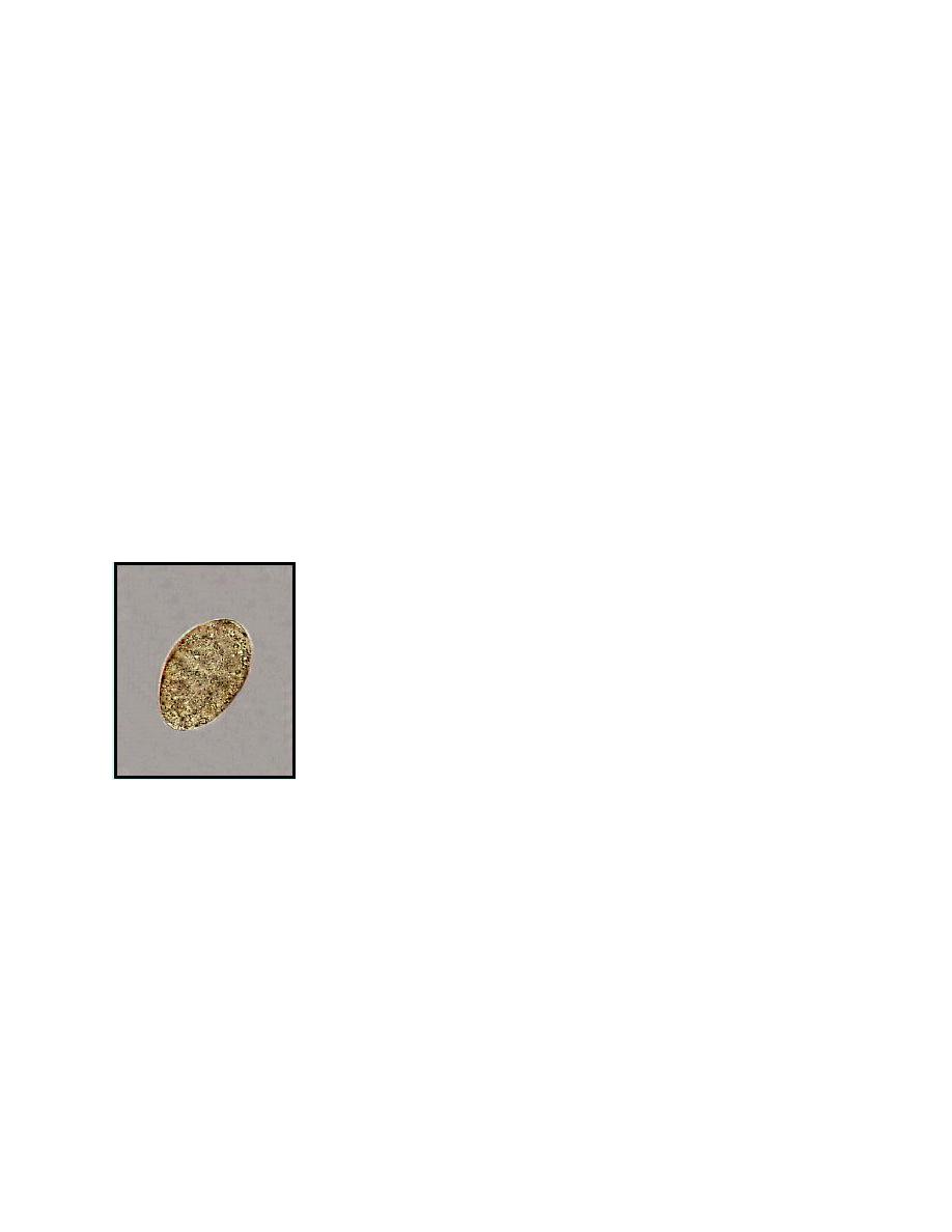
Eggs of F.buski are large, measure 130 to 140 microns by 80 to 85 microns, have a
thin, transparent shell with a small slightly convex operculum at one end, and are
unembryonated when evacuated in the host's feces. They are difficult to
differentiate from eggs of Fasciola hepatica.
2
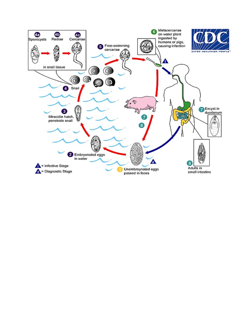
To proceed with their development, eggs of F.buski must reach quiet fresh water.
Here they embryonate in 3 to 7 weeks at a temperature of 26.7 to 32 °C., following
which a miracidium breaks out of the shell through the opened operculum, then
escapes from its embryonic membrane and swims about vigorously in the water.
On contact with an appropriate small planorbid snail (species of Segmentina) the
miracidium penetrates the soft tissues and transforms into a sporocyst. In this
mother spore sac, a generation of rediae is produced.
Usually the redial generation produces a number of vigorous cercariae, which erupt
from the snail and, after swimming about, crawl onto aquatic vegetation and
encyst. Man commonly becomes infected while consuming these aquatic
vegetation, so that some of the encysted metacercariae are set free and swallowed.
After excysting in the duodenum, the larvae become attached to nearby mucosa
and in about 3 months develop into mature worms.
The egg
3
Life Cycle of Fasciolopsis buski
Pathogenicity and Symptomatology
The damage produced by these large fleshy worms is mechanical, obstructive and
toxic. At each site of attachment, a mucosal ulcer is produced. A few worms may
cause no serious intestinal symptoms, but frequently there are dozens to hundreds
in an infection. These embarrass digestion and at times cause acute obstruction.
Toxic metabolites of the parasites are absorbed systemically and produce edema of

4
the face, especially around the eyes, of the abdomen and lower extremities. There
is characteristically a notable eosinophilia.
The early symptoms are diarrhea and hunger pains; those with heavy infections
mimic peptic ulcer. Ascites and asthenia are characteristics, as well as generalized
abdominal pain, anorexia, nausea and vomiting typically occur.
Diagnosis
This is based on recovery of characteristic eggs of F.buski in the stools.
Treatment
Fasciolopsiasis can be treated with prescription medicine taken by mouth, called
praziquantel. It should be taken with liquids during a meal. Praziquantel is
approved by the FDA, but considered investigational for this purpose.