Histology of skin dr. Ahmed Alhuchami
Skin Appendages:
Hair , sweat glands , sebaceous gland and nail

1- Hair:
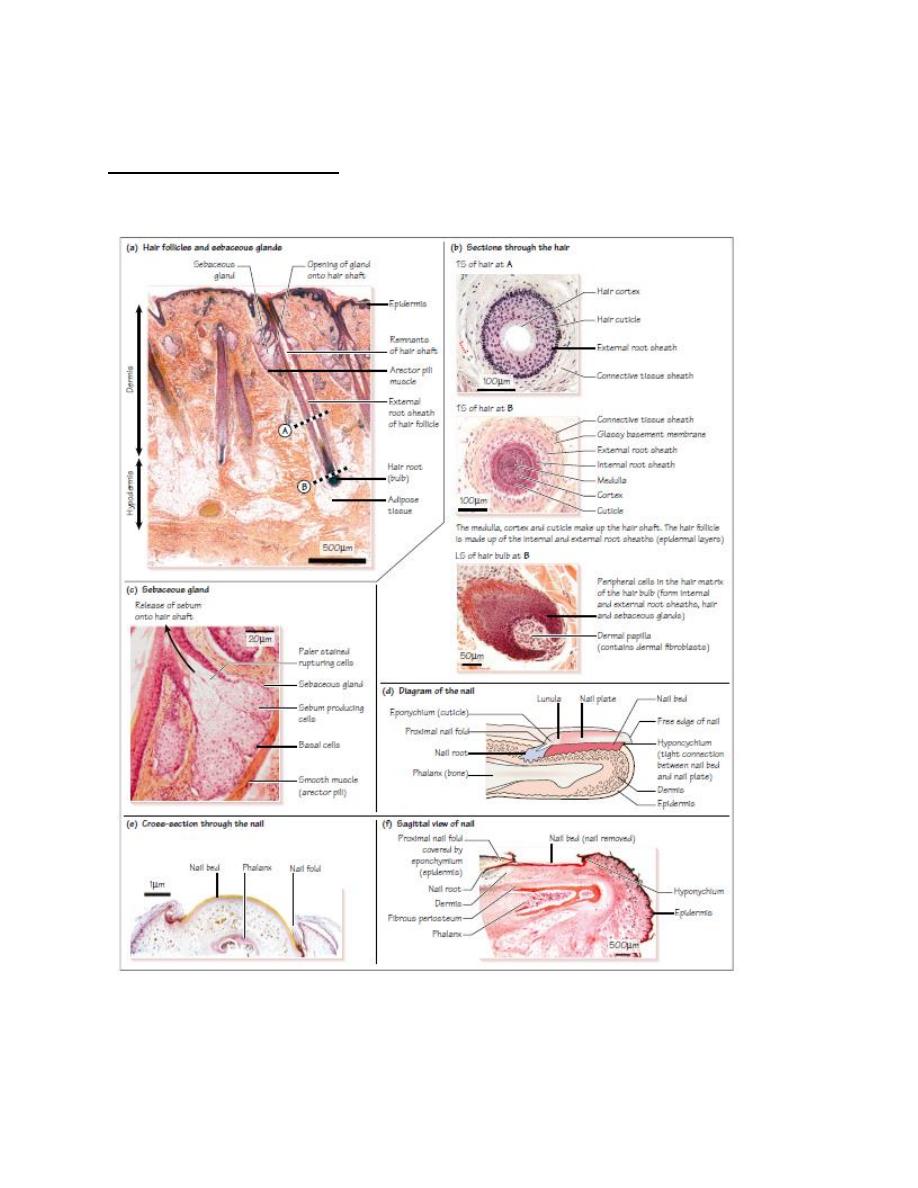
Hairs are made up of hair follicles and hair shafts .The hair shaft is made up of columns of dead
keratinized cells(hard keratin) organized into three layers :
• a central medulla , or core (not seen in fine hairs);
• a keratinized cortex ;
• a thin hard outer cuticle , which is highly keratinized.
Hair follicles are tubular invaginations of the epidermis, which develop as down growths of the
epidermis into the dermis. The hair follicle contains the following.
• An external root sheath ( ERS) , which is continuous with the epidermis. This layer does not
take part in hair formation. A glassy basement membrane separates the ERS from the
surrounding connective tissue.
• An internal root sheath ( IRS ), which lies inside the ERS. The IRS contains keratinized cells
derived from cells in the hair matrix. The type of keratin found here is softer than that found in
the hair itself. The IRS degenerates at the point where the sebaceous gland opens onto the hair.
Hair follicle stem cells in the hair matrix , which is found in the hair bulb , are responsible for
forming hair . The stem cells proliferate, move upwards, and gradually become keratinized to
produce the hair. These stem cells also form the ERS and IRS , and sebaceous glands.
The dermis forms a dermal papilla at the base of the hair follicle/ hair bulb, which provides the
blood supply for the hair. It is separated from the hair matrix by a basement membrane.
Contraction of the arrector pili muscle , a small bundle of smooth muscle cells associated with
the hair follicle, raises the hair, and forms ‘ goose bumps ’ . This helps to release sebum from
the gland into the duct, and to release heat.
2-Sweat glands:
the sweat glands are simple tubular
glands that are found in the superficial
hypodermis bordering on the dermis.
There are eccrine and apocrine sweat glands They
differ in embryology, distribution, and function.
1-
:
They discharge their contents onto the surface of the
skin via coiled secretory ducts). The ducts open out onto epidermal ridges at a sweat
pore. They can be further classified as
. They secrete a watery
fluid which is hypotonic to plasma its evaporation is important for thermoregulation.
Sweat contains water, sodium, potassium, chloride, urea ammonia and lactic acid.

Eccrine sweat glands are simple, coiled, tubular glands present throughout the body, most
numerously on the soles of the feet. Thin skin covers most of the body and contains sweat
glands. Exceptions are the vermillion border of the lips, external ear canal, nail beds, glans penis,
clitoris, and labia minora, which do not contain sweat glands. The thick skin covering the palms
of hands and soles of feet lack all skin appendages except sweat glands.
2-
Apocrine sweat glands
:, also referred to as odoriferous sweat glands, are known for
producing malodorous perspiration. They are large, branched glands, mostly confined to the
axillary and perineal regions, including the perianal region, labia majora in women, and the
scrotum and prepuce in men. Apocrine sweat glands are also present in the nipples and areolar
tissue surrounding the nipples.
3-Sebaceous glands
These glands are branched, acinar holocrine glands found next to hair follicles .The cells rupture
to secrete an oily sebum into the lumen of the hair follicle ( holocrine secretion).
The ruptured cells are continuously replaced by stem cells ( basal cells ), located at the edges of
the gland.
4-Nails
Nails (or nail plates ) consist of a strong plate of hard keratin, and they protect the distal end
of each digit . The nail plate is a specialized layer of stratum corneum . It is formed by the nail
bed ( nail matrix ) underneath the nail plate. Proliferating cells in the basal layer of the nail bed
move upwards continuously. As the cells move upwards they are displaced distally and
gradually transformed into hard keratin, which lengthens and strengthens the nail plate. The
tightly packed, hard, keratinized epidermal cells in the nail plate have lost their nuclei and
organelles. Nails grow at a rate of about 0.1 – 0.2 mm per day.