Ovarian Cycle
Dr. Sumeya

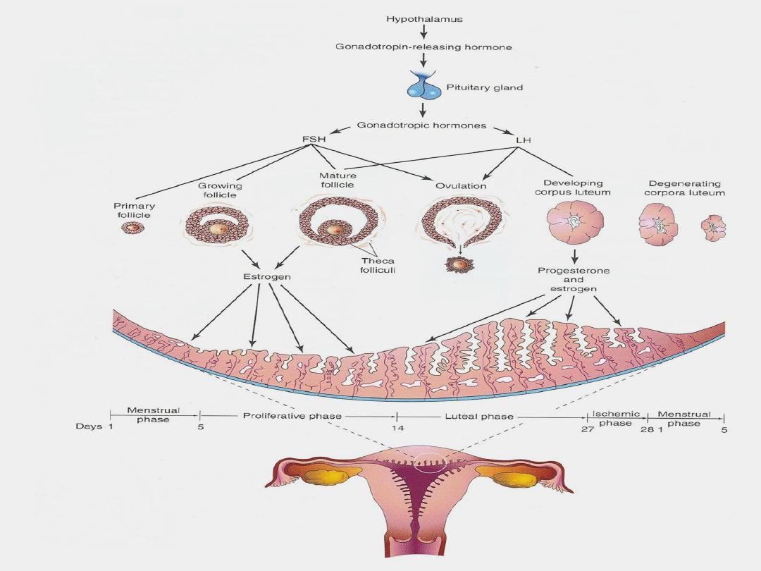
Ovarian Cycle
At puberty,
female undergo regular monthly cycles.
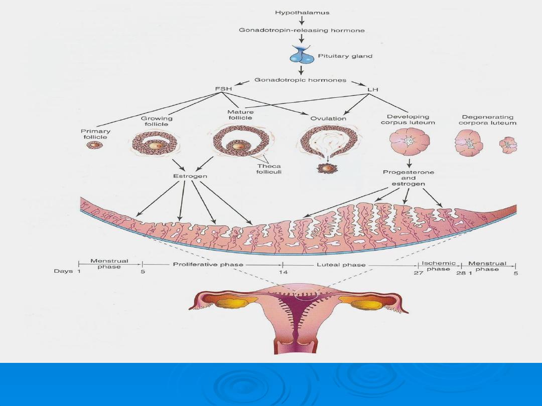
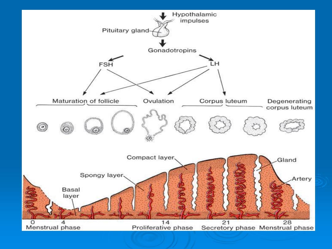
(GnRH),
produced by hypothalamus, acts on anterior pituitary
gland, secrete gonadotropins
(FSH)
&
(LH).
These hormones
stimulate & control cyclic changes in ovary.
At beginning of each ovarian cycle
, 15 to 20 PF are stimulated
to grow .
Normally
, only one of these follicles reaches full maturity, and
others become atretic.
Corpus atreticum
: When a follicle becomes atretic, oocyte
degenerate & replaced by CT.
In the course of a normal menstrual cycle, the
ovary will go through three phases:
1
Follicular phase
2
Ovulation
3
Luteal phase.

In cooperation , theca
interna and granulosa cells
produce
estrogens:
theca interna cells produce androstenedione and
testosterone , and granular cells convert these hormones to
estrone and 17 B-estradiol. As a result of this estrogen
production,
The uterine endometrium enters the follicular or proliferative
phase
Thinnig of the cervical mucous occurs to allow passage of
sperm and
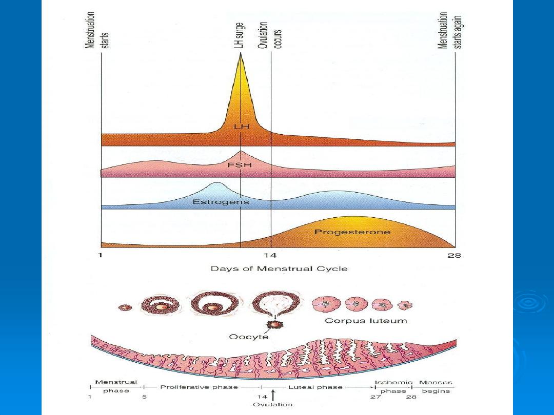
The anterior lobe of the pituitary gland is stimulated to secrete
LH

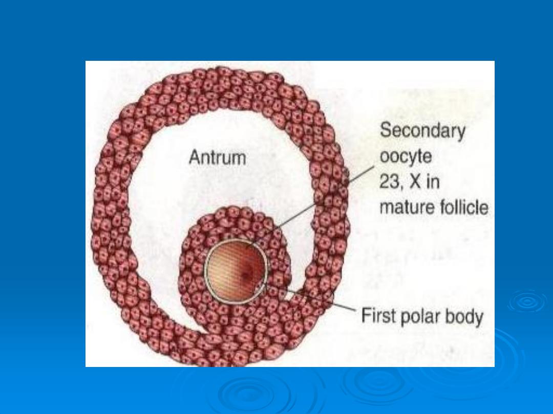
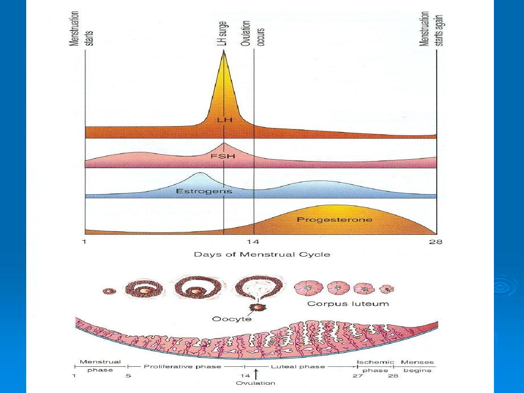
At midcycle, there is an LH surge that :
Elevate concentration of maturation-promoting factor, causing
oocyte to complete meiosis 1 and initiate meiosis 11 :
Stimulate production of progesterone by follicular stromal
cells (lutinization):and
Cause follicular rupture and ovulation.
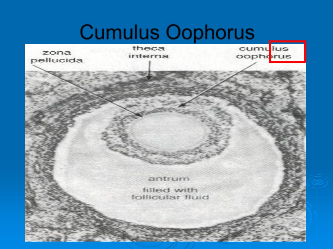
Cumulus Oophorus

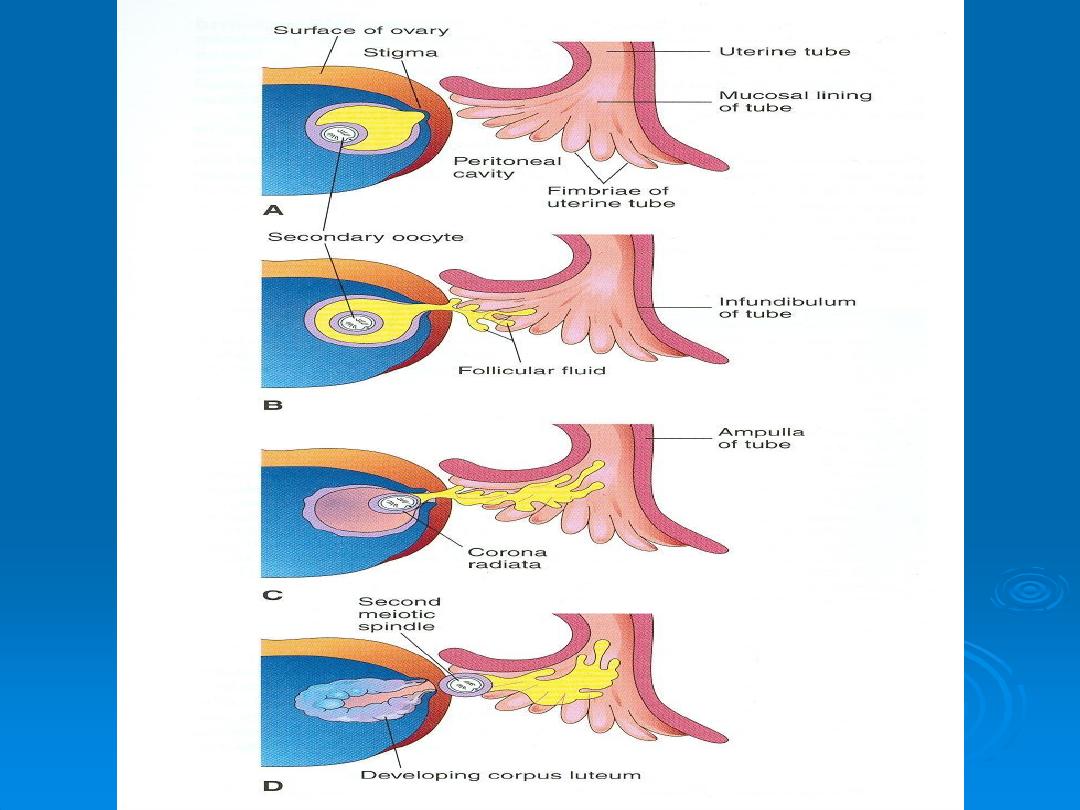
Ovulation
In the meantime, the surface of the ovary begins to bulge locally, and at the
apex, an avascular spot,
the stigma,
appears. The high concentration of LH
increases
•collagenase activity
, resulting in digestion of collagen fibers surrounding the
follicle.
•Prostaglandin levels
and cause local muscular contractions in the ovarian
wall.
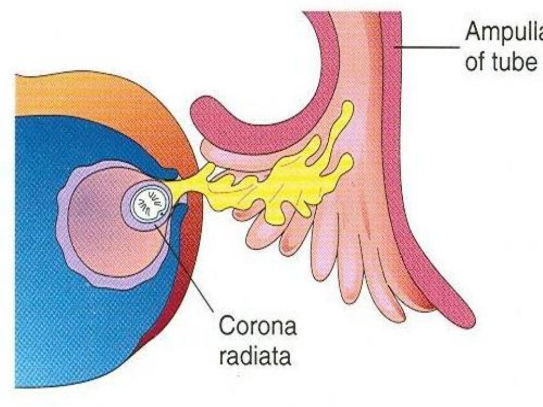
The oocyte, in metaphase of meiosis II, is discharged from the ovary together
with a large number of cumulus oophorus cells. Some of the cumulus
oophorus cells then rearrange themselves around the zona pellucida to form
the corona radiate
During ovulation, some women feel a slight pain “
middle pain
” because it
normally occurs near the middle of the menstrual cycle.
Ovulation is also generally accompanied by
a rise in basal temperature,
which
can be monitored to aid couples in becoming pregnant or preventing
pregnancy.

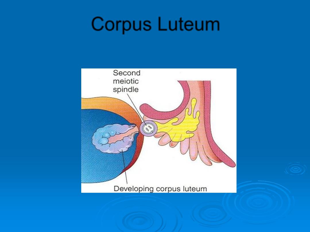
Corpus Luteum
After ovulation, granulosa cells remaining in the wall of the
ruptured follicle, together with cells from the theca interna, are
vascularized
by surrounding vessels.
Under the influence of
LH,
these cells develop a yellowish
pigment and change into lutean cells, which form the corpus
luteum and secrete the hormone
progesterone .
Progesterone, together with estrogenic hormones, causes the
uterine mucosa to enter the progestational or
secretory stage
in
preparation for implantation of the embryo.

Fate of the corpus luteum
If fertilization does not occur:
the corpus luteum reaches maximum development approximately 9 days after
ovulation. Subsequently, the corpus luteum shrinks because of degeneration of
lutean cells and forms a mass of fibrotic scar tissue, the
corpus albicans
.
Fate of the corpus luteum
If the oocyte is fertilized
degeneration of the corpus luteum is prevented by human chorionic gonadotropin
(
hCG)
, a hormone secreted by the syncytiotrophoblast of the developing embryo.
The corpus luteum continues to grow and forms the corpus luteum of pregnancy
(corpus luteum graviditatis).
By the end of the third month, this structure may be one third to one half of the
total size of the ovary. Yellowish luteal cells continue to secrete progesterone
until the end of the fourth month; thereafter, they regress slowly as secretion of
progesterone by the trophoblastic component of the placenta becomes adequate
for maintenance of pregnancy. Removal of the corpus luteum of pregnancy
before the fourth month usually leads to abortion.

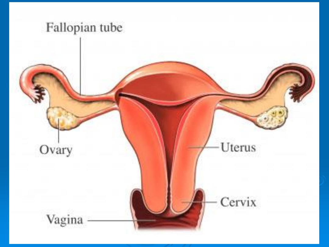
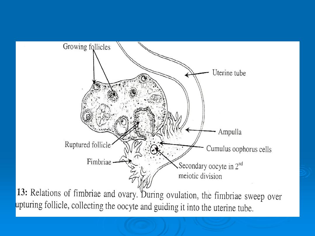
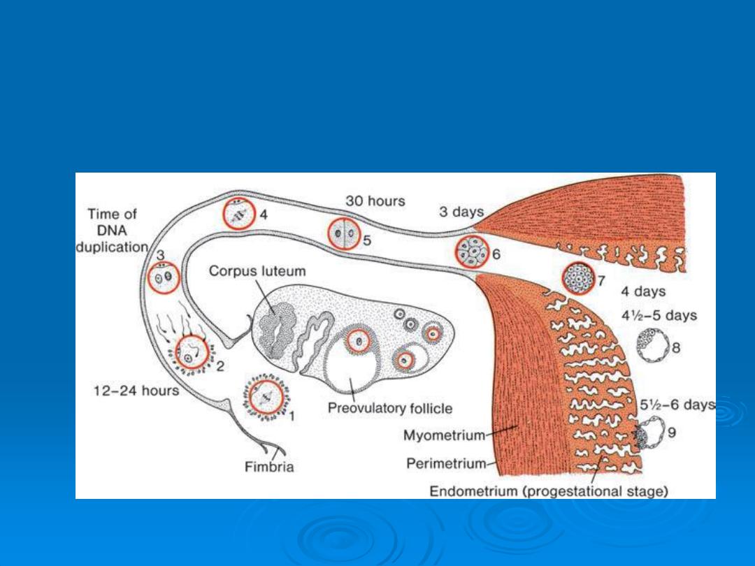
Oocyte Transport
Shortly before ovulation,
fimbriae
of the uterine tube sweep over the
surface of the ovary, and the tube itself begins to contract rhythmically. It
is thought that the oocyte, surrounded by some granulosa cells), is carried
into the tube by these sweeping movements of the fimbriae and by motion
of cilia on the epithelial lining.
Once in the tube, cumulus cells withdraw their cytoplasmic processes from
the zona pellucida and lose contact with the oocyte.
Once the oocyte is in the uterine tube, it is propelled by peristaltic
muscular contractions of the tube and by cilia in the tubal mucosa with the
rate of transport regulated by the endocrine status during and after
ovulation.
In humans, the fertilized oocyte reaches the uterine lumen in
approximately 3 to 4 days.
Corpus Luteum

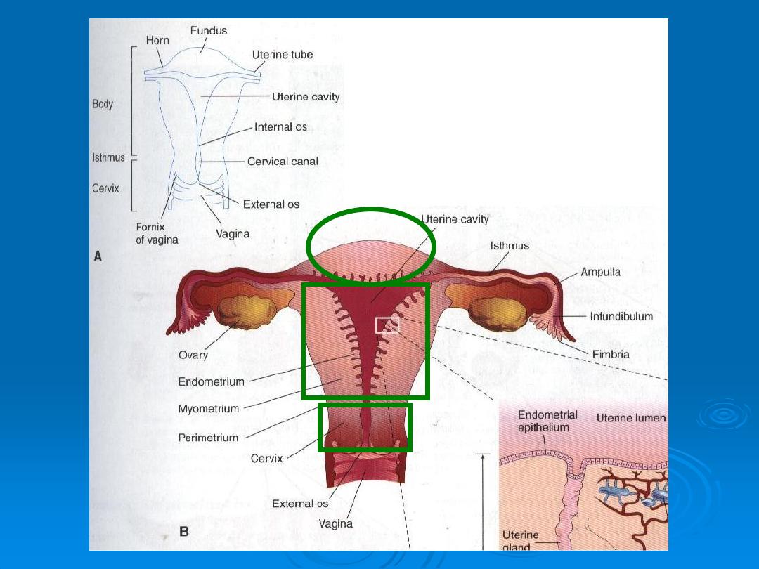
Uterine cycle (uterus at time of implantation)
The wall of the uterus consist of three layers :
1-
Endometrium
or mucosa lining the inside wall.
2-
Myometrium
, athick layer of smooth muscle
3-
Perimetrium
, the peritoneal covering lining the outside wall
From puberty (11-13) until menopause (45 -55years), the endometrium
undergoes changes in a cycle of approximately 28 days under hormonal
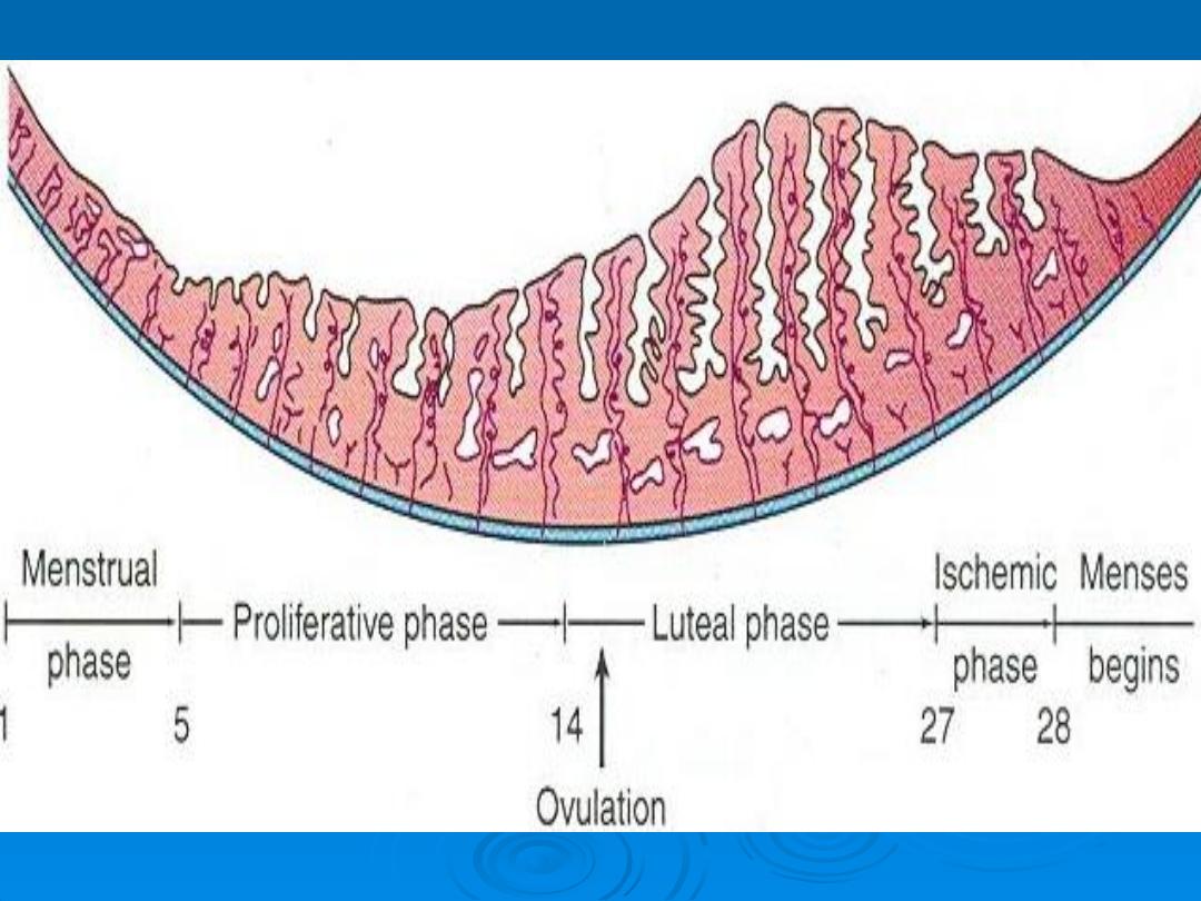
control by the ovaries . During this menstrual cycle , the uterine endometrium
passes through three stages , the
1-Follicullar or proliferative phase
2-Secretory or progestational phase
3-menstrual phase

The proliferative
phase begins at the end of the menstrual phase, is under
the influence of estrogen, and parallels growth of the ovarian follicles.
The secretory
phase begins approximately 2 to 3 days after ovulation in
response to progesterone produced by the corpus luteum.
If fertilization does not occur,
shedding of the endometrium (compact and
spongy layers) marks the beginning of the menstrual phase

.
If fertilization does occur
, the endometrium assists in
implantation and contributes to formation of the placenta.
Later in gestation, the placenta assumes the role of hormone
production, and the corpus luteum degenerates.
At the time of implantation, the mucosa of the uterus is in the
secretory phase .during which time uterine glands and arteries
become coiled and the tissue becomes succulent.
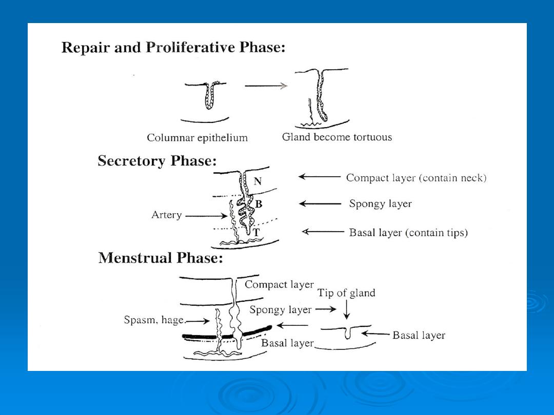

As a result, three distinct layers can be recognized in the endometrium:
a
superficial
compact layer,
an
intermediate spongy
layer,
and a
thin basal
layer .
Normally, the human
blastocyst implants
in the endometrium along
the anterior or posterior wall of the body of the uterus, where it
becomes embedded between the openings of the glands .

If the oocyte is not fertilized, venules and sinusoidal spaces
gradually become packed with blood cells.
When the menstrual phase begins, blood escapes from superfi
cial arteries, and small pieces of stroma and glands break
away.
During the following 3 or 4 days, the compact and spongy
layers are expelled from the uterus, and the basal layer is the
only part of the endometrium that is retained .
This layer, which is supplied by its own arteries, the basal
arteries, functions as the regenerative layer in the rebuilding
of glands and arteries in the proliferative phase .

Thank you