CSF CIRCULATION
Cerebrospinal FluidA clear, colorless fluid that surrounds and permeates the CNS. Offers support, protection and nourishment.
Functions:
– Protection of cranial contents
– Modulates pressure changes (same specific
• gravity as brain)
– Serves as a chemical buffer to maintain constant
• ionic environment
– Serves as a transport medium for nutrients and
• metabolites, endocrine substances and even
• neurotransmitters
Location of CSF
• Two lateral ventricles
• Third ventricle
• Fourth ventricle
• Spinal cord central
• canal
• Subarachnoid space
• Continuous with
• extracellular fluid of
• brain parenchyma
Properties
••
•
•
Volume
: approximately 150 mL
Rate of formation: approximately 0.3 mL/min
Specific gravity : 1.005
Reaction
: Alkaline
In adults, the total excreted CSF is about 400 – 500 ml so it is replaced every 3-4 hours
Composition
Cerebrospinal FluidWater - 99.13%
Solids - 0.87%
Organic substances
Inorganic substances
1.Proteins
2.Amino acids
3.Sugar
1.Sodium
2.Calcium
3.Potassium
4.Magnesium
5.Chlorides
6.Phosphate
7.Bicarbonates
8.Sulfates
4.Cholesterol
5.Urea
6.Uric acid
7.Creatinine
8.Lactic acid
Lymphocytes in CSF : 6/ cu mm
Formation of CSF
• Choroid plexuses of lateral, third and fourth• ventricles
• Ependymal lining of ventricular system
• Blood vessels: Smaller quantities formed from fluid leaking into perivascular spaces surrounding cerebral vessels
Cells are believed to actively secrete Na into the ventricular system in exchange for K. Sodium ions electrically attract Cl and osmotically draw water from the blood vascular system to constitute the CSF.
+
-
The Choroid Plexus
• The choroid plexus also helps to cleanse the• CSF by removing waste products and other
• unnecessary solutes
• Once produced CSF moves freely through the
• ventricles
Choroid Plexus
• Choroid plexushang from the
roof of each
ventricle
• These plexuses
• form CSF
• The plexuses are
• clusters of thin
• walled capillaries
• enclosed by a
• layer of
ependymal cells
The lateral ventricles
• Situated in the cerebral hemispheres, it has a• body, an anterior horn in the frontal pole, posterior
• horn in the occipital pole & inferior horn in the
• temporal pole.
• The two lateral ventricles are interconnected by
• interventricular foramen and it also communicates
• with the 3rd ventricle.
• All ventricles are lined by ependyma.
3rd Ventricle
• It lies below the lateral ventricles.
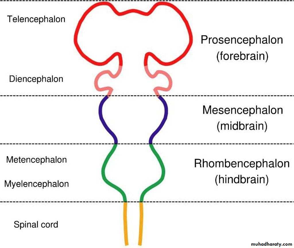
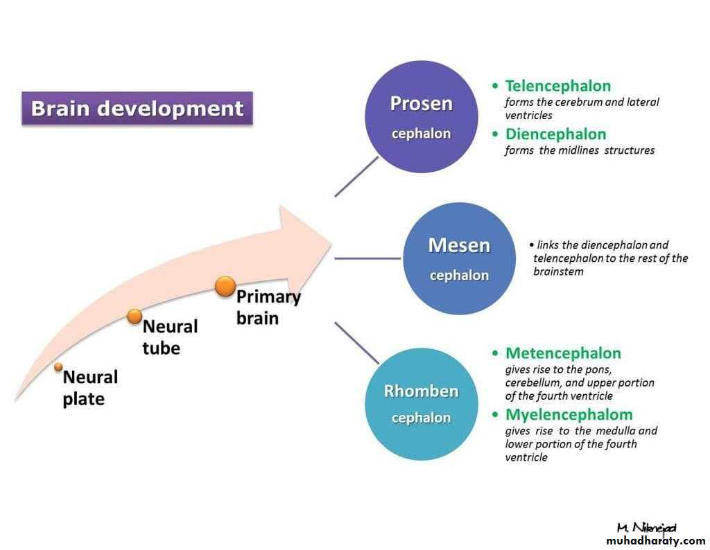
• It is a cavity of the diencephalon. In the roof there is
choroid plexus, that produce CSF.
• Superiorly it communicates with the two lateral
ventricles through the interventricular foramen.
• Inferiorly it communicates with the 4th ventricle
through the cerebral aqueduct.
• The lateral wall is formed by thalamus and
• hypothalamus.
4th Ventricle
It is a cavity of the rhombencephalon• It has a roof and floor.
• The floor is formed by two parts, medullary & pontine part.
• The roof formed by cerebellum
• It has 3 foramina, one is median (foramen of Magendie) located posteriorly and two are lateral (foramina of Lushka)
Ventricles
Lateral
Third
Fourth
Flow of CSF:
• The CSF passes from the lateral ventricles (I and II) through the foramen of Monro into the third ventricle (III) then through the aqueduct of Sylvius into the fourth ventricle (IV) and out into the subarachnoid space through the foramina Lushka & Magendie .Absorption of CSF
• Through the arachnoid villi, a protrusion of arachnoid membrane into the central venous sinus and other sinuses• A valve opens when CSF pressure exceeds venous pressure
• Absorption by veins and capillaries of CNS
arachnoid granulation
Summary
Radiological anatomy
Clinical application
Hydrocephalus
• An abnormal increase in the volume of• CSF
• Symptoms: sleep changes, spastic
• paresis, papilledema, bulging of skull in
• young, seizures, cranial nerve deficits,
• depression.
Hydrocephalus
Lumbar puncture
LP:is a diagnostic procedure that is performed in order to collect a sample of cerebrospinal fluid (CSF) for biochemical, microbiological, and cytological analysis


