Digestive system part -2-Dr: Ezdehar Nassif Ali
DuodenumThe terminal part of the foregut and the cephalic part of the mid gut form the duodenum . The junction of the two parts is directly distal to the origin of the liver bud. As the stomach rotates , the duodenum takes on the form of a C-shaped loop and rotates to the right . This rotation, together with rapid growth of head of pancreas , swings the duodenum from its initial midline position to the right side of the abdominal cavity.
The foregut is supplied by the celiac artery and the mid gut is supplied by the superior mesenteric artery , the duodenum is supplied by branches of both arteries.
• Liver and Gallbladder
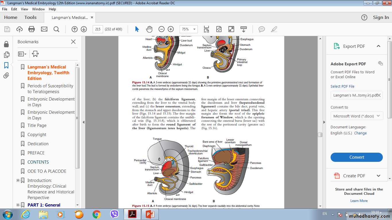
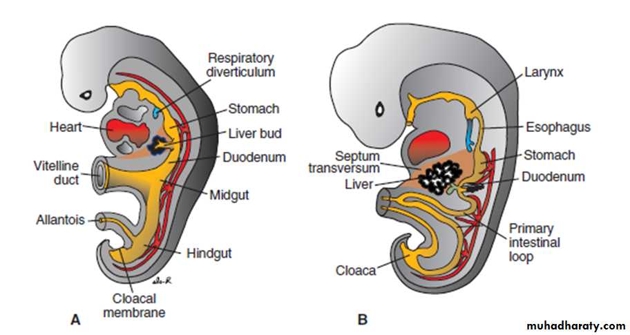
• The liver primordium appears in the middle of the third week as an outgrowth of the endodermal epithelium at the distal end of the foregut.• This outgrowth, the hepatic diverticulum, or liver bud, consists of rapidly
• proliferating cells that penetrate the septum transversum .
The connection between the hepatic diverticulum and the foregut (duodenum) narrows, forming the bile duct.
• A small ventral outgrowth is formed by the bile duct, and this outgrowth gives rise to the gallbladder and the cystic duct. The surface of the liver that is in contact with the future diaphragm is never covered by peritoneum; it is the bare area of the liver.
• Accessory hepatic ducts and duplication of the gallbladder are common and usually asymptomatic.
Pancreas
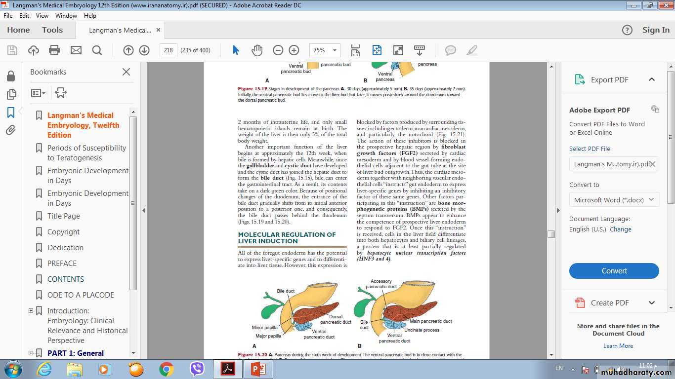
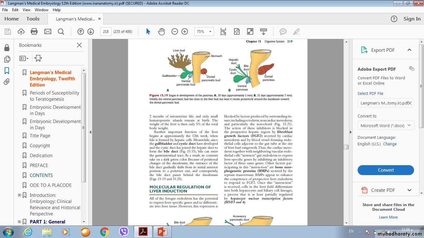
The pancreas is formed by two buds, dorsal and ventral ,originating fromThe endodermal lining of the duodenum . Whereas the dorsal pancreatic bud is in the dorsal mesentery , the ventral pancreatic bud is close to the bile duct . When the duodenum rotates to the right and becomes-C-shaped , the ventral pancreatic bud moves dorsally in a manner similar to the shifting of the entrance of the bile duct . Finally , the ventral bud comes to lie immediately below and behind the dorsal bud.
In the third month of the fetal life ,pancreatic islets of Langerhans develop from the parenchymatous pancreatic tissue and scatter throughout the pancreas.
Insulin secretion begins at approximately the fifth month.
Glucagon- and somatostatin- secreting cells also develop from parenchymal cells.
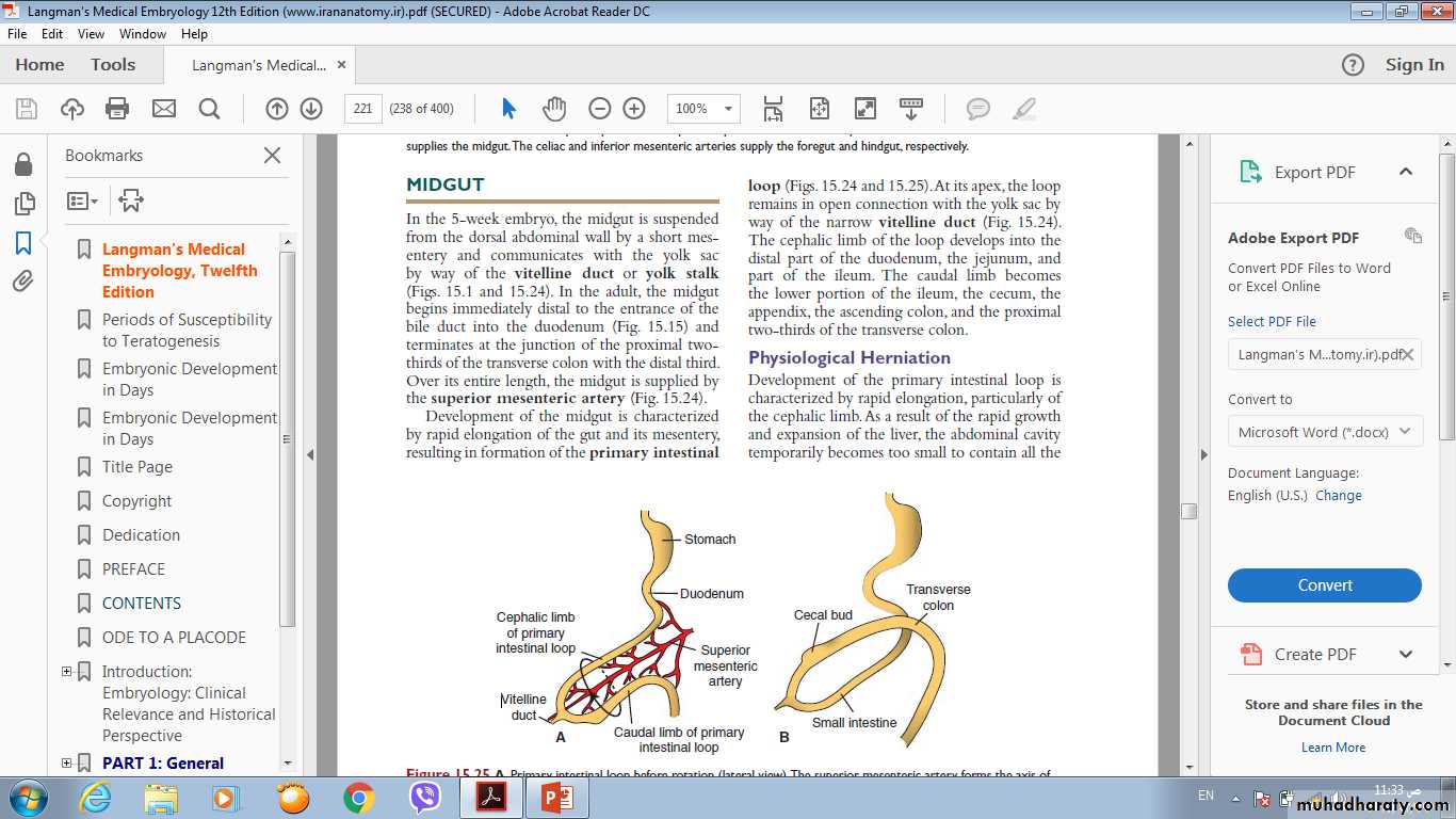
Midgut
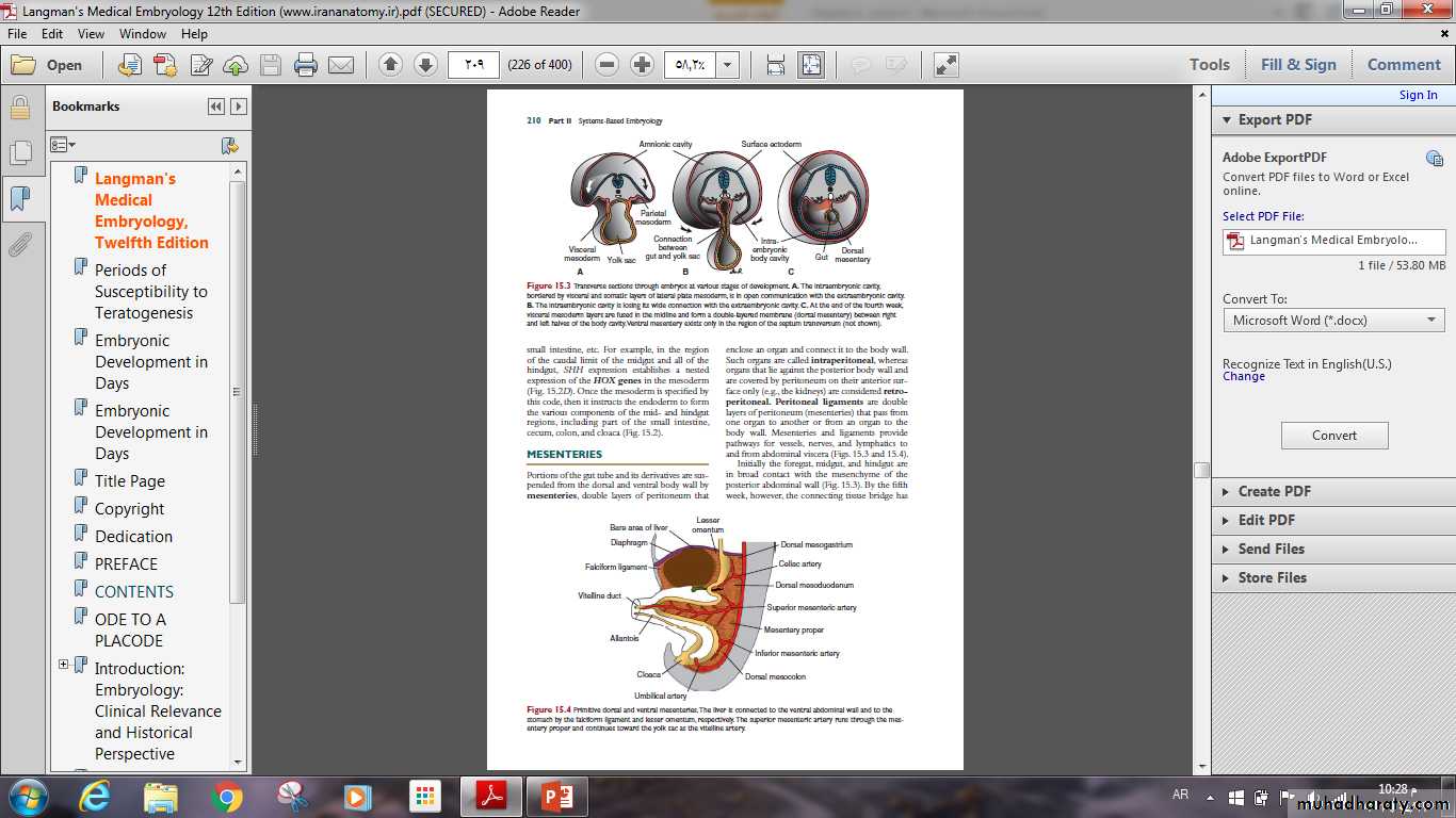
In the 5-week embryo, the midgut is suspended from the dorsal abdominal wall by a short mesentery and communicates with the yolk sac by way of the vitelline duct or yolk stalk . In the adult, the midgut begins immediately distal to the entrance of the bile duct into the duodenum and terminates at the junction of the proximal two thirds of the transverse colon with the distal third.Development of the midgut is characterized by rapid elongation of the
gut and its mesentery, resulting in formation of the primary intestinal loop.
At its apex, the loop remains in open
connection with the yolk sac by way of the narrow vitelline duct.
The cephalic limb of the loop develops into the distal part of the duodenum, the jejunum, and part of the ileum.
The caudal limb becomes the lower portion of the ileum, the cecum, the
appendix, the ascending colon, and
The Proximal two-thirds of the transverse colon.
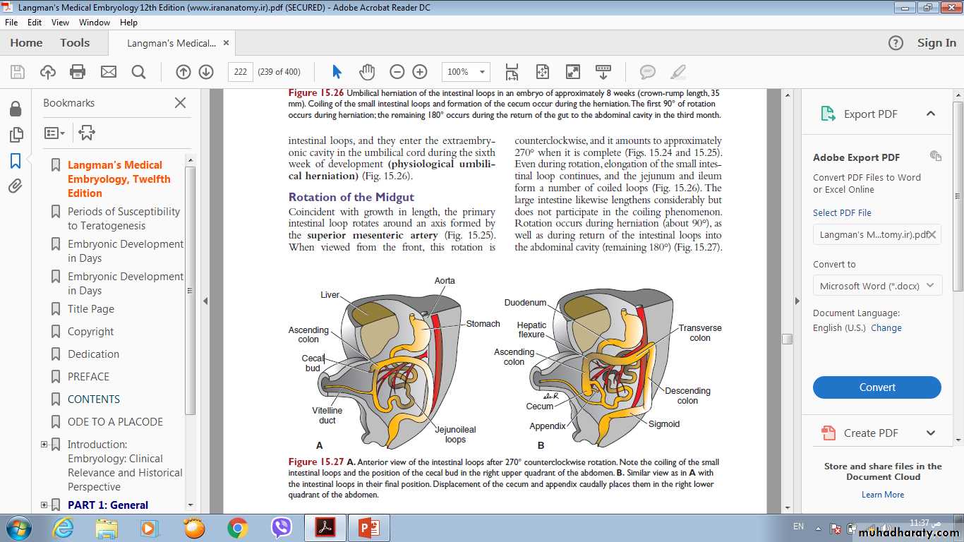
During the sixth week, the loop grows so rapidly that it protrudes into the umbilical cord (physiological herniation) .
During the 10th week, it returns into the abdominal cavity.
While these processes are occurring, the
mid gut loop rotates 270° counterclockwise. Remnants of the vitelline duct, failure of the mid gut to return to the abdominal cavity, malrotation, stenosis ,and duplication of parts of the gut are common mid gut abnormalities.
Hindgut
• The hindgut gives rise to the distal third of the transverse colon , the descending colon, the sigmoid, the rectum, and the upper part of the anal canal.
• The endoderm of the hindgut also forms the internal lining of the bladder and urethra. The terminal portion of the hindgut enters into the posterior region of the cloaca, the primitive anorectal canal; the allantois enters into the anterior portion, the primitive urogenital sinus.
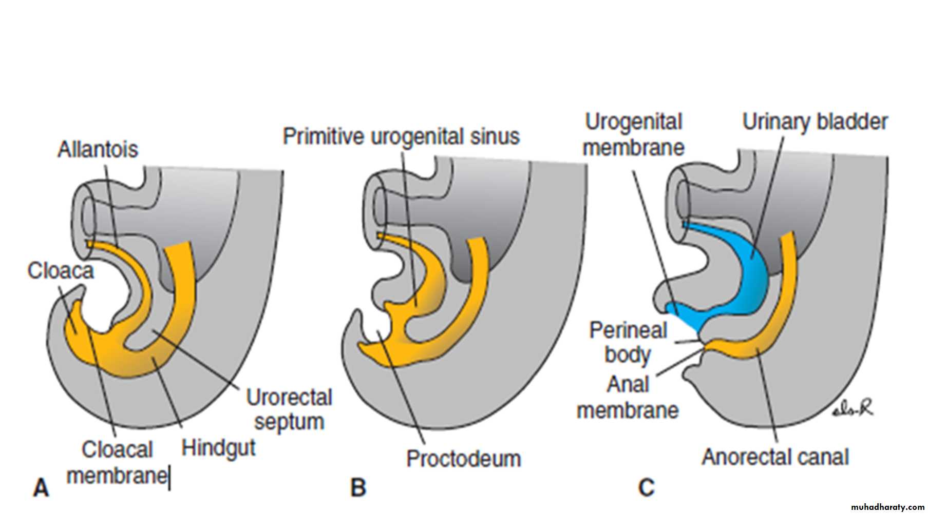
• The cloaca itself is an endoderm-lined cavity covered at its ventral boundary by surface ectoderm. This boundary between the endoderm and the ectoderm forms the cloacal membrane .
• At the end of the seventh week, the cloacal membrane ruptures, creating the anal opening for the hindgut, and a ventral opening for the urogenital sinus. Between the two, the tip of the urorectal septum forms the perineal body.
• The upper part (two-thirds) of the anal canal is derived from endoderm of the hindgut; the lower part (one-third) is derived from ectoderm around the proctodeum.
Hindgut Abnormalities
Abnormalities in the size of the posterior region of the cloaca shift the entrance of the anus anteriorly, causing rectovaginal & rectourethralfistulas and atresia.
Imperforate anus occurs when the anal membrane fails to break down.