Cerebrum -2-

• Cortical Areas
• fronTal lobe
• Frontal lobe
• 3 sulci
• 4 gyri
• Precentral
• Precentral
• sulcus
• gyrus
• Superior
• Superior
• frontal sulcus
• Frontal gyrus
• Inferior
• Middle
• frontal sulcus
• Frontal gyrus
• Inferior
• Frontal gyrus
• Frontal lobe
• Pariteal lobe
• 2 sulci• gyri
• Post-central
• Post-central
• sulcus
• gyrus
• Intra-parietal (arcuate)
• Superior
• sulcus
• parietal lobule
• Inferior
• Parietal lobule
• Superior
• marginal gyrus
• 2nd pareito-
• occipital arcus
• Agnular gyrus
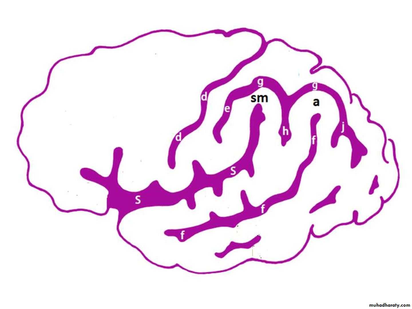
Frontal lobe:
Precentral gyrus.Superior & inferior frontal sulci divide the lobe into superior, middle & inferior frontal gyri.
Superior , middle & inferior frontal gyri
Precentral gyrusSuperior parietal lobule
Inferior parietal lobule
Postcentral gyrus
Intraparietal sulcus
sfs
ifs
• Parietal lobe:
• Postcentral gyrus.
• Intraparietal sulcus divide the lobe into superior & inferior parietal lobules.
SUPEROLATERAL SURFACE

• Temporal lobe
• 2 sulci• Superior
• 3 gyri
• Superior
• Temporal sulcus
• Temporal gyrus
• Inferior
• Middle
• Temporal sulcus
• Temporal gyrus
• Inferior
• Temporal gyrus
• Temporal lobe
• Occipital lobe
• 2 sulci• gyri
• Superior occipital
• Superior
• sulcus
• Occipital gyrus
• Inferior occipital
• Middle
• sulcus
• Occipital gyrus
• Inferior
• Occipital gyrus
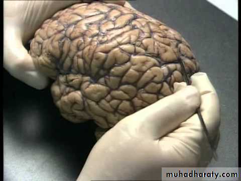
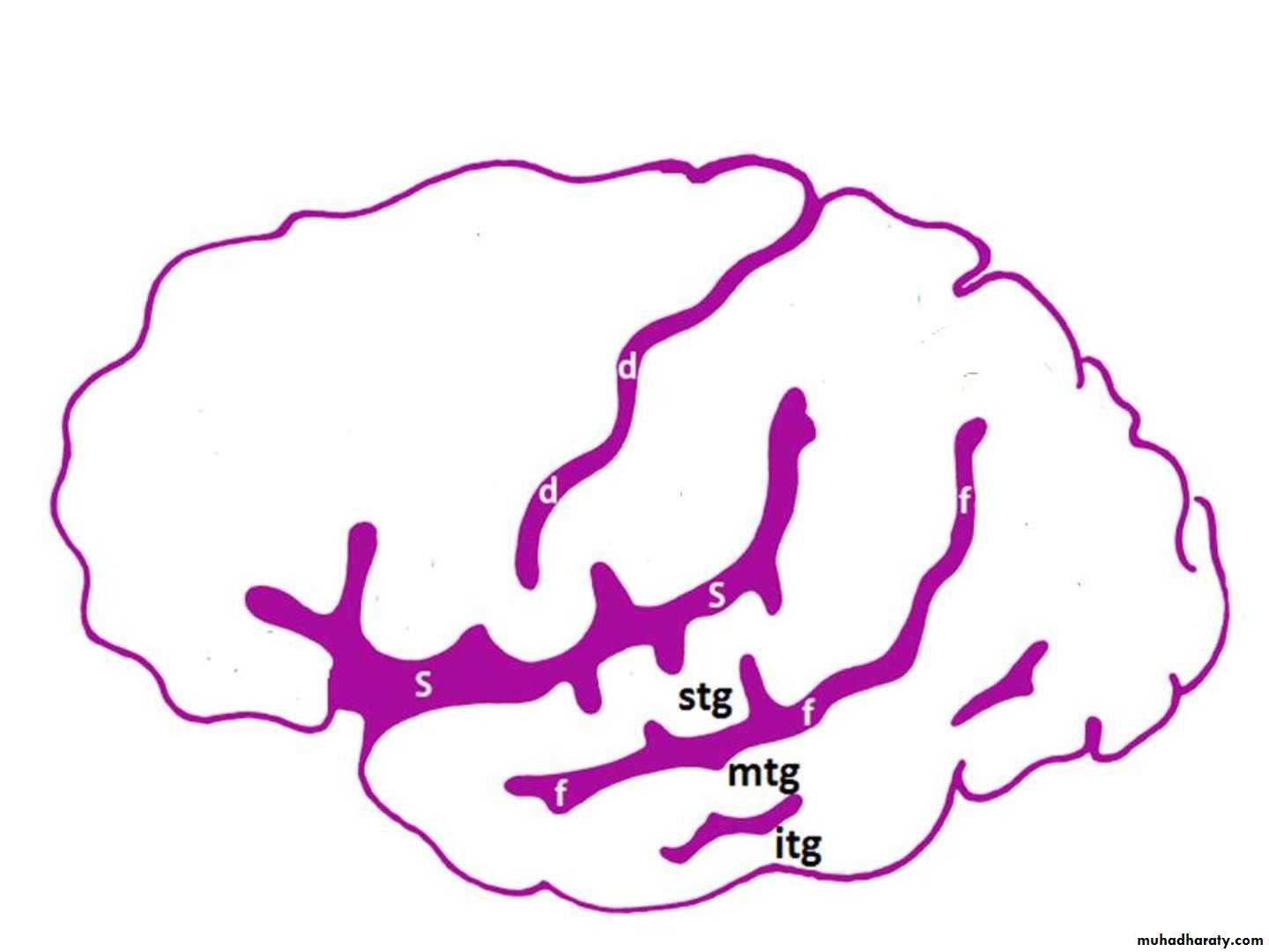
Temporal lobe:
Superior & inferior temporal sulci giving rise to superior, middle & inferior temporal gyri.Insula: the gyrus in the depth of lateral fissure, covered by parts of frontal, parietal & temporal lobes called the opercula (removed in lower picture.).
Superior, middle & inferior temporal gyri
insulasts
its
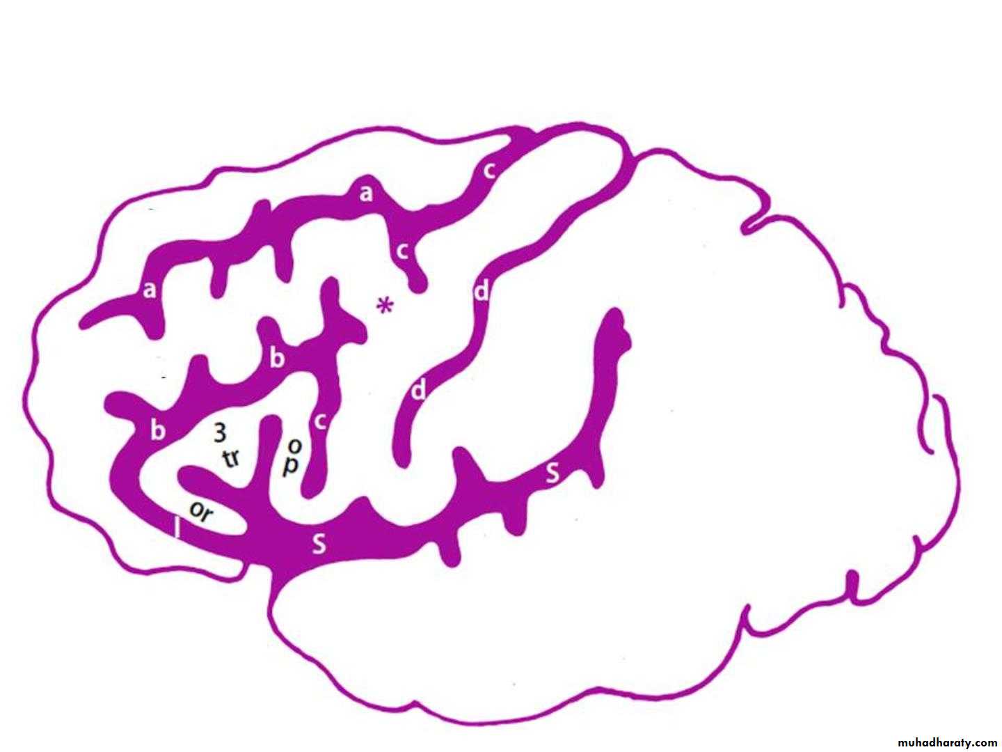
Functional Areas of the Cerebral Cortex
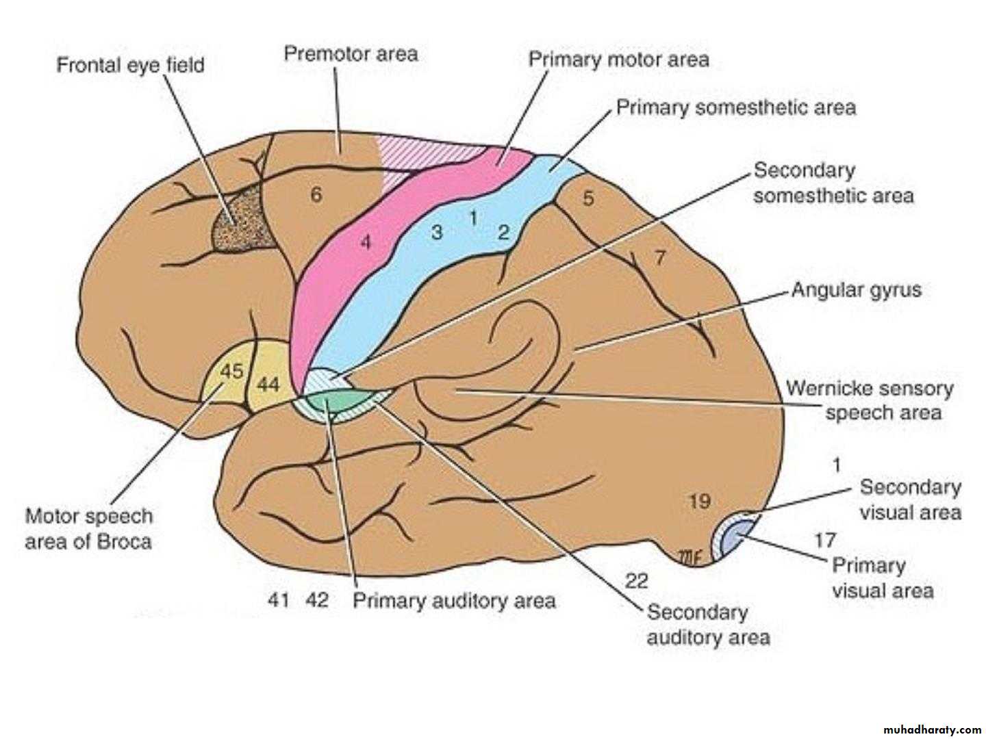
Brodmann produced a numbered, cytological map of cerebral cortex based upon its regional histological characteristicsSubdivisions with similar cellular and laminar structure are called 'areas'
Brodmann's numbering of these cortical locations has become one of the standard ways to identify brain areas.
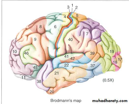
Brodmann’s Map
Frontal Lobe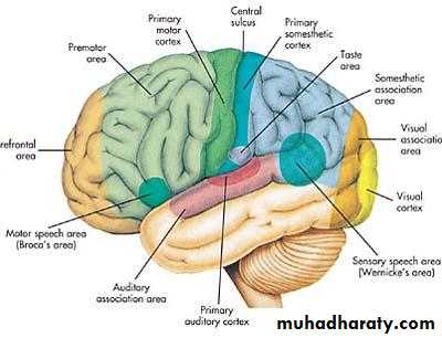
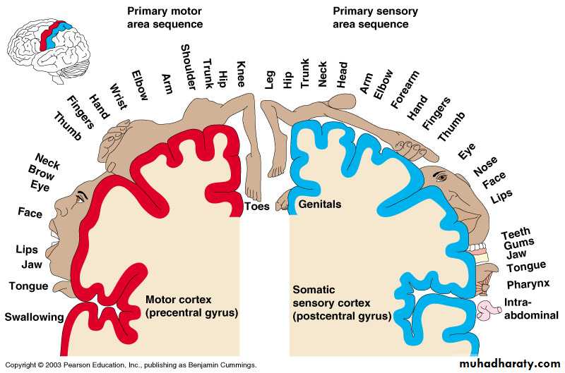
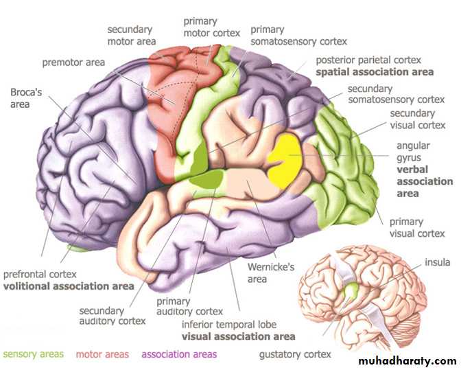
Primary motor cortex: Located in precentral gyrus (Brodmann area 4).
Premotor cortex: Located in the region immediately anterior to the precentral gyrus (Brodmann’s area 6).Frontal eye field: Located in the middle frontal gyrus immediately in front of motor cortex (Brodmann’s area 8).
Broca’s (motor speech) area: Located in the inferior frontal gyrus of the dominant hemisphere, usually left (Brodmann’s area 44 & 45).
Prefrontal cortex: Extensive region of the frontal lobe anterior to premotor area.
Parietal lobe
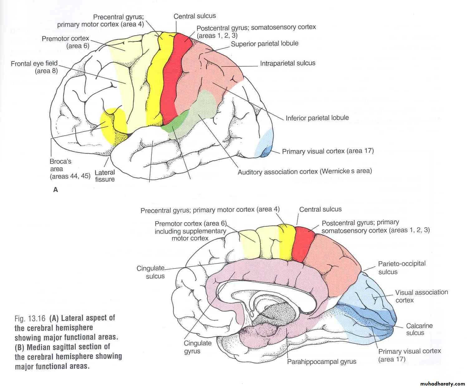
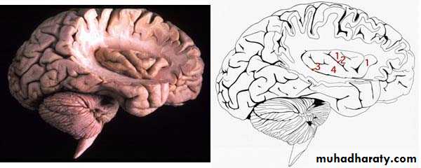
• Primary visual cortex: located on the medial surface of the hemisphere, in the gyri surrounding the calcarine sulcus (Brodmann’s area 17).
Occipital lobe
• Visual association cortex: located around the primary visual cortex.
• Parietal association cortex: located posterior to primary somatosensory cortex.
• Primary somatosensory cortex: located in postcentral gyrus (Brodmann’s area 1, 2, 3).
Temporal Lobe
Auditory association cortex: located immediately around the primary auditory cortex (also includes Wernick’s area)Parahippocampal gyrus: located in the inferomedial part of temporal lobe. Deep to this gyrus lies the hippocampus and the amygdala, which are parts of limbic system
Primary auditory cortex: located in the superior surface of the superior temporal gyrus (Brodmann’s area 41, 42)
Language Area
Organized around the lateral fissure.Broca’s area: concerned with expressive aspects of language.
Wernick’s area: responsible for comprehension of the spoken words.
Nearby regions of temporal lobe and parietal lobe (angular gyrus & supramarginal gyrus of the inferior parietal lobule) are important in naming, reading, writing, and calculation.
The localization of speech centers & mathematical ability is the criterion for defining the dominant cerebral hemisphere.
In 96% of normal right-handed individuals and 70% of normal left-handed individuals, the left hemisphere contains the language centers. These are left hemisphere dominant.
Cerebral dominance becomes established during the first few years after birth.
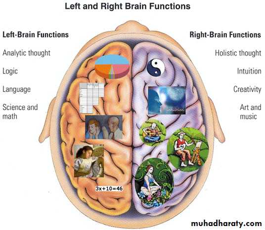
Verbal Memory
Shape MemoryHemispheres communicate via the corpus callosum
Hemispheric Dominance
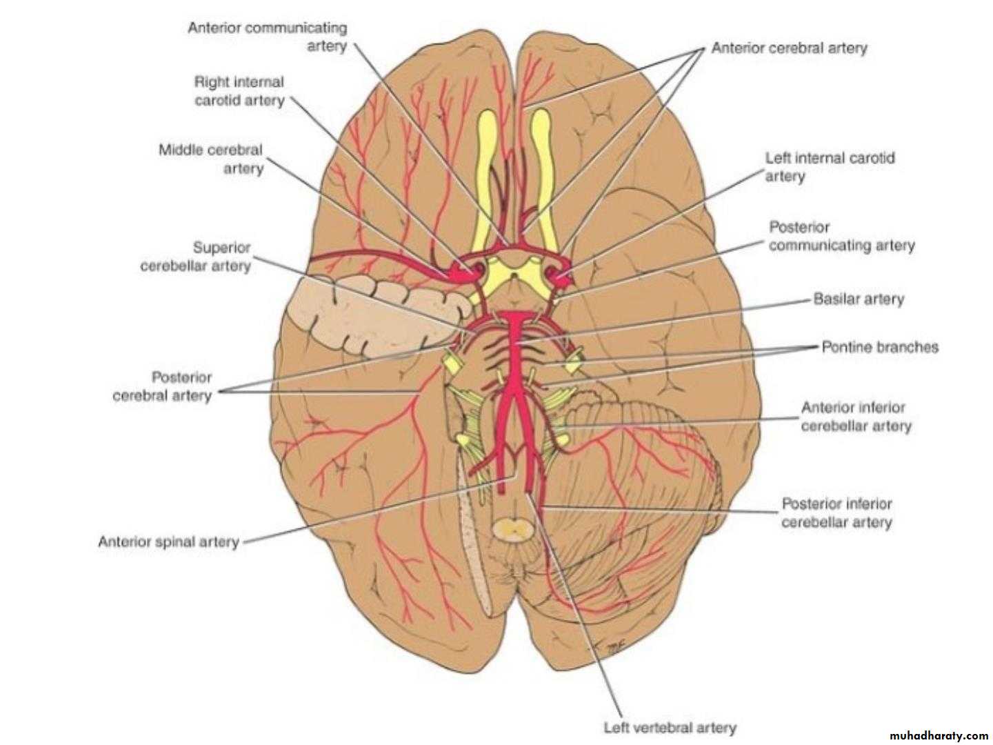
• Blood Supply of the Brain
• . Arteries of the Brain :• •
• •
• •
• The brain is supplied by the two internal carotid
• and the two vertebral arteries.
• The four arteries lie within the subarachnoid
• space,
• their branches anastomose on the inferior
• surface of the brain to form the circle of Willis.
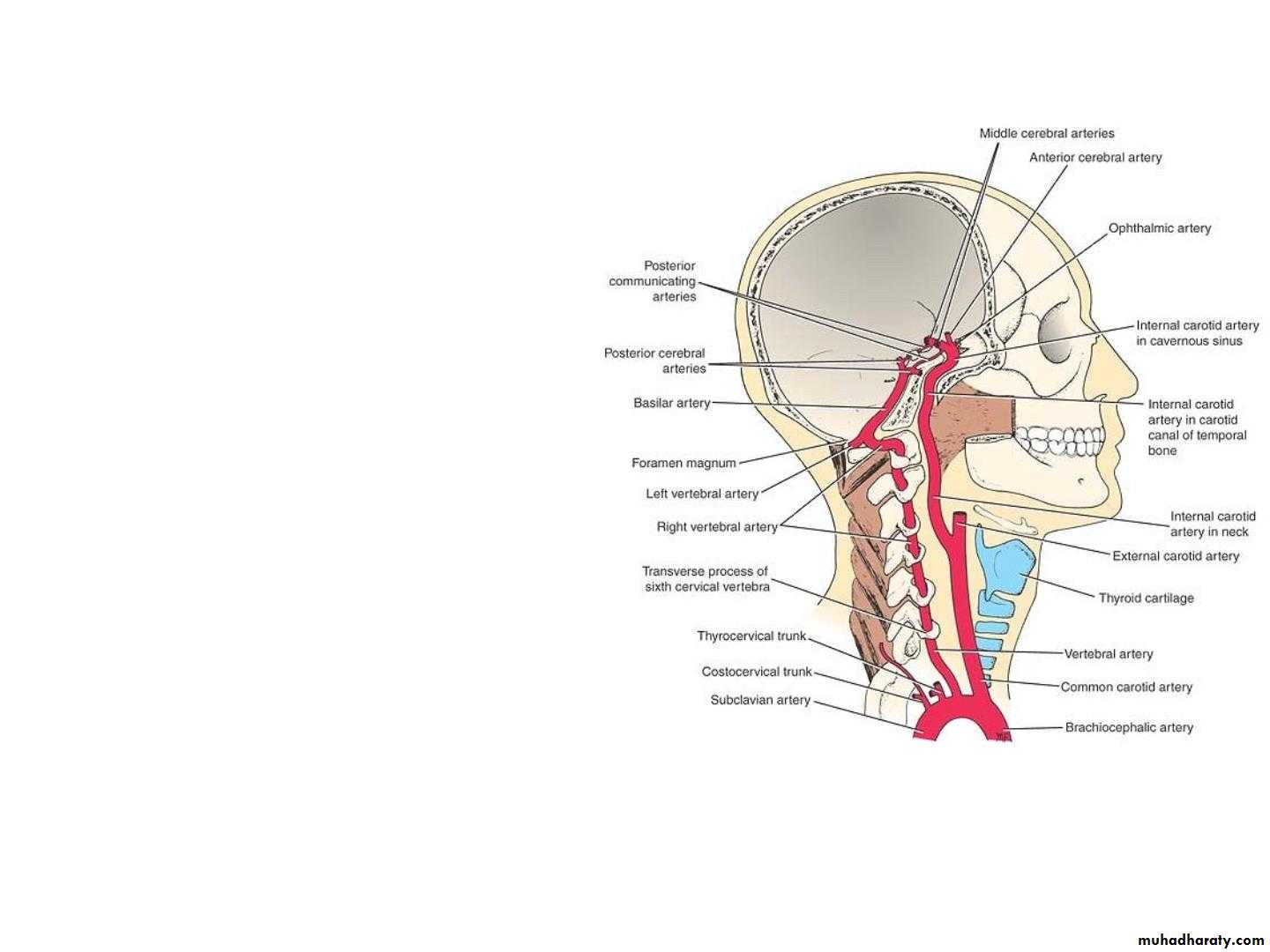
• 1. Internal Carotid Artery :
• •• It ascends the neck
• and perforates the
• base of the skull
• by passing through the
• carotid canal of the
• temporal bone.
• •
• The artery then runs
• horizontally forward
• through the cavernous
• sinus .
• • It now enters the subarachnoid space by
• piercing the arachnoid mater and turns to• the region of the medial end of the lateral
• cerebral sulcus.
• Here, it divides into :
• • Anterior Cerebral arteries
• • Middle Cerebral arteries.

• Branches Of The Cerebral Portion Of
• Internal Carotid artery :• 1. Ophthalmic artery
• 2. Posterior communicating artery
• 3. Choroidal artery
• 4. Anterior Cerebral Artery
• 5. Middle Cerebral Artery
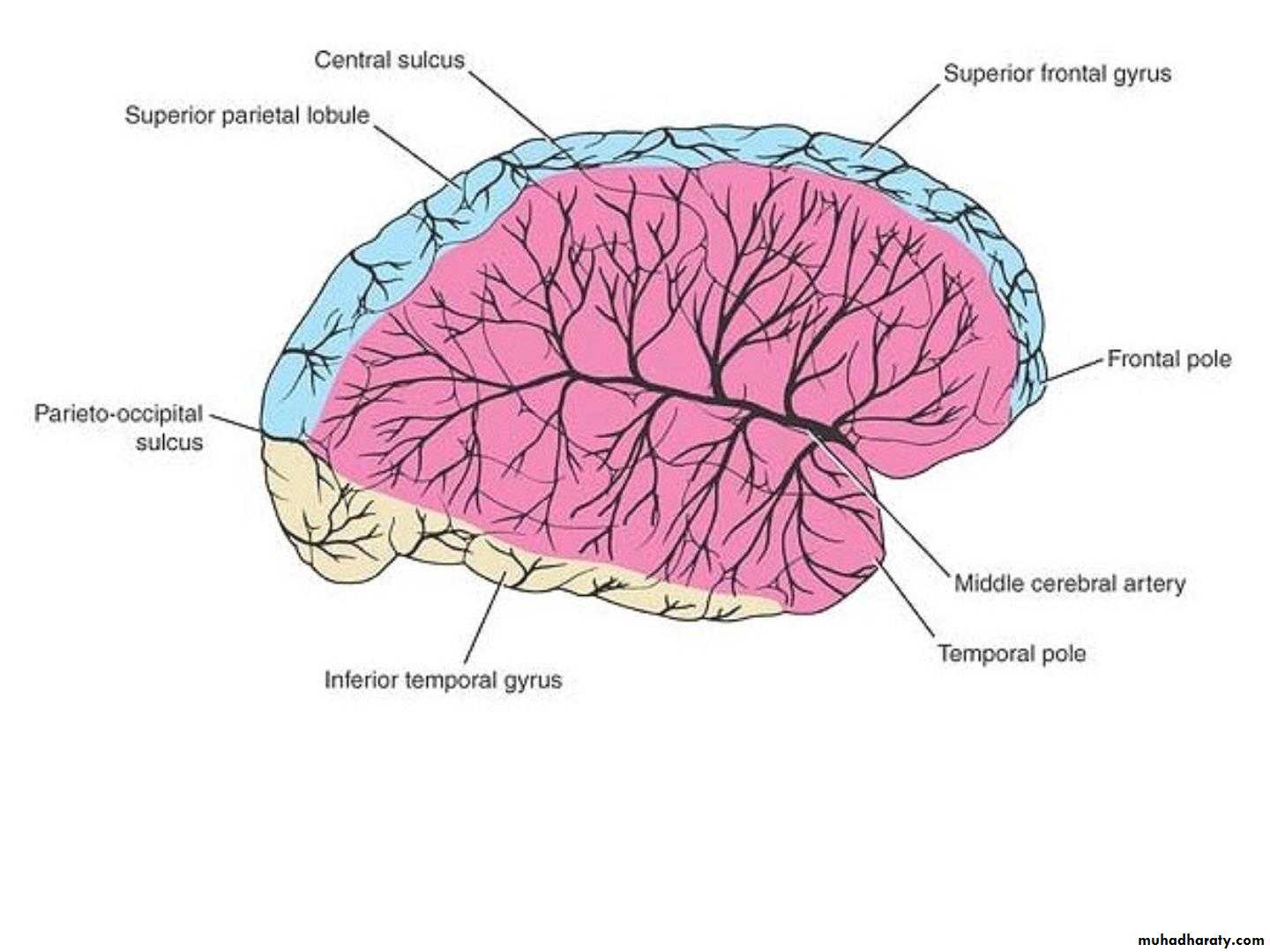
• Anterior Cerebral Artery :
• The cortical branches supply all the medial• surface of the cerebral cortex as far back as
• the parieto-occipital sulcus.
• • They also supply a strip of cortex about 1 inch
• (2.5 cm) wide on the adjoining lateral surface.
• • The anterior cerebral artery thus supplies the
• “leg area” of the precentral gyrus.
• • A group of central branches pierces the anterior
• perforated substance and helps to supply parts
• of the lentiform and caudate nuclei and the
• internal capsule.
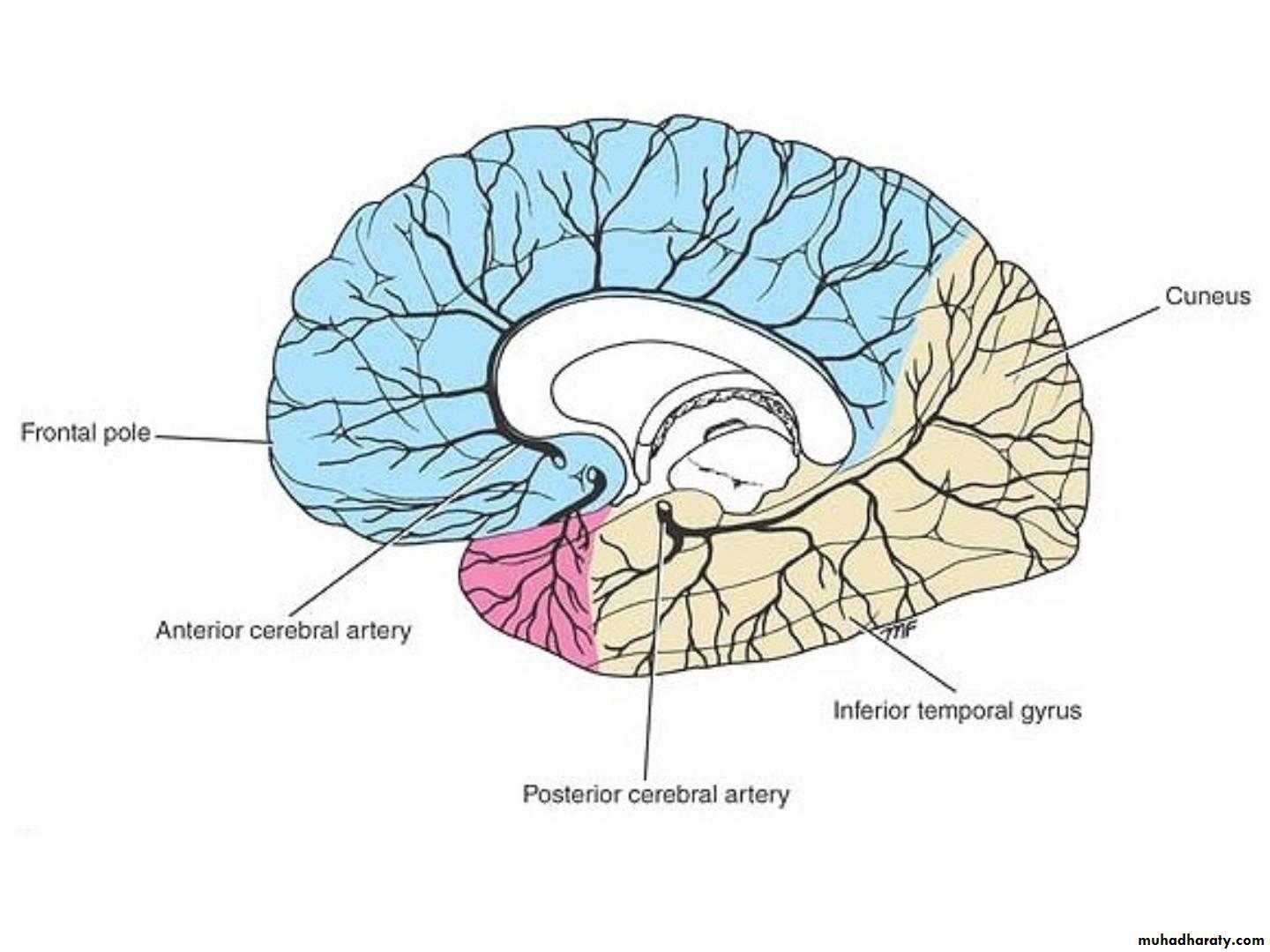
• Me dial Surf ace
• Lat eral Surf ace
• Middle Cerebral Artery :,
• •• •
• the largest branch of the internal carotid, runs
• laterally in the lateral cerebral sulcus.
• Cortical branches supply the entire lateral
• surface of the hemisphere, except for the
• narrow strip supplied by the anterior cerebral
• artery, the occipital pole, and the inferolateral
• surface of the hemisphere, which are supplied
• by the posterior cerebral artery.
• •
• •
• This artery thus supplies all The
• moTor area excepT The “leg
• area.”
• Central branches enter the anterior perforated
• substance and supply the lentiform and
• Me dial Surf ace
• Lat eral Surf ace
• B. Vertebral Artery :
• •• A branch of the first part of the subclavian
• artery,
• •
• It enters the skull through the foramen magnum
• and pierces the dura mater and arachnoid to
• enter the subarachnoid space.
• •
• At the lower border of the pons, it joins the
• vessel of the opposite side to form the basilar
• artery.
• Branches of the Cranial Portion
• 1. The meningeal branches• 2. The posterior spinal artery
• 3. The posterior inferior cerebellar artery
• 4. The medullary arteries
• 5. The basilar artery.
• Basilar Artery :
• • formed by the• union of the two
• vertebral arteries,
• ascends in a
• groove on the
• anterior surface
of pons .
• • At the upper
• border of pons, it divides into
• the two posterior
• cerebral arteries.
• Branches Of Basilar Artery :
• 1. The pontine arteries• 2. The labyrinthine artery
• 3. The anterior inferior cerebellar artery
• 4. The superior cerebellar artery
• 5. The posterior cerebral artery
• The posterior cerebral artery :
• • Curves laterally and backward around the midbrain• and is joined by the posterior communicating
• branch of the internal carotid artery.
• • Cortical branches supply the inferolateral and
• medial surfaces of the temporal lobe and the lateral
• and medial surfaces of the occipital lobe.
• • Thus, the posterior cerebral artery supplies the
• visual cortex.
• • Central branches pierce the brain substance and
• supply parts of the thalamus and the lentiform
• nucleus as well as the midbrain, the pineal, and the
• medial geniculate bodies.
• Me dial Surf ace
• Lat eral Surf ace
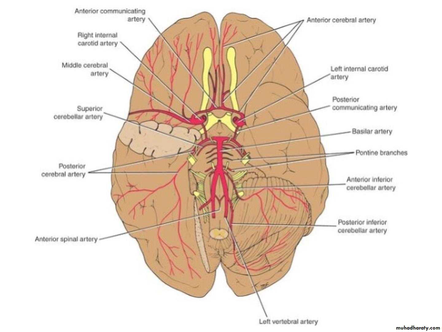
• Circle of Willis
• •• •
• The circle of Willis lies in the interpeduncular
• fossa at the base of the brain. It is formed
• by the anastomosis between the two internal
• carotid arteries and the two vertebral arter-
• ies.
• The circle is formed by :
• 1. anterior cerebral arteries,
• 2. The anterior communicating arteries,
• 3. internal carotid arteries,
• 4. posterior communicating arteries,
• 5. posterior cerebral arteries
• 6. basilar artery
• •
• •• The circle of Willis allows blood that enters by
• either internal carotid or vertebral arteries to be
• distributed to any part of both cerebral
• hemispheres. Cortical and central branches
• arise from the circle and supply the brain
• substance.
• Variations in the sizes of the arteries forming
• the circle are common, and the absence of one
• or both posterior communicating arteries has
• been reported.