
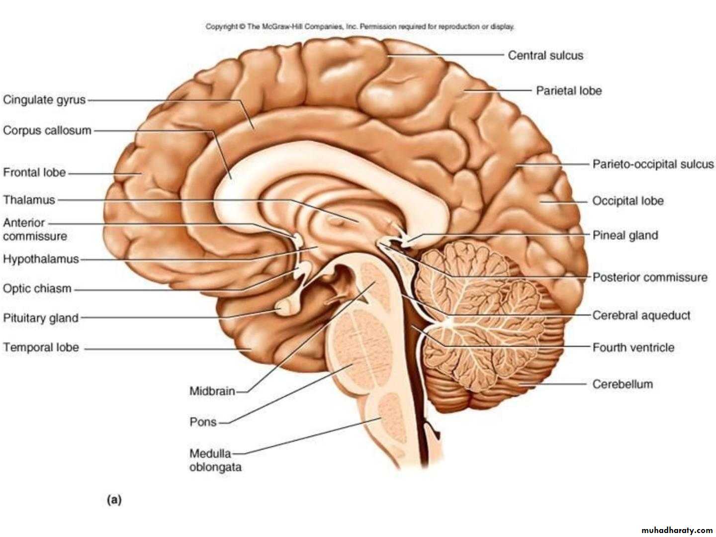
• Diencephalon
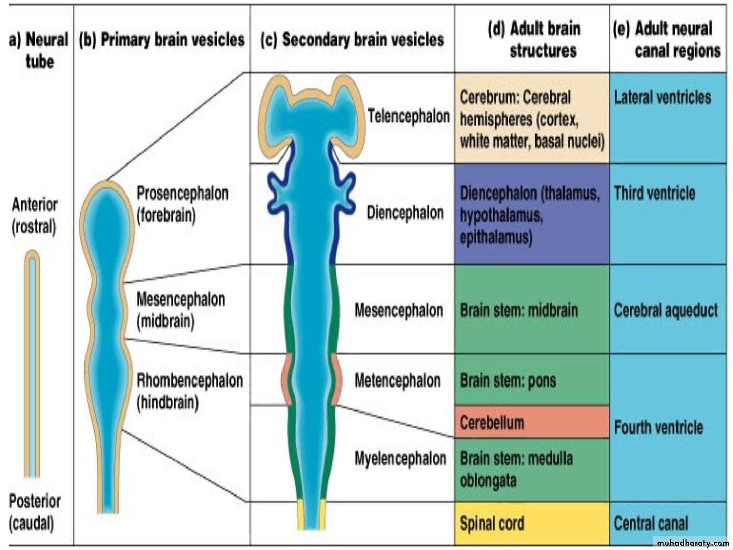
• Diencephalon
• Site:- It is the part of the forebrain which lies above the• midbrain, between the lower parts of the 2 cerebral
• hemispheres.
• • It consists of:
• 1. Thalamus:-the large oval mass of grey matter
• 2. Subthalamus:- it lies directly above midbrain
• 3. Hypothalamus: lies infront of subthalamus
• 4. Metathalamus: formed by lateral & medial geniculate
• body
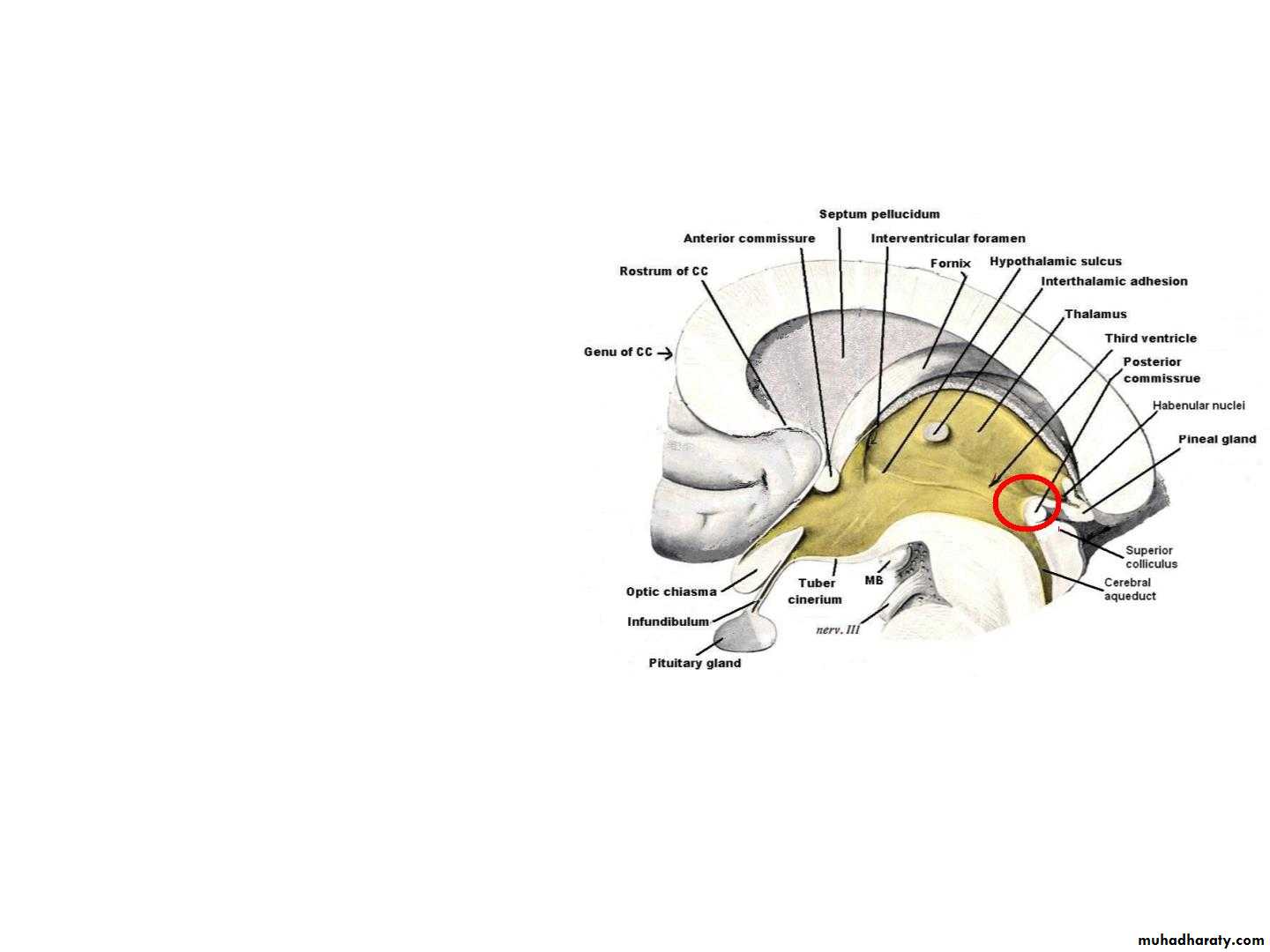
• 5. Epithalamus: Formed of pineal body, 2 habenular
• nuclei & posterior commissure.
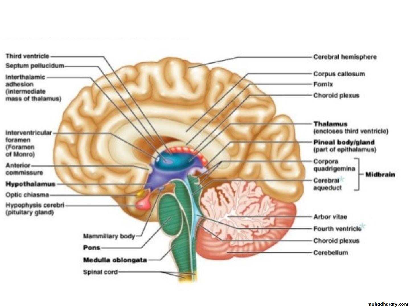
• The third ventricle lies between the 2 halves of the
• diencephalon.
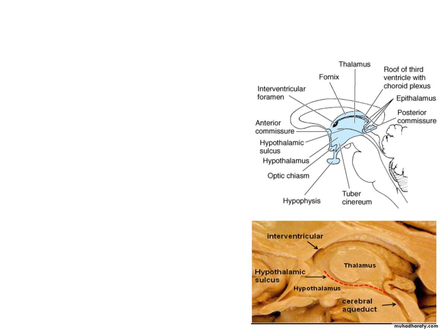
• Dorsal part
• Thalamus & Epithalamus• • On the medial
• surface, the
• diencephalon is
• subdivided, by
• hypothalamic sulcus
• (indicated by black
• line) into:
• CC
• Dorsal
• Ventral
• . Dorsal part:
• . Ventral part:
• Midbrain
• Cerebral
• aqueduct
• Ventral part
• Subthalamus & Hypothalamus
• Anatomy of the thalamus
• • Definition: The thalamus is a large,• paired, ovoid mass of nuclei located
• in the diencephalon, and form the
• upper 2/3 of the lateral wall of the
• third ventricle.
• • Relations :
• • Rostrally: the interventricular
• foramen.
• • Ventrally: the hypothalamic sulcus.
• • Posteriorly: the posterior
• commissure
• • Medially: the third ventricle
• • Laterally: the posterior limb of the
• internal capsule
• Relations
• Dorsal: lateral ventricle• Rostrally
• interventricular
• foramen
• Medial: 3rd
• ventricle
• Lateral :
• Internal
• capsule
• Ventral: Subthalamus & Hypothalamus
• Caudal: midbrain
• • Thalamus has :
• - 2 ends Anterior and posterior• - 4 surfaces Medial, lateral,
• superior (dorsal) and inferior
• (ventral)
• • Anterior end forms a forward
• projection (anterior
• tubercle). It forms the
• posterior boundaries of the
• interventricular foramen
• • Posterior end (pulvinar of
• the thalamus)
• Lies just above the superior
• colliculus and medial and
• lateral geniculate bodies
• Surfaces of the thalamus
• 4 Surfaces:• •
• •
• •
• •
• Superior
• Inferior
• Medial
• Lateral
• S
• L M
• l
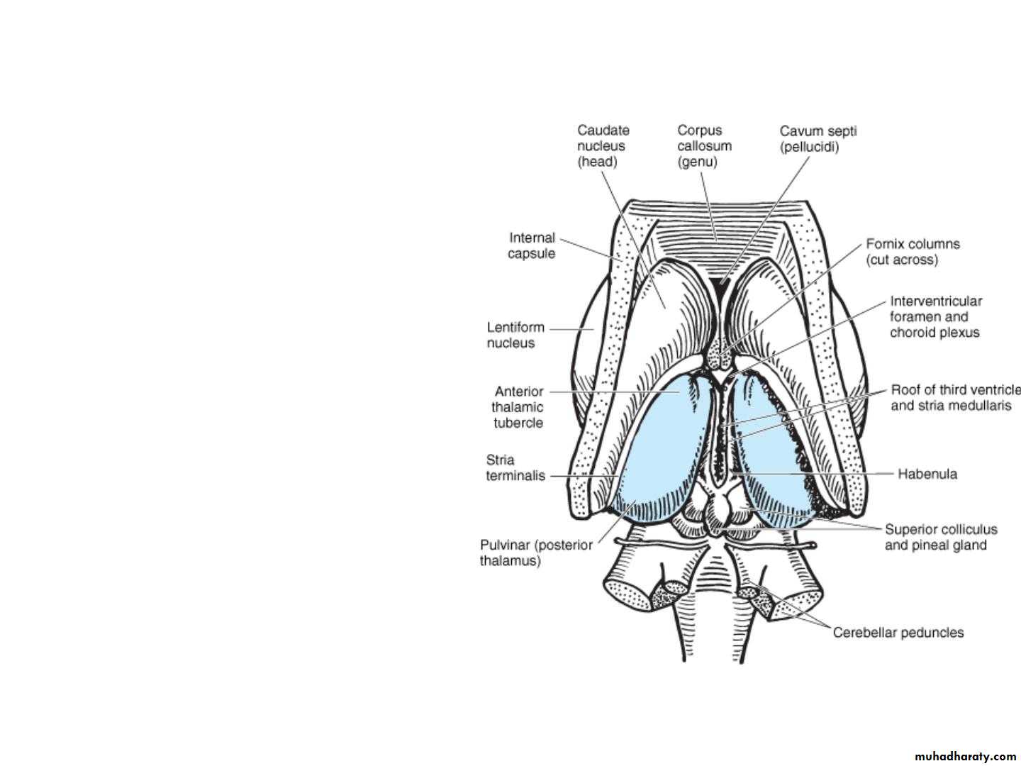
• Superior Surface
• caudate nucleus• stria terminalis
• • It is covered by thin layer
• of white matter called
• stratum zonale
• • Bounded laterally by
• LV
• caudate
• nucleus,
• thalamostriate vein and a
• nerve fiber bundle called
• stria terminalis
• thalamo-
• striate vein
• • Lateral part lies in the
• floor of the lateral
• ventricle and covered by
• ependyma
• ependyma
• choroid plexus
• Medial Surface
• • forms the upper 2/3 of the• lateral wall of the third
• ventricle.
• • It connects the medial
• surface of the other
• thalamus on the opposite
• side by a band of gray
• matter, the interthalamic
• connection (interthalamic
• adhesion).
• • It is covered by ependyma
• •
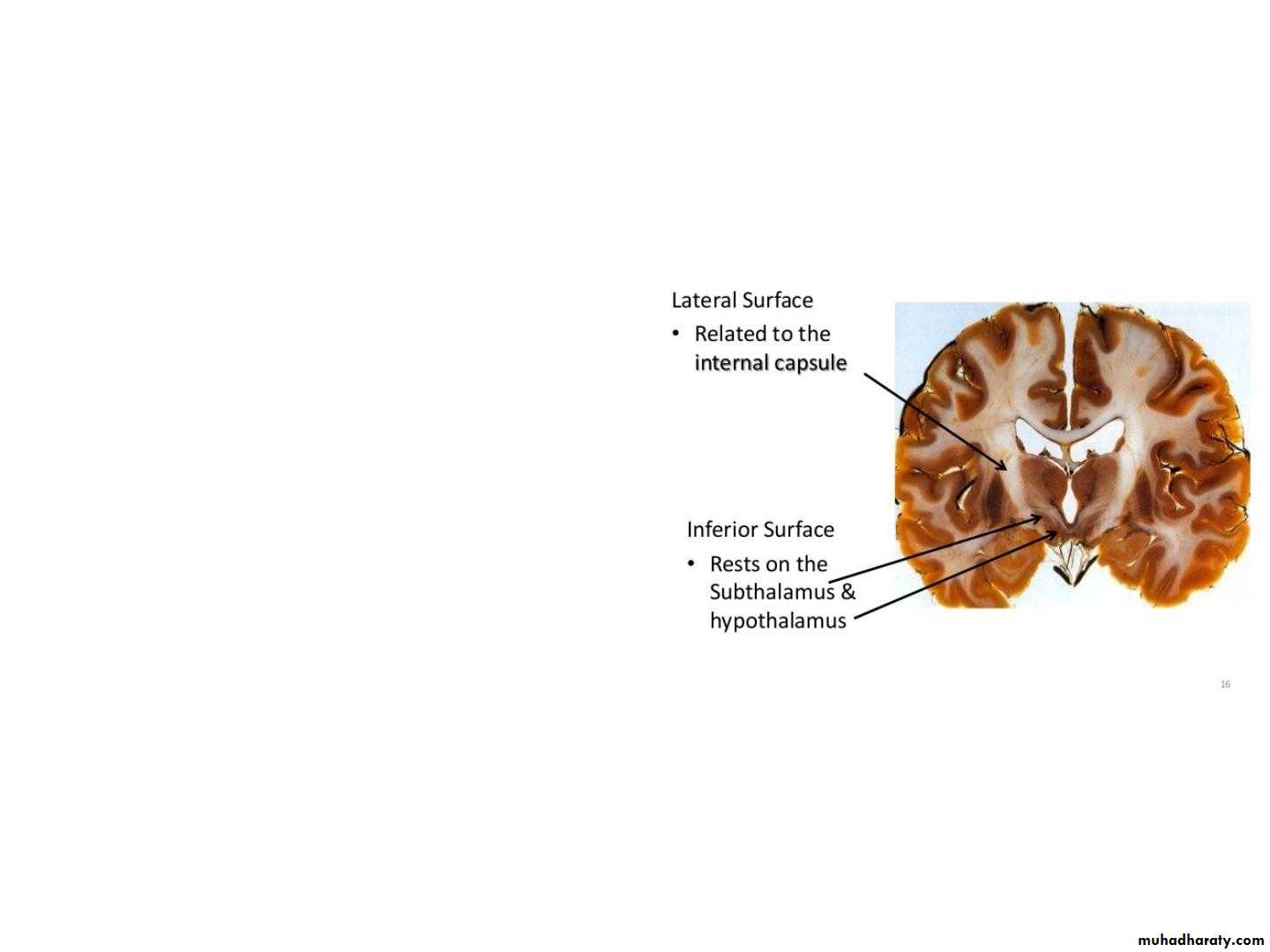
• Lateral surface
• • It is covered by a layer of white
• matter called external medullary
• lamina (a narrow band of
• myelinated fibres).
• • It related to the posterior limb of
• the internal capsule
• Inferior surface (ventral)
• The inferior surface is continuous
• with the tegmentum of the
• midbrain.
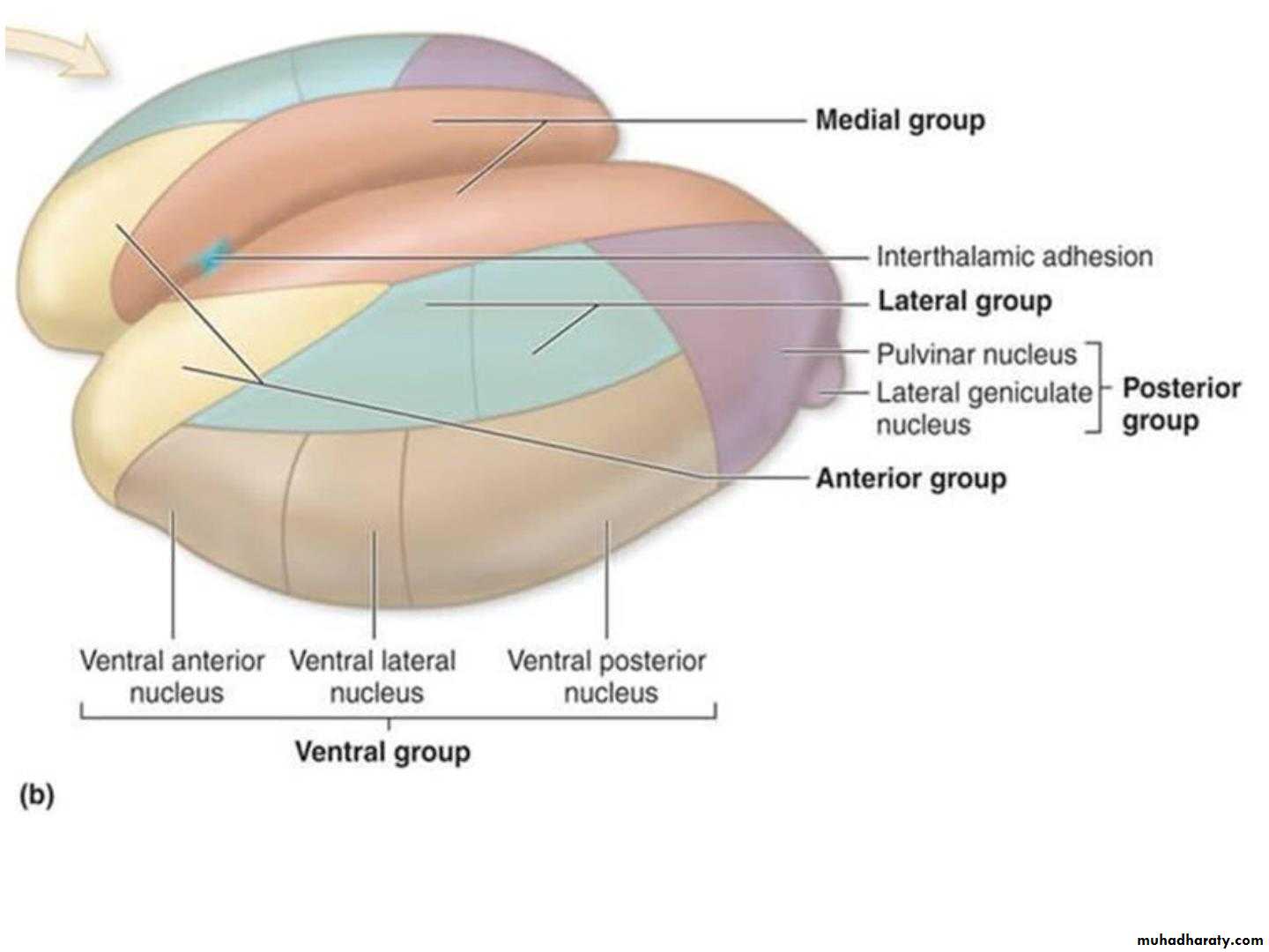
• Internal Organization
• • Thalamus is composed of grey matter,• interrupted by 2 vertical sheaths of
• white matter called medullary laminae.
• • External medullary lamina:
• .
• Located laterally, separates reticular
• nucleus from the rest of the thalamic
• mass . It contains thalamocortical &
• corticothalamic fibers
• • Internal medullary lamina
• .
• Y shaped complex of nuclei and fibers,
• separates the thalamus into anterior
• group between the 2 limbs of Y shaped
• lamina and two tiers of nuclei medial
• and lateral group on each side of the
• stem of Y shaped lamina.
• Internal structure of the thalamus
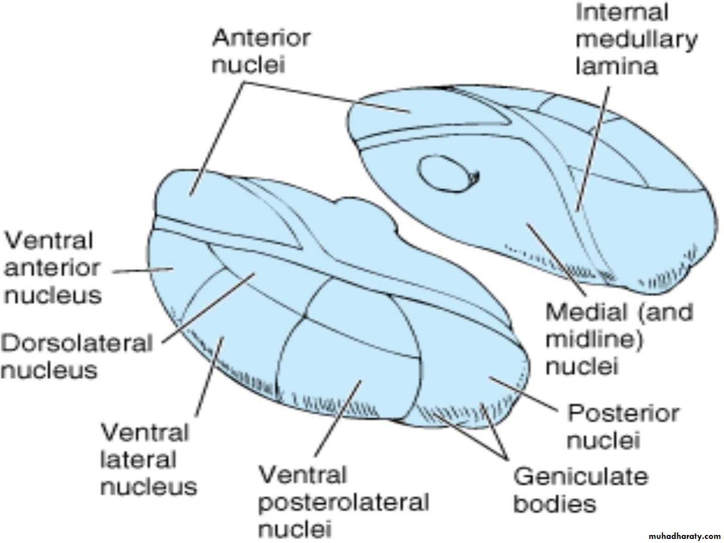
• • The anterior part contains anterior nuclear group is in between• the bifurcated fibers of the internal medullary lamina
• • The medial part of the thalamus consists of; the dorsomedial
• nucleus (DM) (or mediodorsal nucleus).
• • The lateral part of the thalamus divided into 2 parts ventral and
• dorsal parts.
• • ventral group includes;
• .
• .
• .
• ventral anterior (VA)
• ventral lateral (VL) and
• ventral posterior nuclei (VP) that includes ventral posterolateral
• and ventral posteromedial nuclei.
• • Dorsal group includes
• .
• .
• .
• lateral dorsal nucleus (LD)
• lateral posterior nucleus (LP)
• the pulvinar (P).
• Anterior Part of the thalamus
• •• •
• The anterior part of the thalamus contains the
• anterior thalamic nuclei.
• Afferent: 1. from the mammillary nuclei through the
• mammillothalamic tract.
• 2. from the hypothalamus and cingulate gyrus
• •
• •
• Efferent: to the cingulate gyrus and hypothalamus.
• Function: of the anterior thalamic nuclei is closely
• associated with that of the limbic system and is
• concerned with emotional tone and the mechanisms
• of recent memory.
• Medial Part of the thalamus
• •• •
• The medial part of the thalamus contains the large
• dorsomedial nucleus and several smaller nuclei.
• Afferent: from the olfactory cortex, amygdaloid
• nucleus and hypothalamic nuclei
• •
• •
• Efferent : to prefrontal cortex
• Function: The medial part of the thalamus is
• responsible for the integration of a large variety of
• sensory information including somatic, visceral,
• and olfactory information, and the relation of this
• information to one's emotional feelings and
• subjective states.

• Lateral part of the thalamus
• .• The lateral group divided into 2 parts ventral and
• dorsal parts.
• .
• .
• .
• .
• Ventral group includes;
• ventral anterior (VA)
• ventral lateral (VL) and
• ventral posterior nuclei (VP) that includes ventral
• posterolateral and ventral posteromedial nuclei.
• .
• .
• .
• .
• Dorsal group includes
• lateral dorsal nucleus (LD)
• lateral posterior nucleus (LP)
• the pulvinar (P).
• Dorsal group of the Nuclei
• •• Dorsal Tier (group) of the Nuclei includes:
• the lateral dorsal nucleus,
• .
• .
• .
• •
• the lateral posterior nucleus, and
• the pulvinar.
• They receive inputs from the other thalamic
• nuclei and integrate these inputs
• •
• They project the integrated information into
• sensory association areas in the cerebral cortex in
• the parietal, temporal and occipital lobes.
• Ventral Tier (group) of the Nuclei
• • It consists of the following nuclei in a craniocaudal sequence:• • Ventral anterior nucleus.
• • Afferent: from the reticular formation, the substantia nigra, the corpus striatum
• and other thalamic nuclei
• • Efferent to the motor areas and the premotor cortex.
• • Function: it probably influences the activities of the motor cortex.
• • Ventral lateral nucleus.
• • Afferent: similar to those of the ventral anterior nucleus but, in addition, has a
• major input from the cerebellum and a minor input from the red nucleus.
• • Efferent: to the motor and premotor regions of the cerebral cortex.
• • Function: it probably influences motor activity.
• • Ventral posterior nucleus.
• • This nucleus is subdivided into the ventral posteromedial nucleus and the ventral
• posterolateral nucleus .
• • Afferent: The ventral posteromedial nucleus receives the ascending trigeminal
• and gustatory pathways, while the ventral posterolateral nucleus receives the
• important ascending sensory tracts, the medial and spinal lemnisci.
• • Efferent The thalamocortical projections from these important nuclei pass
• through the posterior limb of the internal capsule and corona radiata to the
• primary somatic sensory areas of the cerebral cortex in the postcentral gyrus
• (areas 3, 1, and 2).
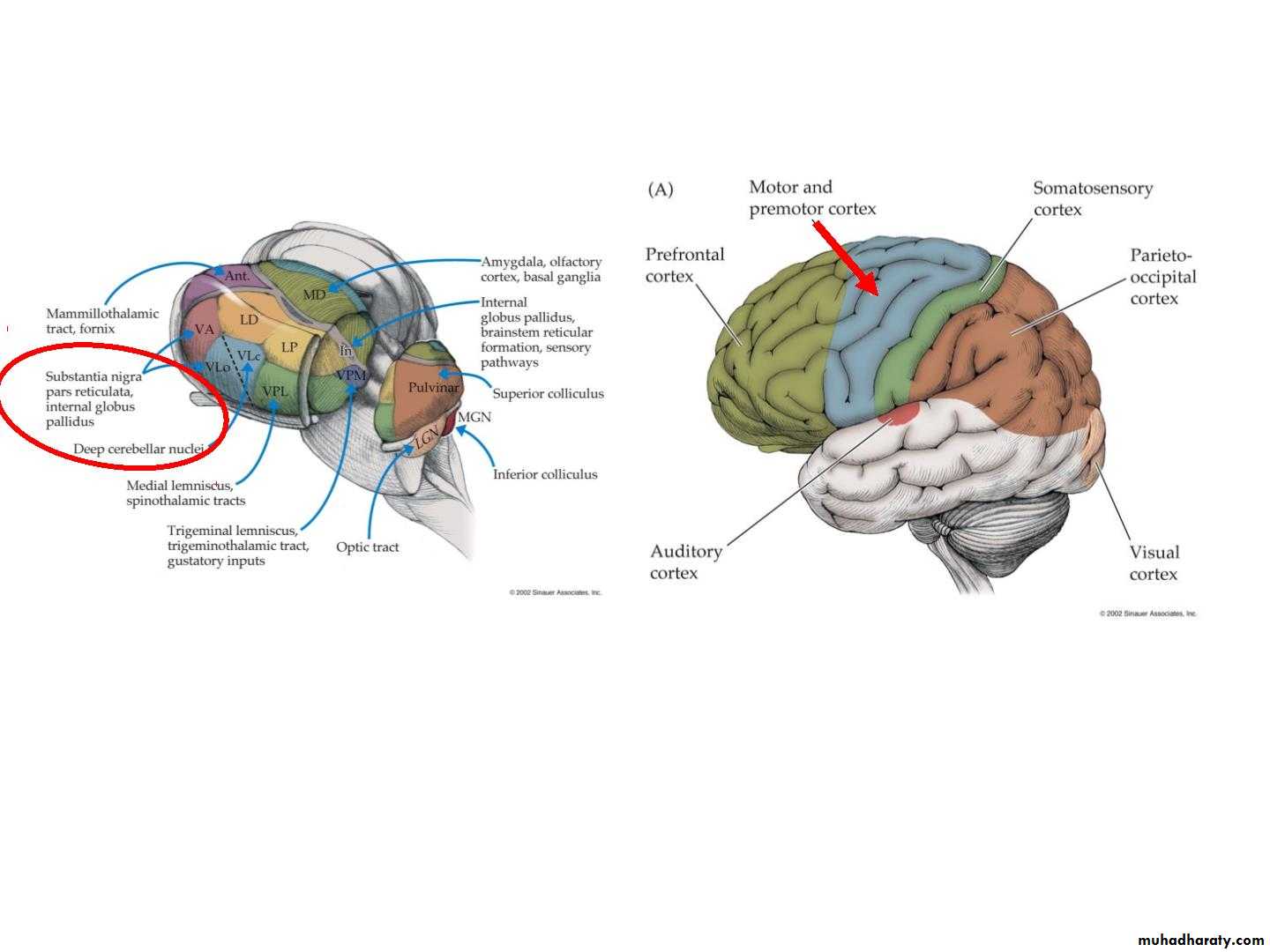
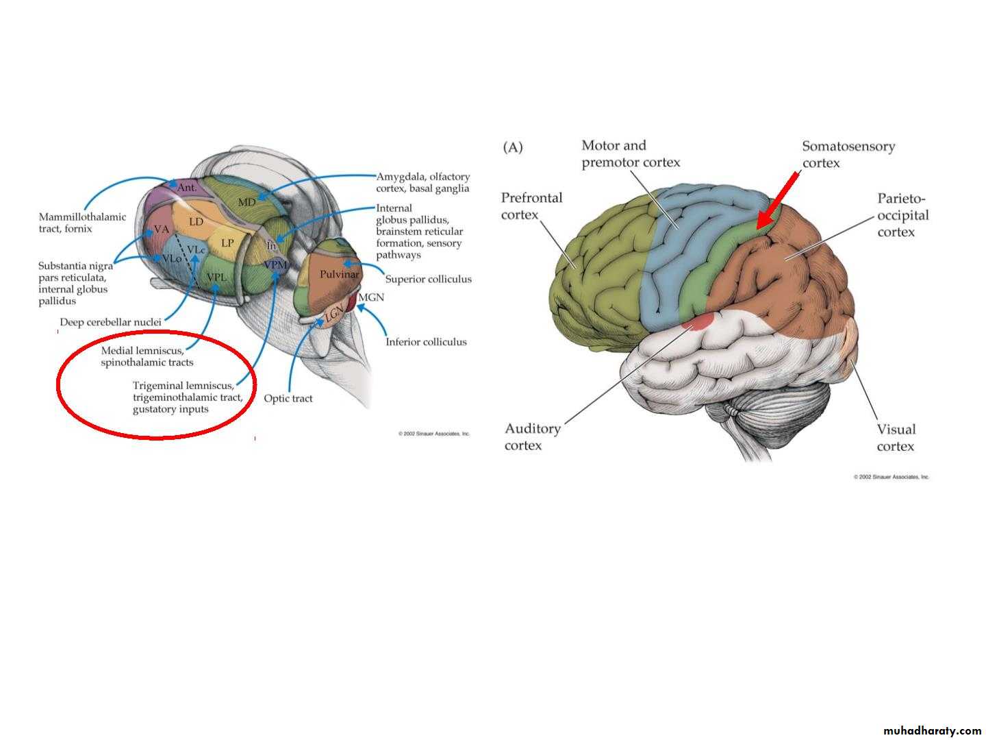
• Sensory relay
• Ventral posterior group• all sensation from body and
• head, including pain
• Other Nuclei of the Thalamus
• • The intralaminar nuclear group are small collections of nerve cells within the
• internal medullary lamina one of these nuclei, the centromedian nucleus .
• .
• .
• Afferent: from the reticular formation, the spinothalamic and trigeminothalamic
• tracts;
• Efferent: to other thalamic nuclei, which in turn project to the cerebral cortex, and
• fibers to the corpus striatum.
• • The midline group, also known as the periventricular nuclei, are on the medial
• surface of the thalamus and in the massa intermedia (absent in 30% of human
• brains),
• • Reticular nucleus of thalamus is a thin layer of nerve cells between the external
• medullary lamina and the posterior limb of the internal capsule. Afferent from the
• cerebral cortex and the reticular formation, and its efferent is mainly to other
• thalamic nuclei.
• • The medial geniculate body (MGB) forms part of the auditory pathway
• • lateral geniculate body (LGB) forms part of the visual pathway
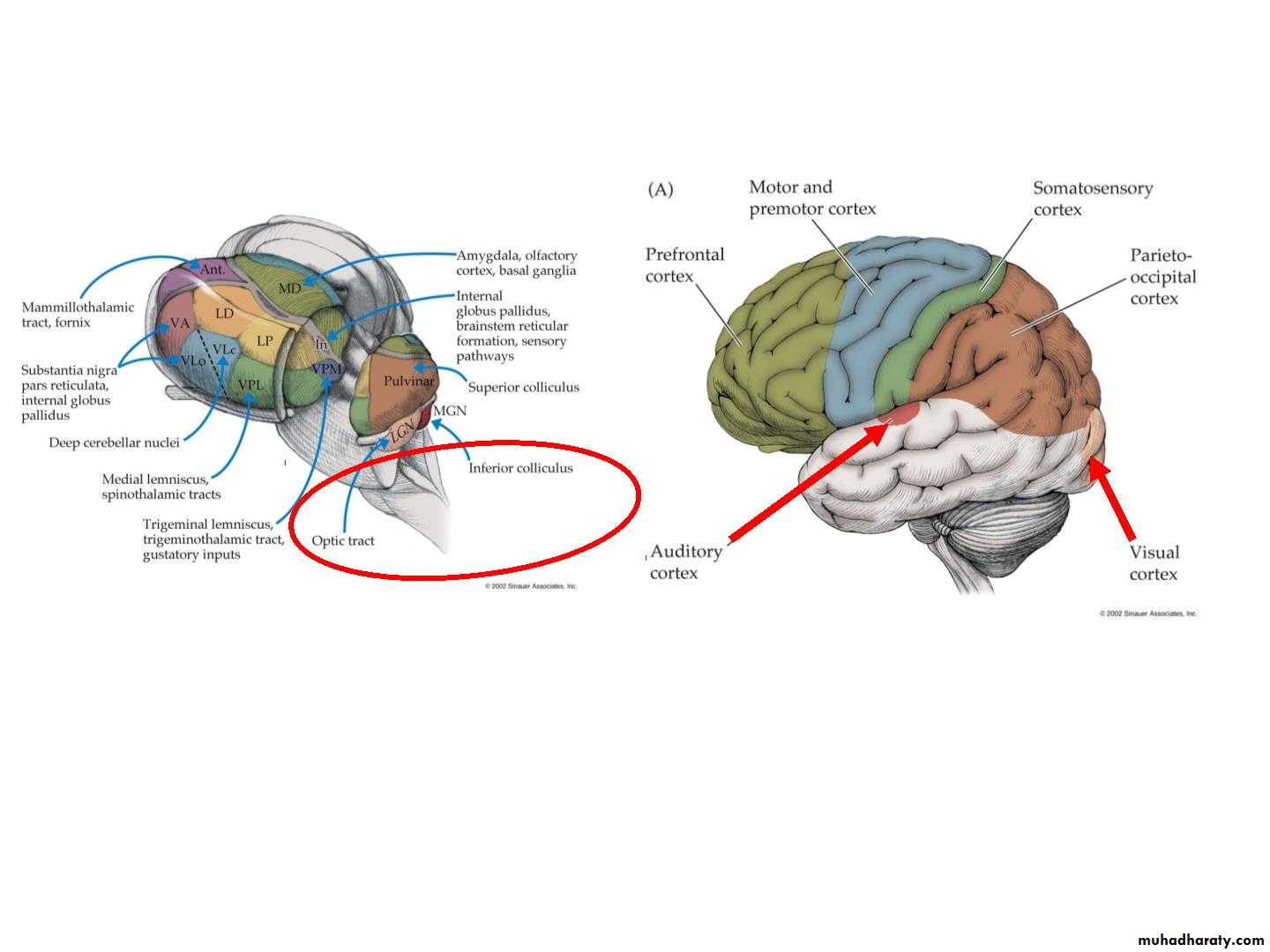
• Metathalamus
• Vision and Hearing• Blood supply of the thalamus
• The thalamus is supplied by 2 sets of arteries derived from• the posterior cerebral artery
• 1. Thalamo-perforating arteries: supply the anterior and
• medial parts of the thalasmus
• 2. Thalamo-geniculate arteries: supply the lateral and
• posterior parts of the thalamus
• Venous drainage of the thalamus by the thalamic veins
• which join the thalamostriate vein
• The thalamostriate vein with the choroidal vein form the
• internal cerebral vein
• Function of the thalamus
• 1. Thalamic nuclei process, integrate, and relay information for the• sensory, motor, limbic, and motivational systems.
• 2. Play a critical role in sensation and motor control.
• • The ventroanterior and the ventrolateral nuclei of the thalamus
• form part of the basal nuclei circuit and thus are involved in the
• performance of voluntary movements.
• • The large dorsomedial nucleus has extensive connections with the
• frontal lobe cortex and hypothalamus. There is considerable
• evidence that this nucleus lies on the pathway that is concerned
• with subjective feeling states and the personality of the individual.
• • The intralaminar nuclei are closely connected with the activities of
• the reticular formation and are able to influence the levels of
• consciousness and alertness in an individual.
• • 3. Thalamic nuclei also appear important for transferring
• information from one part of the cerebral cortex to another.
• Thalamic Lesions
• Cerebrovascular lesions or tumors of thalamus lead to:• • Loss of sensation in the contralateral side of face and body
• followed by distressing discomfort and burning and diffuse
• pain in the anaesthetic areas (thalamic pain)
• •Thalamic syndrome: Abnormal voluntary movements
• (chorea or hemiballismus) with hemisensory disturbance
• •Thalamic Hand
• •The contralateral hand is held in an abnormal posture in
• some patients with thalamic lesions. The wrist is pronated and
• flexed, the metacarpophalangeal joints are flexed, and the
• interphalangeal joints are extended. The fingers can be moved
• actively, but the movements are slow. The condition is due to
• altered muscle tone in the different muscle groups.
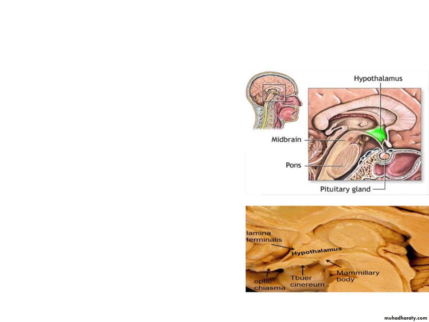
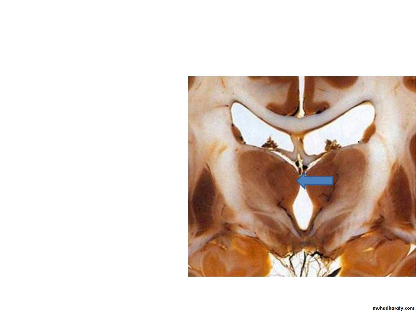
• Hypothalamus
• • The hypothalamus is the part of the• diencephalon forming the floor and
• the lower part of the lateral wall of
• the third ventricle. It extends from
• the region of the optic chiasma to
• the caudal border of the
• mammillary bodies.
• • Relations
• • Above: the thalamus.
• • Below: the hypothalamus merges
• into the tegmentum of the
• midbrain.
• • Laterally: the internal capsule
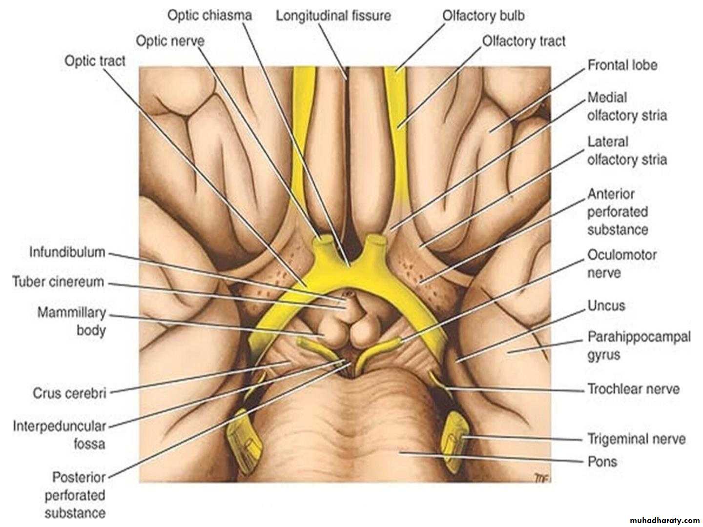
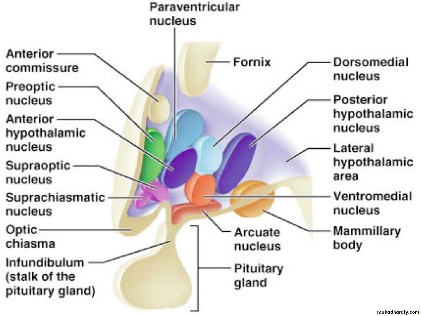
• Structures forming the hypothalamus
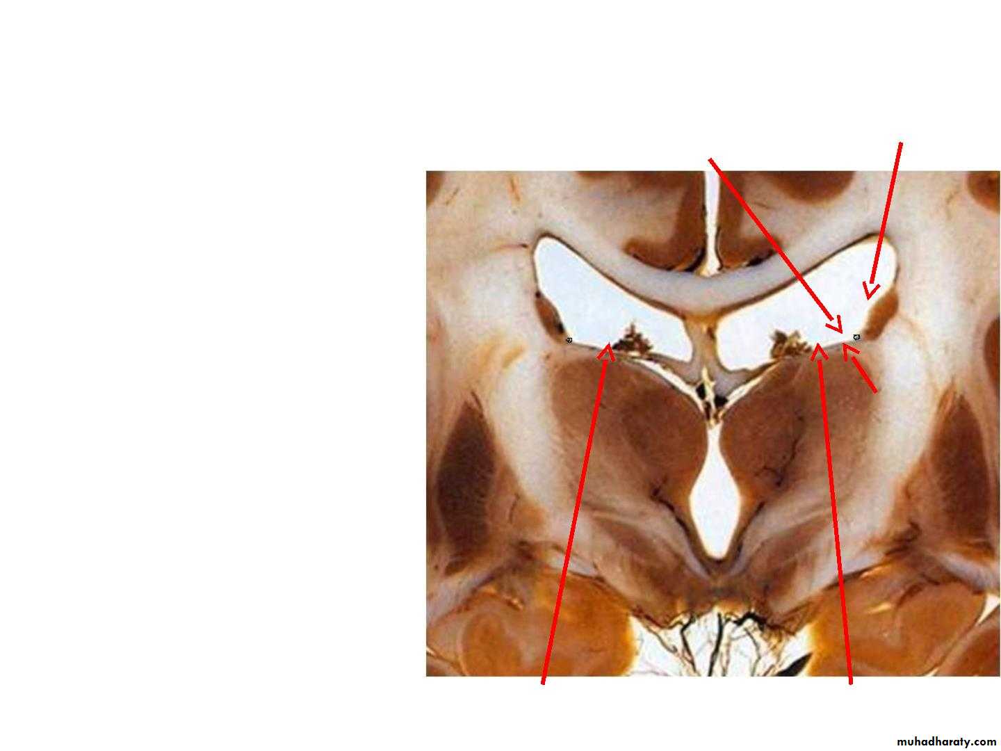
• The structures forming the• hypothalamus lie in the
• interpeduncular fossa, these
• structures are: optic chiasma,
• 1
• tuber cinerum and infundibulum,
• and mammillary bodies.
• 2
• 3
• Anterior to the hypothalamus is
• 4
• an area that, for functional
• reasons, is often included in the
• hypothalamus, it is referred to as
• the preoptic area.
• 4
• 4
• 1. optic chiasma
• 2. infundibulum
• 3. tuber cinereum
• 4. mamillary bodies
• Hypothalamic Nuclei
• The hypothalamic nuclei are divided by an imaginary parasagittal plane into• medial and lateral zones. Lying within the plane are the columns of the fornix
• and the mammillothalamic tract, which serve as markers.
• Medial Zone
• Lateral Zone
• •It includes the following nuclei
• arranged from anterior to posterior:
• •It includes the following nuclei
• arranged from anterior to
• posterior:
• (1) part of the preoptic nucleus;
• (2) the anterior nucleus,
• (1) part of the preoptic nucleus,
• (3) part of the suprachiasmatic
• nucleus;
• (2) part of the suprachiasmatic
• nucleus,
• (4) the paraventricular nucleus;
• (5) the dorsomedial nucleus;
• (6) the ventromedial nucleus;
• (7) the infundibular (arcuate) nucleus
• (8) the posterior nucleus.
• (3) the supraoptic nucleus,
• (4) the lateral nucleus,
• (5) the tuberomammillary nucleus,
• (6) the lateral tuberal nuclei.
• Hypothalamic Lines of Communication
• •• The hypothalamus receives information from the
• rest of the body through:
• •
• •
• •
• •
• (1) nervous connections,
• (2) the bloodstream, and
• (3) cerebrospinal fluid.
• The neurons of the hypothalamic nuclei respond
• and exert their control via the same routes.
• •
• The cerebrospinal fluid may serve as a conduit
• between the neurosecretory cells of the
• hypothalamus and distant sites of the brain.
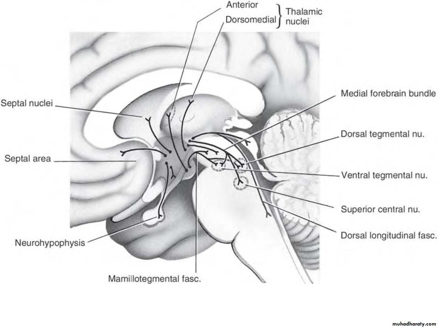
• Afferent Nervous Connections of the Hypothalamus
• • The main afferent pathways are :• • 1. Somatic and visceral afferents. General somatic sensation and gustatory
• and visceral sensations reach the hypothalamus through collateral
• branches of the lemniscal afferent fibers and the tractus solitarius and
• through the reticular formation.
• • 2. Visual afferents leave the optic chiasma and pass to the
• suprachiasmatic nucleus.
• • 3. Olfaction travels through the medial forebrain bundle.
• • 4. Corticohypothalamic fibers arise from the frontal lobe of the cerebral
• cortex and pass directly to the hypothalamus.
• • 5. Hippocampohypothalamic fibers pass from the hippocampus through
• the fornix to the mammillary body.
• • 6. Amygdalohypothalamic fibers pass from the amygdaloid complex to the
• hypothalamus through the stria terminalis
• • 7. Thalamohypothalamic fibers arise from the dorsomedial and midline
• thalamic nuclei.
• • 8. Tegmental fibers arise from the midbrain
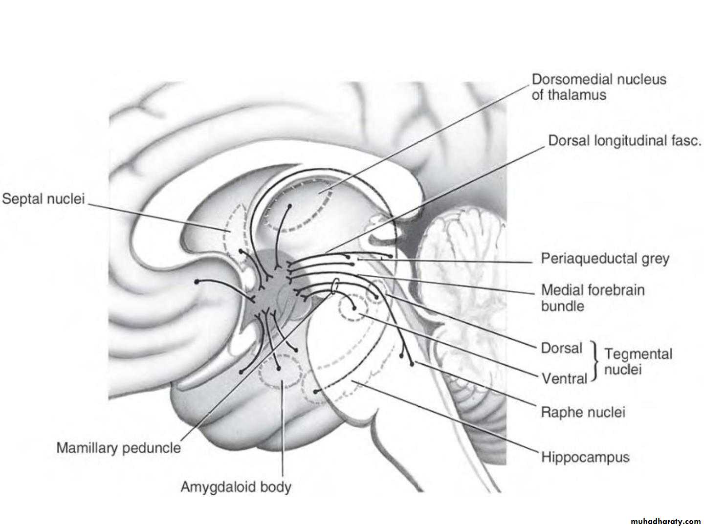
• Connections of the hypothalamus
• The major tracts conveying input to the hypothalamus.• Efferent Nervous Connections of the
• Hypothalamus• • the main efferent pathways are:
• • 1. Descending fibers to the brainstem and spinal cord influence the
• peripheral neurons of the autonomic nervous system. They descend
• through a series of neurons in the reticular formation.
• • The hypothalamus is connected to the parasympathetic nuclei of the
• oculomotor, facial, glossopharyngeal, and vagus nerves in the brainstem. In
• a similar manner, the reticulospinal fibers connect the hypothalamus with
• sympathetic cells of origin in the lateral gray horns of the first thoracic
• segment to the second lumbar segment of the spinal cord and the sacral
• parasympathetic outflow at the level of the second, third, and fourth sacral
• segments of the spinal cord.
• • 2. The mammillothalamic tract arises in the mammillary body and
• terminates in the anterior nucleus of the thalamus. Here, the pathway is
• relayed to the cingulate gyrus.
• • 3. The mammillotegmental tract arises from the mammillary body and
• terminates in the cells of the reticular formation in the tegmentum of the
• midbrain.
• • 4. Multiple pathways to the limbic system.
• The major tracts conveying output from the hypothalamus.
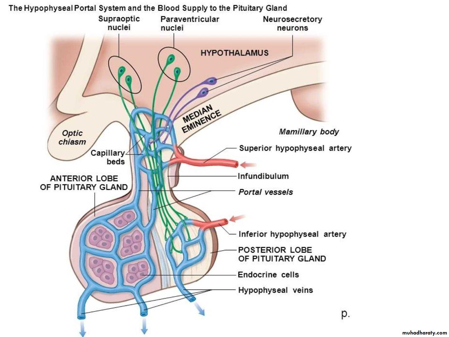
• Connections of the Hypothalamus With
• the Hypophysis Cerebri (pituitary gland(• •
• •
• The hypothalamus is connected to the hypophysis
• cerebri (pituitary gland) by two pathways:
• (1) nerve fibers that travel from the supraoptic
• and paraventricular nuclei to the posterior lobe of
• the hypophysis
• •
• •
• (2) long and short portal blood vessels that
• connect sinusoids in the median eminence and
• infundibulum with capillary plexuses in the
• anterior lobe of the hypophysis.
• These pathways enable the hypothalamus to
• influence the activities of the endocrine glands.
• Hypothalamohypophyseal Tract
• • From the supraoptic and paraventricular nuclei of the hypothalamus to• the posterior lobe of the pituitary gland
• • The hormones vasopressin and oxytocin are synthesized in the nerve cells
• of the supraoptic and paraventricular nuclei. The hormones are passed
• along the axons together with carrier proteins called neurophysins and
• are released at the axon terminals. Here, the hormones are absorbed into
• the bloodstream in fenestrated capillaries of the posterior lobe of the
• hypophysis.
• • The hormone vasopressin (antidiuretic hormone) is produced mainly in
• the nerve cells of the supraoptic nucleus. Its function is to cause
• vasoconstriction. It also has an important antidiuretic function.
• • The oxytocin hormone is produced mainly in the paraventricular nucleus.
• It stimulates the contraction of the smooth muscle of the uterus and
• causes contraction of the myoepithelial cells that surround the alveoli and
• ducts of the breast and assists in the expression of the milk from the
• breasts.
• Hypophyseal Portal System
• • The hypophyseal portal system is formed on each side from the superior• hypophyseal artery, which is a branch of the internal carotid artery.
• • The artery enters the median eminence and divides into tufts of
• capillaries. These capillaries drain into long and short descending vessels
• that end in the anterior lobe of the hypophysis by dividing into vascular
• sinusoids that pass between the secretory cells of the anterior lobe.
• • Neurosecretory cells situated mainly in the medial zone of the
• hypothalamus are responsible for the production of the releasing
• hormones and release-inhibitory hormones. The hormones are packaged
• into granules and are transported along the axons of these cells into the
• median eminence and infundibulum. Here, the granules are released by
• exocytosis onto fenestrated capillaries at the upper end of the
• hypophyseal portal system
• • The portal system carries the releasing hormones and the release-
• inhibiting hormones to the secretory cells of the anterior lobe of the
• hypophysis.
• • Releasing Hormones
• • 1. Gonadotropin-releasing hormone (GnRH) that regulates the release of follicle• stimulating hormone (FSH, follitropin) and luteinizing hormone (LH, lutropin) from the
• hypophysis.
• • 2. Thyrotropin-releasing hormone (TRH) that regulates the release of thyrotropin
• (thyroid-stimulating hormone, TSH) and prolactin from the hypophysis.
• • 3. Corticotropin-releasing hormone (CRH) that regulates the release of
• adrenocorticotropin (adrenocorticotropic hormone, ACTH)
• • 4. Growth hormone-releasing hormone (GRH or GHRH) that regulates the release
• of growth hormone (somatotropin) from the hypophysis.
• • 5. Prolactin-releasing factor (PRF) regulates the release of prolactin (lactogenic
• hormone, mammotropic hormone) from the hypophysis.
• • 6. Melanocyte-stimulating hormone-releasing factor (MRF) stimulates the release of
• melanocyte stimulating hormone which stimulates the formation of melanin pigment
• and its dispersion in melanocytes.
• • Inhibiting Hormones
• • 1. Growth hormone release-inhibiting hormone (GIH, GHRIH) also called somatostatin
• (SS) or somatotropin release-inhibiting hormone (SRIH) acts to inhibit the release of
• growth hormone and thyrotropin from the hypophysis.
• • 2. Prolactin release-inhibiting hormone (PIH) or dopamine (DA) acts to inhibit the
• release of prolactin from the hypophysis.
• • 3. Melanocyte-stimulating hormone release inhibiting factor (MIF) acts to inhibit the
• release of melanocyte-stimulating hormone
• Functions of the Hypothalamus
• • 1. The hypothalamus has a controlling influence on the autonomic• nervous system and appears to integrate the autonomic and
• neuroendocrine systems, thus preserving body homeostasis. The anterior
• hypothalamic area and the preoptic area influence parasympathetic
• responses; posterior and lateral nuclei influnce sympathetic responses,
• • 2. Endocrine Control: The nerve cells of the hypothalamic nuclei, by
• producing the releasing factors or release-inhibiting factors control the
• hormone production of the anterior lobe of the hypophysis (pituitary
• gland).
• • 3. Neurosecretion the supraoptic and paraventricular nuclei secrete the
• vasopressin and oxytocin hormones
• • 4. Temperature Regulation
• • The anterior portion of the hypothalamus controls the mechanisms that
• lower the body temperature. Stimulation of the posterior portion of the
• hypothalamus results in production of heat.
• Function of the hypothalamus
• • 5. Regulation of Food and Water Intake• • Stimulation of the lateral region of the hypothalamus initiates the feeling of
• hunger and results in an increase in food intake. Stimulation of the medial region
• of the hypothalamus inhibits eating and reduces food intake.
• • Experimental stimulation of other areas in the lateral region of the hypothalamus
• causes an immediate increase in the desire to drink water.
• • 6. Emotion and Behavior
• • Emotion and behavior are a function of the hypothalamus, the limbic system, and
• the prefrontal cortex. Some authorities believe that the hypothalamus is the
• integrator of afferent information received from other areas of the nervous
• system and brings about the physical expression of emotion;
• • 7. Control of Circadian Rhythms
• • The hypothalamus controls many circadian rhythms, including body temperature,
• adrenocortical activity, eosinophil count, and renal secretion. Sleeping and
• wakefulness, although dependent on the activities of the thalamus, the limbic
• system, and the reticular activating system, are also controlled by the
• hypothalamus.
• Clinical Disorders Associated With
• Hypothalamic Lesions• • Causes : inflammation, neoplasm, or vascular disorder. Because of its deep-seated central
• position,
• • Obesity Severe obesity can occur as the result of hypothalamic lesions. It is generally
• associated with genital hypoplasia or atrophy.
• • Sexual Disorders
• • In children, there may be sexual retardation and. After puberty, the patient with
• hypothalamic disease may have impotence or amenorrhea.
• • Hyperthermia and Hypothermia
• • Diabetes Insipidus
• • Diabetes insipidus results from a lesion of the supraoptic nucleus or from the interruption
• of the nervous pathway to the posterior lobe of the hypophysis. Characteristically, the
• patient passes large volumes of urine of low specific gravity. As a result, the patient is
• extremely thirsty and drinks large quantities of fluids. The condition must be distinguished
• from diabetes mellitus, in which there is glucosuria.
• • Disturbances of Sleep
• • The occurrence of either frequent short periods of sleep during the waking hours or
• insomnia has been observed in patients with hypothalamic lesions.
• • Emotional Disorders
• • Attacks of unexplained weeping or laughter, uncontrollable rage, depressive reactions,
• and even maniacal outbursts all have been observed in patients with hypothalamic
• lesions.
• SUBTHALAMUS
• •• •
• It lies between the thalamus and tegmentum of
• the midbrain
• It contains 3 nuclei
• 1) upper end of red nucleus,
• 2) upper end of substantia nigra
• 3) subthalamic nuclei
• Epithalamus
• •• Relatively small part,
• located in most caudal
• and dorsal region
• • Lies immediately rostral
• to superior colliculus
• • Consists of:
• .
• .
• Pineal gland &
• Habenular nuclei